
The prospect of an ultrasound at seven weeks pregnant is often met with a whirlwind of emotions: excitement, anticipation, and perhaps a touch of apprehension. While the primary purpose of this early scan is to confirm the viability of the pregnancy and establish a due date, it also offers a fascinating glimpse into the nascent stages of human development. For expectant parents, this appointment is a significant milestone, transforming the abstract concept of pregnancy into a tangible reality. Beyond the emotional impact, understanding what to expect technically can help demystify the process and enhance the viewing experience. This article delves into the technological marvels behind a 7-week ultrasound, focusing on the sophisticated equipment and the intricate processes that allow us to witness this early stage of life. We will explore the fundamental principles of ultrasound technology, the specific adaptations for early pregnancy imaging, and the data processing that transforms sound waves into visual representations of the developing embryo.

The Technological Underpinnings of Early Gestation Imaging
At its core, an ultrasound, or sonography, is a non-invasive medical imaging technique that utilizes high-frequency sound waves to create images of internal body structures. The technology behind a 7-week pregnancy scan, while seemingly simple in its output, is a sophisticated interplay of physics, engineering, and advanced software. The fundamental principle is echo-location, a concept familiar from bats and dolphins, applied to medical diagnostics.
The Transducer: The Heart of the Ultrasound System
The key piece of equipment in any ultrasound examination is the transducer, often referred to as the probe. For early pregnancy scans, a transvaginal transducer is typically used. This is a slender, wand-like device that is inserted into the vagina. The reason for this approach is critical for visualizing structures within the pelvis at this early stage. The vaginal wall is thin, allowing the transducer to be placed very close to the uterus and the developing embryo, minimizing the distance sound waves need to travel and thus producing a clearer, higher-resolution image.
The transducer contains piezoelectric crystals, which are the workhorses of ultrasound technology. When an electric current is applied to these crystals, they vibrate at extremely high frequencies, generating sound waves beyond the range of human hearing (typically between 2 to 18 megahertz for medical applications). These sound waves are then directed into the pelvic region.
The Journey of Sound Waves: Reflection and Detection
Once emitted, these high-frequency sound waves travel through the body’s tissues. When they encounter structures of different densities and acoustic impedances (e.g., fluid, tissue, bone), they are reflected back towards the transducer. The piezoelectric crystals, in a reverse of their transmitting function, now vibrate when these returning echoes strike them. These vibrations are converted back into electrical signals.
The speed at which the sound waves travel through tissue is relatively constant. The ultrasound machine measures the time it takes for an echo to return to the transducer. By knowing the speed of sound in tissue, the machine can accurately calculate the distance from the transducer to the structure that reflected the sound wave. This fundamental principle of time-of-flight is the basis for all distance measurements in ultrasound imaging.
Signal Processing: From Echoes to Images
The electrical signals generated by the returning echoes are then processed by the ultrasound machine’s sophisticated computer system. This processing involves several crucial steps:
- Amplification: The returning echoes are often very weak. The machine amplifies these signals to make them detectable and measurable.
- Filtering: Various filters are applied to remove unwanted noise and artifacts that could obscure the image.
- Digitization: The analog electrical signals are converted into digital data that the computer can manipulate and display.
- Beamforming: The ultrasound machine can electronically steer the sound beam in different directions, allowing the sonographer to scan across the entire area of interest without physically moving the transducer excessively. This is achieved by precisely controlling the timing and frequency of the electrical pulses sent to the individual piezoelectric crystals.
- Image Reconstruction: The processed digital data is then used to reconstruct a two-dimensional image. Each pixel in the displayed image represents a specific point in the body, with its brightness corresponding to the strength of the returning echo. Stronger echoes, returning from denser structures, appear brighter, while weaker echoes from less dense structures appear darker.
Visualizing the Early Embryo: What to Expect on Screen
At seven weeks of gestation, the developing embryo is incredibly small, typically measuring around 10 to 15 millimeters (about half an inch) in crown-rump length. While the image may not be as detailed as later ultrasounds, the technology is capable of revealing remarkable insights. The primary focus of a 7-week scan is to confirm the presence of a gestational sac, the yolk sac, and the developing embryo itself.
The Gestational Sac and Yolk Sac: Early Indicators of Pregnancy
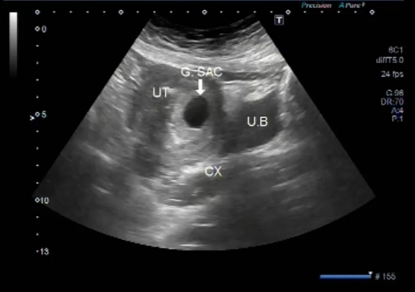
The gestational sac is the first structure to become visible on an ultrasound in early pregnancy, typically around 4.5 to 5 weeks. By seven weeks, it will be clearly defined. It appears as a round or oval, fluid-filled sac within the uterus. Inside the gestational sac, you will see the yolk sac. The yolk sac is a crucial structure that provides nourishment to the early embryo. It appears as a small, circular or oval structure within the gestational sac. Its presence is a strong indicator of a healthy, progressing pregnancy.
The Embryonic Pole: The First Glimpse of Life
The most exciting element of a 7-week ultrasound is the visualization of the embryonic pole. This is the earliest detectable form of the embryo. It will appear as a small, bright, linear or slightly curved structure attached to the wall of the yolk sac. While it may be very small, its presence is a definitive sign of an intrauterine pregnancy.
Detecting Cardiac Activity: The Heartbeat’s Confirmation
One of the most significant findings at a 7-week ultrasound is the detection of embryonic cardiac activity. The tiny heart begins to beat around week 5.5 to 6 of gestation. The ultrasound machine, through its sensitive detection of movement and blood flow, can pick up this rhythm. On screen, this may appear as a flickering within the embryonic pole. In some advanced machines, Doppler ultrasound can be used to visualize the blood flow within the fetal heart, providing a clearer confirmation of the heartbeat. The detection of a strong, consistent heartbeat is a major reassurance for expectant parents and healthcare providers.
Measuring Growth and Establishing a Due Date
Accurate measurements are a cornerstone of early pregnancy ultrasounds. The sonographer will measure the crown-rump length (CRL) of the embryo. This measurement is the most accurate way to date a pregnancy in the first trimester. By comparing the CRL to established growth charts, the gestational age can be determined with a high degree of accuracy, allowing for the establishment of an estimated due date. Other measurements may include the diameter of the gestational sac and the yolk sac.
Advanced Features and Considerations in Early Gestation Imaging
Modern ultrasound machines are not just basic imaging devices; they are complex diagnostic tools that incorporate advanced software and hardware to enhance image quality, provide additional diagnostic information, and improve the overall patient experience. Understanding these features can shed light on the sophistication involved in capturing those precious early glimpses of the developing baby.
Doppler Ultrasound: Visualizing Blood Flow
While not always standard at a 7-week scan, Doppler ultrasound technology can be employed. This technique uses the Doppler effect to assess blood flow within the embryonic heart, umbilical cord, and uterine arteries. By bouncing sound waves off moving red blood cells, Doppler ultrasound can detect the speed and direction of blood flow. This provides valuable information about the oxygenation and nourishment being supplied to the developing embryo and can help assess the health of the placenta’s connection to the uterus. The visual representation of Doppler data typically appears as colored hues (e.g., red for flow towards the transducer, blue for flow away) superimposed on the grayscale ultrasound image.
Image Optimization and Digital Enhancement
The raw data captured by the transducer is processed by sophisticated algorithms to optimize the ultrasound image. This includes:
- Gain Control: Adjusting the overall brightness of the image.
- TGC (Time Gain Compensation): A crucial feature where the machine selectively amplifies echoes based on their depth. Deeper structures return weaker echoes, so TGC compensates for this attenuation to ensure uniform brightness across the entire image.
- Focusing: The ultrasound beam can be electronically focused at specific depths to achieve the sharpest possible image resolution.
- Edge Enhancement and Smoothing: Software algorithms can be applied to sharpen the outlines of structures and reduce image graininess, making it easier to discern fine details.
- Image Archiving and Review: Modern ultrasound machines are equipped with robust digital storage capabilities, allowing images and video clips to be saved, reviewed, and shared. This is invaluable for tracking pregnancy progression and for consultation with other medical professionals.

The Role of the Sonographer and the Ultrasound Machine’s Intelligence
It’s important to remember that while the technology is advanced, the interpretation of the ultrasound images relies heavily on the skill and expertise of the sonographer. They are trained to operate the equipment, maneuver the transducer to obtain optimal views, and identify normal and abnormal findings. The ultrasound machine itself incorporates elements of “intelligence” through its software, which assists in measurements, provides anatomical labels, and can even flag potential anomalies for further review. However, the final diagnosis and clinical interpretation are always made by a qualified medical professional. The technology serves as a powerful tool in their hands, enabling them to peer into the earliest stages of life with remarkable clarity.
aViewFromTheCave is a participant in the Amazon Services LLC Associates Program, an affiliate advertising program designed to provide a means for sites to earn advertising fees by advertising and linking to Amazon.com. Amazon, the Amazon logo, AmazonSupply, and the AmazonSupply logo are trademarks of Amazon.com, Inc. or its affiliates. As an Amazon Associate we earn affiliate commissions from qualifying purchases.