Spongiotic dermatitis, frequently referred to by dermatologists as “eczematous dermatitis,” is a descriptive term rather than a single, standalone disease. In the realm of clinical dermatology, the term refers to a specific pattern of inflammation observed under a microscope when a skin biopsy is performed. When pathologists examine tissue samples showing this condition, they observe an intercellular edema—essentially, fluid accumulation between skin cells—that causes the cells to pull apart, creating a sponge-like appearance.
Understanding this condition requires navigating the intersection of clinical presentation and microscopic pathology. While the term is technical, it serves as the underlying histological foundation for a variety of common skin conditions that patients encounter in daily life. By deconstructing what occurs at a cellular level, individuals can better understand the root causes of their chronic skin irritations and the targeted strategies required to manage them effectively.
The Pathological Mechanism of Spongiosis
To grasp why skin behaves the way it does during an episode of spongiotic dermatitis, one must look at the structural integrity of the epidermis. The epidermis acts as a barrier, held together by proteins that maintain tight junctions between cells. Spongiotic dermatitis occurs when this barrier is compromised, either due to external triggers or internal immune responses.
Intercellular Edema: The “Sponge” Effect
The hallmark of this condition is the migration of fluid into the spaces between keratinocytes, which are the primary cells of the epidermis. As this fluid accumulates, the cells do not necessarily lose their connections, but they are pushed further apart. Under a microscope, the resulting stretched-out appearance—with the desmosomes (the protein bridges connecting the cells) looking like thin strands stretching across the gaps—resembles a kitchen sponge.
The Role of Inflammatory Mediators
This swelling is not a passive event; it is driven by an active inflammatory response. When the immune system detects a threat—whether it is a contact allergen, an irritant, or an autoimmune signal—it releases cytokines and other signaling molecules. These chemicals increase vascular permeability, allowing serum and inflammatory cells, such as lymphocytes or eosinophils, to infiltrate the epidermal layers. This influx of fluid is what causes the visible symptoms of clinical eczema: redness, swelling, and the formation of tiny, fluid-filled blisters known as vesicles.
Clinical Presentations and Common Triggers
While “spongiotic dermatitis” is a histological diagnosis, it correlates with several well-known clinical conditions. A dermatologist will often use the term as a bridge to explain why a patient is experiencing recurring rashes, regardless of whether the underlying trigger is allergic or environmental.
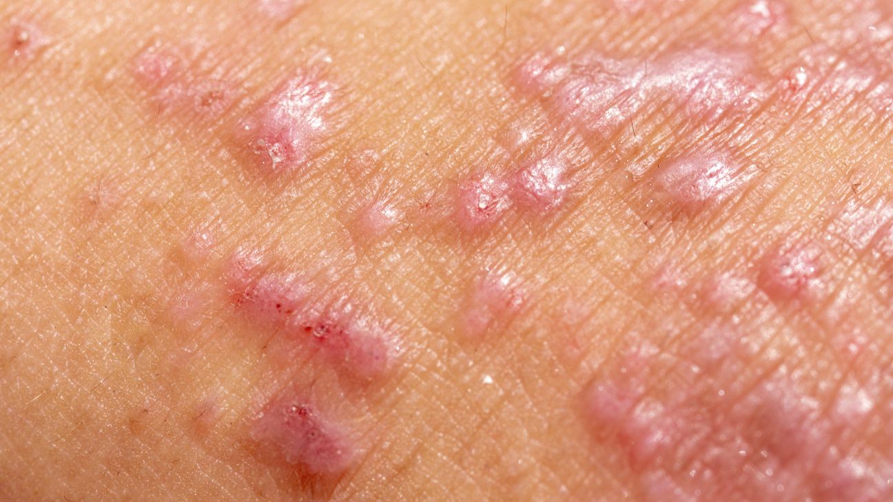
Allergic Contact Dermatitis
This is perhaps the most classic manifestation of the spongiotic pattern. It occurs when the immune system develops a hypersensitivity to a specific substance. When the skin comes into contact with an allergen—such as nickel, fragrances, or rubber additives—T-cells in the skin are activated, triggering the characteristic inflammatory cascade that leads to spongiosis.
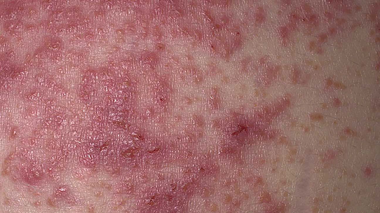
Atopic Dermatitis and Chronic Irritation
Atopic dermatitis, often referred to simply as eczema, is frequently characterized by a chronic spongiotic pattern. In these cases, the skin barrier is often genetically compromised, making it easier for environmental irritants to penetrate the skin and trigger the sponge-like swelling. Similarly, irritant contact dermatitis—caused by harsh soaps, cleaning agents, or constant friction—can lead to the same histological pattern, even if the immune pathway differs slightly from a true allergy.
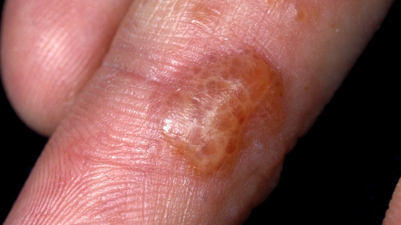
Nummular Eczema and Other Variants
Nummular eczema, which presents as coin-shaped patches of irritated skin, is another common clinical form of spongiotic dermatitis. These lesions often begin as small, intensely itchy vesicles that eventually coalesce into dry, scaly patches. The persistence of these patches is a testament to the chronic nature of the underlying spongiotic process, which can continue as long as the trigger remains present or the skin barrier function remains impaired.
Diagnostic Procedures and Clinical Evaluation
Because many skin conditions share similar visual appearances, the diagnostic process is critical for effective management. A dermatologist does not simply rely on the visual appearance of a rash; they must determine the underlying etiology to provide a long-term solution.
The Necessity of Skin Biopsies
A biopsy is the gold standard for confirming a diagnosis of spongiotic dermatitis. During the procedure, a small sample of skin is removed under local anesthesia and sent to a dermatopathologist. The pathologist examines the tissue for the classic signs: epidermal edema, widened intercellular spaces, and the presence of inflammatory cells. While this confirms the “what,” it does not always specify the “why,” which is why it is often paired with clinical history.
Patch Testing and Differential Diagnosis
To determine if the spongiotic dermatitis is caused by an allergy, dermatologists often utilize patch testing. This involves applying various suspected allergens to the back under specialized adhesive patches for 48 hours. If the skin reacts to a specific substance, it provides a clear roadmap for avoidance. Other differential diagnoses must also be considered, such as fungal infections or rare skin conditions that can mimic spongiosis. By systematically ruling out these alternatives, clinicians can tailor a treatment plan that addresses the specific cause of the inflammatory state.
Management and Therapeutic Strategies
Treating spongiotic dermatitis is rarely about “curing” it, as the condition is often a reaction to external factors or an expression of a chronic immune state. Instead, the focus is on calming the inflammation, restoring the barrier, and preventing future flares.
Topical Corticosteroids and Calcineurin Inhibitors
The first line of defense is almost always topical therapy. Corticosteroids remain the standard for reducing inflammation, as they act quickly to shut down the immune response that leads to the sponge-like swelling of the epidermis. For sensitive areas like the face or skin folds, doctors may prescribe calcineurin inhibitors, which manage inflammation through a different mechanism that does not involve the thinning risks associated with long-term steroid use.
Barrier Repair and Environmental Control
Since spongiosis is fundamentally a failure of the skin barrier, repairing that barrier is essential. This involves the liberal use of emollients and ceramides that “fill the gaps” in the lipid layer of the skin. By keeping the skin hydrated and protected, the patient can prevent the ingress of irritants that keep the spongiotic process active. Furthermore, identifying and removing the trigger—whether it is a specific brand of laundry detergent, a nickel-plated watch, or an occupational hazard—is the most effective way to achieve long-term remission.
Lifestyle Adjustments and Long-term Maintenance
Living with a condition defined by spongiotic dermatitis requires a shift in daily habits. This includes opting for fragrance-free personal care products, using lukewarm water for bathing, and wearing breathable fabrics to minimize friction. Because this condition can become chronic, patients often benefit from a maintenance regimen that continues even when the skin appears clear. Understanding the cyclical nature of the rash—recognizing early signs of itching or redness before they turn into full-blown lesions—allows for proactive treatment, which can significantly improve the quality of life for those managing this persistent inflammatory skin pattern.
In conclusion, while spongiotic dermatitis sounds like a complex medical term, it serves as a vital clue for both patients and doctors. It signifies that the skin is in a state of reactive inflammation, requiring not just temporary relief, but a comprehensive approach to barrier health and environmental awareness. Through proper diagnosis and diligent care, the symptoms associated with this common histological pattern can be managed successfully, allowing the skin to regain its protective integrity.
aViewFromTheCave is a participant in the Amazon Services LLC Associates Program, an affiliate advertising program designed to provide a means for sites to earn advertising fees by advertising and linking to Amazon.com. Amazon, the Amazon logo, AmazonSupply, and the AmazonSupply logo are trademarks of Amazon.com, Inc. or its affiliates. As an Amazon Associate we earn affiliate commissions from qualifying purchases.