
Magnetic Resonance Imaging (MRI) stands as one of the most powerful and insightful diagnostic tools in modern medicine, a testament to technological ingenuity. Unlike X-rays or CT scans that utilize ionizing radiation, MRI harnesses the power of strong magnetic fields and radio waves to generate detailed images of organs, soft tissues, bone, and virtually all internal body structures. At the core of its diagnostic capabilities lie various imaging sequences, with T1-weighted and T2-weighted images being the fundamental building blocks. Understanding these two primary types is crucial for appreciating the technological sophistication of MRI and its unparalleled ability to differentiate between healthy and diseased tissues.
The Technological Marvel of MRI
MRI represents a confluence of physics, engineering, and digital processing, offering a non-invasive window into the human body. Its development revolutionized diagnostics, providing unprecedented detail of soft tissues like the brain, spinal cord, muscles, and internal organs, which are often poorly visualized by other imaging modalities. The technology relies on the inherent magnetic properties of hydrogen protons, abundant in the water molecules found throughout the body. When a patient enters the powerful magnetic field of an MRI scanner, these protons align themselves with the field. A brief burst of radiofrequency energy is then pulsed into the patient, knocking these aligned protons out of alignment. When the radiofrequency pulse is turned off, the protons relax back into alignment with the main magnetic field, releasing energy in the process. This emitted energy is detected by the MRI scanner and converted into signals that a computer uses to construct detailed cross-sectional images.
How MRI Works: A Brief Overview
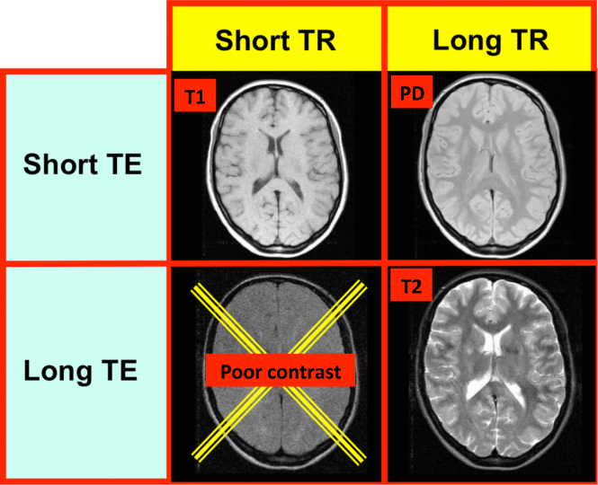
The journey from a patient lying in the scanner to a detailed anatomical image involves several intricate steps. First, the main magnetic field (B0) aligns the hydrogen protons. Then, a radiofrequency (RF) pulse, tuned to the specific resonant frequency of hydrogen, is applied. This pulse excites the protons, causing them to absorb energy and flip their spin orientation. When the RF pulse is removed, the protons begin to “relax” back to their original alignment and energy state, releasing the absorbed energy as a radio signal. The rate at which these protons relax is not uniform across all tissues; different tissues have distinct relaxation properties, specifically T1 (longitudinal relaxation) and T2 (transverse relaxation) times. By manipulating the timing of the RF pulses and the signal reception, MRI technologists can create images that emphasize either T1 or T2 characteristics, thereby highlighting different tissue properties and pathologies. Gradient coils are also used to spatially encode the signals, allowing the system to pinpoint the origin of each signal and construct a 3D image slice by slice. The digital data is then processed by powerful computers, transforming the complex signals into the high-resolution images clinicians use for diagnosis.
Decoding T1-Weighted Images
T1-weighted images are one of the fundamental sequences in MRI, primarily used for anatomical detail and for demonstrating the presence of fat and proteinaceous fluid. In a T1-weighted sequence, the timing parameters (specifically the repetition time, TR, and echo time, TE) are set to primarily reflect the T1 relaxation times of tissues. The T1 relaxation time describes how quickly the longitudinal magnetization (the alignment of protons with the main magnetic field) recovers after the RF pulse. Tissues with short T1 times appear bright, while those with long T1 times appear dark.
What T1 Reveals
The characteristic appearance of tissues on T1-weighted images is often summarized by “fat is bright, water is dark.”
- Fat: Due to its short T1 relaxation time, fat appears very bright (hyperintense). This makes T1 sequences excellent for visualizing fatty tissues, such as subcutaneous fat, bone marrow, and fat within organs.
- Water/Cerebrospinal Fluid (CSF): Water and CSF have long T1 relaxation times, causing them to appear dark (hypointense). This means that structures like ventricles in the brain and the spinal canal, which are filled with CSF, will appear dark. Edema (swelling) and most cysts, which are high in water content, also appear dark on T1.
- Muscle: Muscle tissue typically appears intermediate in signal intensity.
- Bone Cortex: The dense outer layer of bone (cortical bone) contains very little water and hydrogen protons, so it appears dark on all MRI sequences, including T1.
- Hemorrhage (acute): Acute blood products can appear bright on T1, particularly due to the presence of methemoglobin. This can be crucial in identifying fresh bleeds.
- Gadolinium Contrast: Gadolinium-based contrast agents, often administered intravenously, significantly shorten the T1 relaxation time of tissues where they accumulate. This causes these areas to appear dramatically brighter on T1-weighted images, making them invaluable for detecting tumors, inflammation, and vascular abnormalities that enhance with contrast.
Clinical Applications of T1
T1-weighted images are the workhorse for anatomical visualization and are often the first sequence acquired in many MRI protocols.
- Anatomical Detail: They provide excellent gray-white matter differentiation in the brain and good delineation of organ boundaries, making them ideal for assessing overall anatomy.
- Tumor Detection: While tumors often appear darker or isointense to surrounding tissue without contrast, their enhancement after gadolinium administration makes T1 crucial for detecting and characterizing neoplasms.
- Fat-Containing Lesions: Lipomas (benign fatty tumors) or dermoid cysts, which contain fat, are brightly hyperintense on T1-weighted images.
- Hemorrhage Dating: The signal characteristics of blood products change over time, and T1 can help differentiate between acute, subacute, and chronic hemorrhage.
- Pre- and Post-Contrast Imaging: T1 sequences are almost always acquired both before and after contrast administration to evaluate for enhancement, a key indicator of pathology.

Unpacking T2-Weighted Images
T2-weighted images constitute the other foundational sequence in MRI, known for their ability to highlight pathology and fluid. In a T2-weighted sequence, the TR and TE parameters are adjusted to primarily reflect the T2 relaxation times of tissues. The T2 relaxation time describes how quickly the transverse magnetization (the coherent spinning of protons in phase) decays after the RF pulse. Tissues with long T2 times appear bright, while those with short T2 times appear dark.
What T2 Highlights
The characteristic appearance of tissues on T2-weighted images is often summarized by “water is bright, fat is bright.”
- Water/Cerebrospinal Fluid (CSF): Water and CSF have long T2 relaxation times, causing them to appear very bright (hyperintense). This makes T2 sequences excellent for visualizing CSF-filled spaces, edema, inflammation, and most cysts.
- Fat: Fat also has a relatively long T2 relaxation time and can appear bright on T2-weighted images. However, often a technique called “fat suppression” is applied to T2 sequences to make fat appear dark, which helps to further highlight water-containing pathology against a dark fat background.
- Muscle: Muscle tissue typically appears intermediate to dark in signal intensity.
- Bone Cortex: Similar to T1, cortical bone appears dark due to its low water content.
- Pathology (General): The vast majority of pathological processes, such as tumors, infection, inflammation, and infarction, involve an increase in tissue water content (edema). Since water appears bright on T2, these pathologies are typically hyperintense and thus easily detectable.
- Fluid-Filled Structures: Cysts, effusions, and areas of necrosis (tissue death) are generally very bright on T2.
Clinical Applications of T2
T2-weighted images are primarily used for pathology detection and characterization due to their sensitivity to changes in water content.
- Detection of Edema and Inflammation: T2 sequences are exquisitely sensitive to increased water content, making them ideal for identifying swelling, inflammation, and infection in virtually any part of the body.
- Brain Pathology: They are crucial for detecting brain tumors, strokes (ischemic lesions), demyelinating diseases like multiple sclerosis (MS) plaques, and other white matter lesions.
- Spinal Cord Lesions: Spinal cord edema, tumors, and syrinxes (fluid-filled cavities) are well-visualized on T2.
- Musculoskeletal Imaging: T2 sequences are vital for assessing ligament tears, muscle strains, joint effusions, and bone marrow edema.
- Abdominal and Pelvic Imaging: Used to detect cysts, abscesses, and fluid collections in organs like the liver, kidneys, and reproductive organs.
- Fluid-Attenuated Inversion Recovery (FLAIR): This is a specialized T2 sequence where the signal from normal CSF is suppressed, making pathologies near the ventricles (like MS plaques) more conspicuous.
Beyond T1 and T2: Advanced MRI Techniques
While T1 and T2 form the cornerstone of MRI, the field has continuously evolved, introducing numerous advanced sequences and techniques that build upon these fundamental principles to extract even more specific diagnostic information. These innovations further underscore MRI’s status as a leading-edge diagnostic technology.
FLAIR, DWI, and More
- FLAIR (Fluid-Attenuated Inversion Recovery): As mentioned, FLAIR is a modified T2 sequence where the signal from free water (like CSF) is nullified or suppressed. This makes periventricular and cortical lesions, which contain increased water but are not CSF, appear bright against a dark CSF background. FLAIR is particularly valuable in neuroimaging for detecting multiple sclerosis plaques, subarachnoid hemorrhage, and certain types of brain tumors.
- DWI (Diffusion-Weighted Imaging): DWI measures the microscopic movement (diffusion) of water molecules within tissues. The restriction of water diffusion, as seen in acute stroke or certain highly cellular tumors, results in a bright signal on DWI. This sequence is critically important for detecting acute ischemic stroke within minutes of onset, far earlier than conventional T1 or T2 sequences.
- Perfusion-Weighted Imaging (PWI): PWI assesses blood flow to tissues by tracking the passage of a contrast agent. It helps evaluate tissue viability, particularly in stroke, by identifying areas of reduced blood supply.
- MRA (Magnetic Resonance Angiography): MRA visualizes blood vessels. It can be performed with or without contrast, providing detailed images of arteries and veins to detect stenoses (narrowing), aneurysms, or dissections.
- MRS (Magnetic Resonance Spectroscopy): MRS is a specialized technique that detects and quantifies specific metabolites (chemical compounds) within tissues. It provides biochemical information, which can help differentiate tumor types, assess tumor aggressiveness, and diagnose metabolic disorders.
- Functional MRI (fMRI): fMRI measures small changes in blood flow that occur with brain activity. By detecting these changes, fMRI can map brain functions, identifying areas responsible for motor skills, language, or vision, crucial for pre-surgical planning.

The Future of Diagnostic Imaging
The ongoing evolution of MRI technology continues to push the boundaries of non-invasive diagnostics. Faster scanning times, higher field strength magnets, and more sophisticated pulse sequences are leading to even finer anatomical detail and more precise physiological information. The integration of AI and machine learning is revolutionizing image reconstruction, quantitative analysis, and even automated pathology detection, promising to enhance diagnostic accuracy and efficiency. From molecular imaging that targets specific cellular processes to real-time image guidance for minimally invasive procedures, MRI remains at the forefront of medical technology, continually redefining what is possible in understanding and treating the human body. As the digital age progresses, the interplay of physics, computer science, and clinical insight will ensure that MRI continues to be a cornerstone of advanced medical care, offering unparalleled insights that were once unimaginable.
aViewFromTheCave is a participant in the Amazon Services LLC Associates Program, an affiliate advertising program designed to provide a means for sites to earn advertising fees by advertising and linking to Amazon.com. Amazon, the Amazon logo, AmazonSupply, and the AmazonSupply logo are trademarks of Amazon.com, Inc. or its affiliates. As an Amazon Associate we earn affiliate commissions from qualifying purchases.