
For over a century, the Mantoux Tuberculin Skin Test (TST) has been the gold standard for detecting latent tuberculosis. The process is deceptively simple: a small amount of purified protein derivative (PPD) is injected into the skin, and 48 to 72 hours later, a healthcare provider looks for a reaction. However, the question of “what does a positive tuberculin skin test look like” is no longer just a clinical query for a human doctor; it has become a complex challenge for software engineers, data scientists, and medical technology firms.
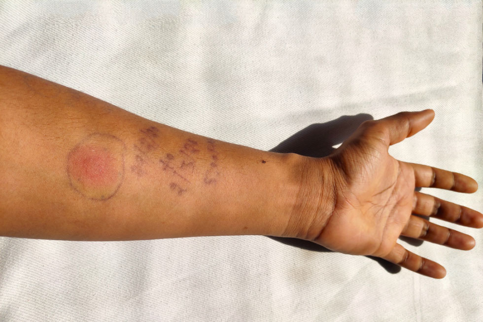
In the modern era, the interpretation of a “positive” result is shifting from the subjective human eye to the objective precision of artificial intelligence and advanced digital imaging. As we move toward a more digitized healthcare landscape, technology is solving the age-old problem of diagnostic variability, ensuring that the “look” of a positive test is captured with unprecedented accuracy.
From Manual Measurement to Digital Precision: The Evolution of Diagnostic Hardware
Historically, determining if a TB skin test was positive relied on the “ballpoint pen and ruler” method. A clinician would feel for induration—the hard, raised area of the skin—and measure its diameter. This method is notoriously prone to human error, with significant variability between different practitioners. Technology is stepping in to replace this tactile guesswork with sophisticated optical sensors.
The Limitations of Human Interpretation
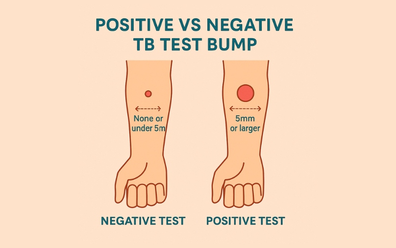
Human vision is susceptible to environmental factors like lighting, skin tone contrast, and fatigue. When a clinician asks what a positive test looks like, they are often looking for a raised bump of 5mm, 10mm, or 15mm depending on the patient’s risk factors. However, redness (erythema) is often mistaken for induration (the actual swelling), leading to false positives. The “tech” solution involves filtering out these visual distractions to focus on the underlying dermal structure.
High-Resolution Optical Sensors and 3D Imaging
Modern gadgets are now being developed to perform 3D topographical mapping of the injection site. Using structured light sensors—similar to the technology used in facial recognition software on smartphones—these devices can measure the exact volume and height of an induration. By capturing a three-dimensional model of the skin’s surface, the technology can distinguish between a flat red patch and a medically significant raised bump, providing a definitive numerical value that removes the “it looks like…” ambiguity.
Automated Calipers and Digital Integration
Beyond simple imaging, some med-tech startups are developing digital calipers that sync via Bluetooth to Electronic Health Records (EHR). When the device measures the diameter of the skin reaction, the data is instantly uploaded, time-stamped, and verified. This ensures that the visual evidence of a positive test is backed by a digital audit trail, reducing the risk of clerical errors that can occur during manual data entry.
AI-Powered Algorithms: “Seeing” Induration Beyond the Naked Eye
The most significant leap in understanding what a positive TB test looks like comes from the field of Computer Vision (CV). By training machine learning models on tens of thousands of images of injection sites, software can now identify a positive result with a level of nuance that exceeds human capability.
Training Models on Diverse Dermatological Datasets
One of the historical challenges in diagnosing TB skin tests is the variation in how reactions appear on different skin tones. A positive result on lighter skin may show distinct redness, while on darker skin, the reaction might be more subtle or present differently. Tech developers are addressing this by training AI on diverse datasets to ensure that algorithms can identify a positive test regardless of the patient’s ethnicity. This “algorithmic fairness” is a trending topic in medical AI, ensuring that software provides equitable diagnostic accuracy.
Neural Networks and Sub-Dermal Analysis
Advanced neural networks are being designed to analyze not just the surface of the skin, but the patterns of inflammation. By using multi-spectral imaging—taking photos under different wavelengths of light—AI can “see” blood flow patterns and fluid accumulation under the skin surface. This allows the software to confirm a positive test even before the induration is fully visible to the naked eye, or to rule out a “pseudo-positive” caused by a simple allergic reaction to the needle.
Reducing False Positives through Predictive Analytics
Software tools are now capable of cross-referencing a digital image of a skin test with a patient’s digital health history. By using predictive analytics, the AI can weigh the “look” of the skin test against the patient’s risk profile (such as previous exposure or immunocompromised status). This holistic tech approach ensures that the “positive” designation is not just a visual measurement, but a data-driven diagnosis.
The Role of Mobile Apps and Telehealth in TB Monitoring
In the wake of the global shift toward telehealth, mobile software has become a critical tool in monitoring TB tests. Patients no longer necessarily need to return to a clinic for a reading; instead, apps are allowing them to document their own results under the guidance of intelligent software.
Smartphone-Based Diagnostic Interfaces
What does a positive test look like through a smartphone camera? Several apps now guide users to take a photo of their arm next to a reference object (like a credit card or a specialized calibration sticker) to provide scale. The app’s internal software then processes the image, correcting for shadows and lens distortion, to provide a preliminary reading. This democratization of diagnostics is particularly vital in remote areas where access to specialized clinics is limited.
Enhancing Data Security and Privacy
With the rise of mobile diagnostic apps comes the critical need for digital security. The images of a positive tuberculin test are sensitive medical data. Developers are implementing end-to-end encryption and biometric locks to ensure that the visual record of a patient’s TB status remains private. Furthermore, blockchain technology is being explored as a way to store these diagnostic “snapshots” securely, allowing for a tamper-proof history of a patient’s respiratory health.
Real-Time Reporting and Cloud Synchronization
When an app identifies what looks like a positive result, it can immediately trigger a series of automated actions. Through cloud-based platforms, the image can be flagged for a remote specialist’s review. This instant connectivity ensures that if a test “looks” positive, the patient is moved into the next phase of the care continuum—such as a chest X-ray or blood work—without the delays inherent in traditional bureaucratic healthcare systems.
Future Tech: Beyond the Surface of the Skin
As we look toward the future of technology in tuberculosis diagnostics, the very definition of “looking at a test” is evolving. We are moving away from reactive visual checks toward proactive, continuous monitoring via wearables and molecular tech.
Wearable Biosensors for Latent Infection
Imagine a wearable patch that doesn’t just look for a reaction once, but monitors the skin’s physiological response in real-time. Future gadgets may utilize micro-needles and biosensors that detect the chemical markers of a TB reaction (such as cytokines) the moment they are released. In this scenario, the “positive look” is a digital signal sent to a smartwatch, rather than a physical bump on the skin.
Augmented Reality (AR) in Clinical Training
To train the next generation of healthcare providers, AR tools are being used to simulate what a positive tuberculin skin test looks like across a variety of scenarios. Using AR headsets, students can view “digital overlays” on real human arms or mannequins, helping them learn to distinguish between different grades of induration. This tech-driven education ensures a higher standard of manual reading when digital tools are unavailable.
The Integration of Genomics and Diagnostic Software
The final frontier of “what a test looks like” involves the merging of visual data with genomic sequencing. Future software platforms will likely combine the image of a positive skin test with the genetic profile of the TB strain involved. By analyzing the visual characteristics of the skin’s inflammation alongside the bacteria’s DNA, tech tools will be able to predict how a patient will respond to specific antibiotics, turning a simple skin test into a cornerstone of personalized medicine.

Conclusion: A New Visual Standard in the Digital Age
The question “what does a positive tuberculin skin test look like” is being answered with increasing complexity by the world of technology. No longer is the answer merely “a raised bump of a certain size.” Today, the answer involves high-resolution 3D mapping, AI-driven computer vision, and secure mobile platforms that ensure every reaction is measured with clinical precision.
As software continues to eat the world of diagnostics, the subjectivity of the human eye is being supplemented—and in some cases, replaced—by algorithms that do not tire and sensors that do not blink. For patients and providers alike, this technological shift means fewer false positives, more reliable data, and a faster path to treatment. In the intersection of health and technology, the “look” of a positive test is now a data point in a much larger, life-saving digital ecosystem.
aViewFromTheCave is a participant in the Amazon Services LLC Associates Program, an affiliate advertising program designed to provide a means for sites to earn advertising fees by advertising and linking to Amazon.com. Amazon, the Amazon logo, AmazonSupply, and the AmazonSupply logo are trademarks of Amazon.com, Inc. or its affiliates. As an Amazon Associate we earn affiliate commissions from qualifying purchases.