In the rapidly advancing landscape of health technology, the tools used for internal diagnostics have undergone a radical transformation. What was once a rudimentary mechanical process has evolved into a sophisticated synergy of fiber optics, high-definition digital imaging, and artificial intelligence. The flexible sigmoidoscopy represents a critical intersection of mechanical engineering and digital innovation. Far from being just a medical procedure, it is a showcase of how hardware miniaturization and software integration are redefining preventive medicine.
By exploring the technical architecture of the flexible sigmoidoscope, we can better understand the trajectory of modern medical gadgets and the digital systems that support them. This article examines the technological breakthroughs that make this diagnostic tool possible, the integration of AI in real-time screening, and the future of gastrointestinal tech.
1. The Engineering of Flexibility: Hardware and Optical Innovation
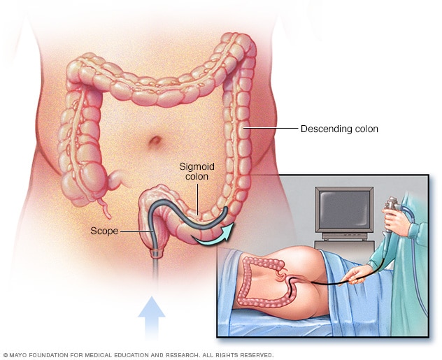
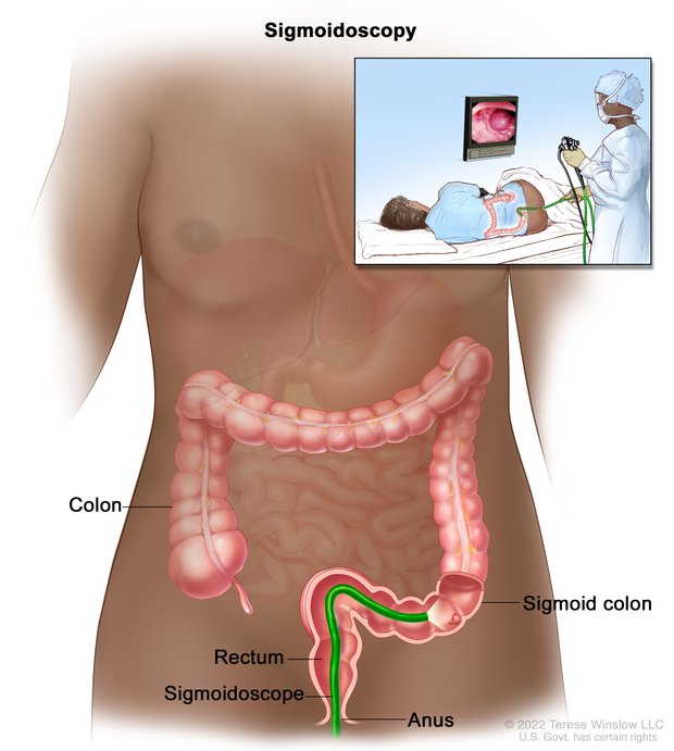
At its core, a flexible sigmoidoscopy relies on a device called a sigmoidoscope. To the layperson, it may appear to be a simple flexible tube, but from a tech perspective, it is a complex assembly of micro-components designed to navigate the intricate topography of the human body while transmitting high-bandwidth data.
High-Definition Imaging and CMOS Sensors
The transition from analog fiber-optic bundles to digital “chip-on-the-tip” technology marked a turning point in diagnostic tech. Modern sigmoidoscopes utilize CMOS (Complementary Metal-Oxide-Semiconductor) sensors, the same fundamental technology found in high-end smartphone cameras. These sensors are miniaturized to fit within the distal end of the scope, allowing for 1080p or even 4K resolution images of the intestinal lining.
The challenge for engineers is balancing resolution with light sensitivity. Because the internal environment of the colon is devoid of natural light, the tip of the scope must house high-intensity LEDs that produce minimal heat to avoid tissue damage. The synchronization between these LED pulses and the sensor’s shutter speed is managed by a dedicated image processor, ensuring a flicker-free, crystal-clear feed for the specialist.
Mechanical Maneuverability and Materials Science
The “flexibility” in flexible sigmoidoscopy is a result of advanced materials science. The insertion tube is constructed from multi-layered polymers that provide a specific “torque-stable” property. This means that when a technician rotates the handle, the tip responds with a 1:1 ratio, a feat of mechanical engineering that requires the perfect balance of rigidity and elasticity.
Inside the tube, a series of ultra-thin stainless steel wires connect the control knobs to the articulating tip. These wires must be durable enough to withstand thousands of bends without snapping or stretching. Recent iterations in this tech niche have introduced “variable stiffness” scopes, which allow the operator to adjust the rigidity of the tube via a digital interface, optimizing the device for different anatomical challenges.
2. AI Integration: Computer-Aided Detection (CADe) Systems
The most significant recent trend in endoscopic technology is the integration of Artificial Intelligence (AI). While the hardware captures the image, the software is now beginning to interpret it in real-time. This is often referred to as “Augmented Endoscopy.”
Real-Time Pattern Recognition
AI algorithms, specifically deep learning neural networks, are being trained on millions of frames of endoscopic footage. During a flexible sigmoidoscopy, these AI tools run in the background, scanning the video feed for subtle irregularities that the human eye might overlook. When the software identifies a potential polyp or lesion, it places a visual “bounding box” around the area on the monitor, alerting the technician.
This technology, known as Computer-Aided Detection (CADe), significantly reduces the “miss rate” of screenings. From a software development standpoint, the challenge lies in latency. The AI must process high-definition video frames in milliseconds to provide real-time feedback without lagging behind the physical movement of the scope.
Computer-Aided Characterization (CADx)
Beyond just finding an abnormality, the next frontier in this tech niche is CADx, or Computer-Aided Characterization. This technology uses optical maneuvers like Narrow Band Imaging (NBI)—which uses specific wavelengths of light to highlight vascular patterns—and analyzes the data through an AI lens. The software can then provide a probability score of whether a growth is benign or precocious. This “optical biopsy” reduces the need for physical tissue sampling, showcasing how software can augment or even replace traditional laboratory processes.
3. The Digital Ecosystem: Data Management and Telehealth
A flexible sigmoidoscopy does not exist in a vacuum; it is part of a broader digital health ecosystem. The data generated during a ten-minute procedure can amount to several gigabytes of high-definition video and metadata, necessitating robust digital infrastructure.
Cloud Integration and DICOM Standards
Modern sigmoidoscopy suites are integrated into the hospital’s Picture Archiving and Communication System (PACS) using DICOM (Digital Imaging and Communications in Medicine) standards. This ensures that the high-resolution video captured is securely encrypted and uploaded to the cloud or local servers.
For tech professionals, the interest lies in the interoperability of these systems. The ability for a specialist in one part of the world to access a high-definition recording of a procedure performed elsewhere allows for “telementoring.” In this scenario, an expert can provide a secondary consultation in real-time or asynchronously, facilitated by high-speed 5G networks and secure cloud gateways.
Cybersecurity in Diagnostic Tech
As diagnostic tools become more connected, they become targets for digital threats. A sigmoidoscope connected to a network is an IoT (Internet of Things) device. Manufacturers are now prioritizing cybersecurity, implementing “secure boot” protocols and encrypted data transmission to ensure that patient video feeds cannot be intercepted or tampered with. This focus on digital security is essential for maintaining the integrity of the diagnostic process and protecting sensitive personal data.
4. The Future of Internal Imaging: From Tubes to Robotics
The trajectory of flexible sigmoidoscopy tech is moving toward less invasive and more automated solutions. We are currently witnessing a shift from manual controls to robotic-assisted platforms and wireless diagnostics.
Robotic Endoscopy
Traditional sigmoidoscopy requires a high level of manual dexterity. New robotic platforms are changing this by decoupling the operator from the device. Instead of holding the scope, the technician uses a joystick or a digital interface to guide a robotic arm that controls the insertion tube. This technology provides sub-millimeter precision and eliminates the physical strain on the operator, leading to more consistent results. Furthermore, robotic systems can “map” the path taken, creating a 3D digital twin of the patient’s anatomy for future reference.
Wireless Capsule Alternatives
While the flexible sigmoidoscope remains the gold standard for targeted diagnostics and interventions (like removing polyps), the tech for “pill cameras” or capsule endoscopy is rapidly advancing. These are essentially micro-computers contained within a swallowable pill, equipped with cameras, batteries, and wireless transmitters.
The current limitation of capsule tech is the lack of “locomotion”—the pill simply moves with the body’s natural processes. However, experimental tech involves “active locomotion,” where external magnetic fields are used to “steer” the capsule inside the body. Once perfected, this could represent the ultimate evolution of the flexible sigmoidoscopy: a fully wireless, digitally controlled, non-invasive diagnostic tool.

Conclusion
The “what” of a flexible sigmoidoscopy is far more than a medical definition; it is a complex tapestry of modern technology. From the CMOS sensors and fiber-optic lighting that capture the internal world in high definition to the AI algorithms that serve as a second pair of expert eyes, the procedure is a testament to technical ingenuity.
As we look forward, the convergence of robotics, cloud computing, and miniaturized sensors promises to make these tools even more precise and accessible. For those in the tech and digital security sectors, the evolution of such diagnostic tools provides a fascinating case study in how specialized hardware and sophisticated software can work in tandem to solve some of the most critical challenges in human health. The future of the flexible sigmoidoscope is not just flexible in its physical form, but in its ability to adapt to the ever-changing digital landscape.
aViewFromTheCave is a participant in the Amazon Services LLC Associates Program, an affiliate advertising program designed to provide a means for sites to earn advertising fees by advertising and linking to Amazon.com. Amazon, the Amazon logo, AmazonSupply, and the AmazonSupply logo are trademarks of Amazon.com, Inc. or its affiliates. As an Amazon Associate we earn affiliate commissions from qualifying purchases.