In the vast and rapidly evolving landscape of modern medicine, diagnostic imaging stands as a critical pillar, empowering clinicians with the ability to peer inside the human body without invasive procedures. Among the most widely recognized and indispensable tools in this arena are Magnetic Resonance Imaging (MRI) and Computed Tomography (CT) scans. While both serve the overarching goal of providing detailed anatomical views to aid in diagnosis, treatment planning, and monitoring, they operate on fundamentally different technological principles and, consequently, excel in visualizing different types of tissues and conditions. Understanding these distinctions is not merely an academic exercise; it’s crucial for patients, healthcare providers, and anyone interested in the technological marvels that underpin contemporary healthcare. This article delves into the core mechanics, applications, advantages, and limitations of MRI and CT scans, offering a comprehensive comparative analysis of these two revolutionary technologies.

Understanding CT Scans: A Glimpse into X-ray Technology
The CT scan, often simply referred to as a “CAT scan” (Computed Axial Tomography), has been a cornerstone of diagnostic imaging since its introduction in the early 1970s. It represents a significant leap forward from conventional X-rays, transforming two-dimensional shadowgrams into detailed cross-sectional views.
How CT Scans Work: Principles of X-ray Imaging
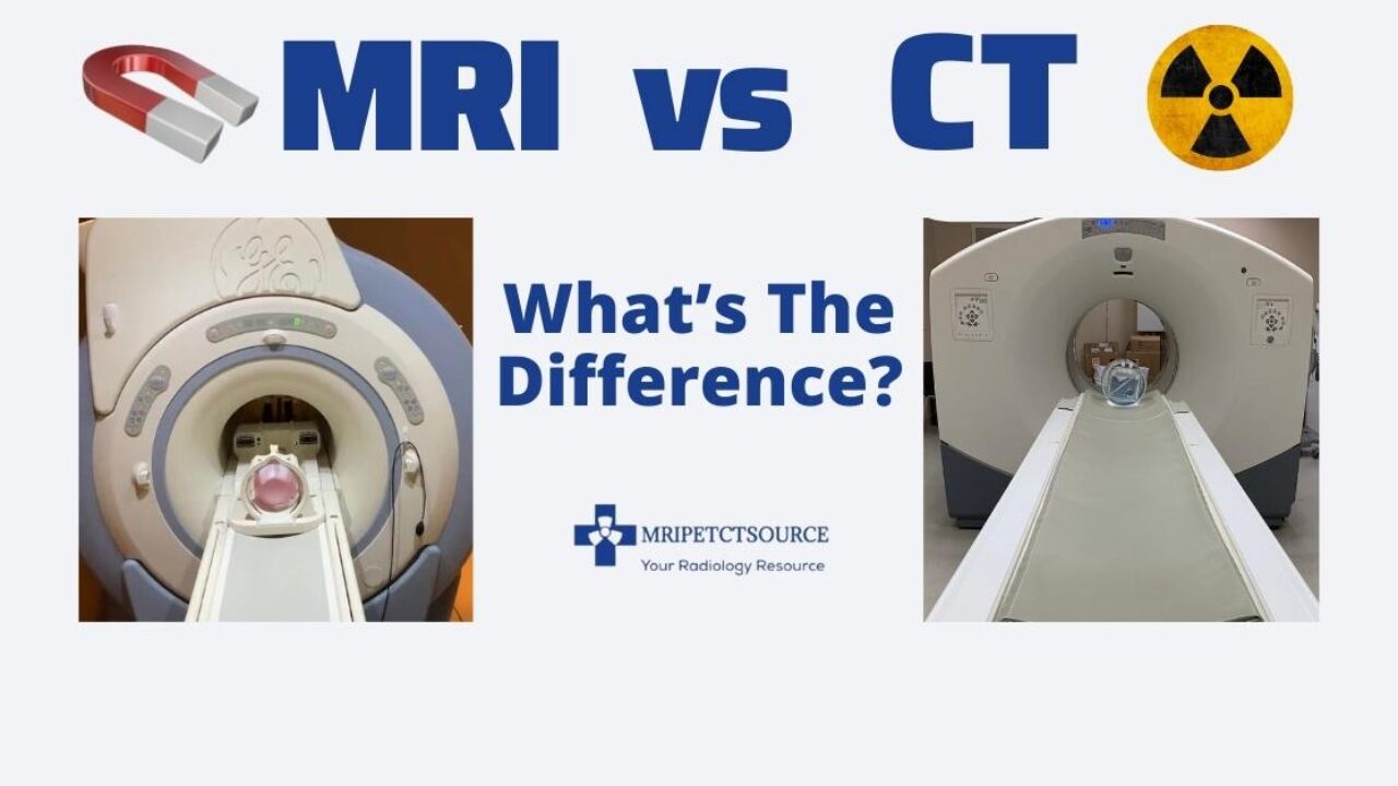
At its heart, a CT scan employs sophisticated X-ray technology. The patient lies on a motorized table that slides through a large, donut-shaped machine called a gantry. Inside this gantry, an X-ray tube rotates around the patient, emitting a narrow beam of X-rays. As these X-rays pass through the body, different tissues absorb them to varying degrees—bones, being dense, absorb more X-rays than soft tissues, which in turn absorb more than air.
On the opposite side of the X-ray tube, an array of detectors captures the X-rays that have passed through the body. As the X-ray tube and detectors rotate, they collect thousands of individual X-ray measurements from multiple angles. A powerful computer then processes these measurements using complex algorithms to reconstruct detailed cross-sectional images, or “slices,” of the body. These slices can then be stacked together to create sophisticated 3D renderings of organs, bones, and blood vessels. The core principle here is the differential attenuation of X-rays, where the density of tissues dictates how much radiation passes through them, creating the contrast needed for image formation. A key aspect to note is the use of ionizing radiation in this process.
Key Applications of CT Scans
CT scans are renowned for their speed, availability, and excellent depiction of bone and acute conditions. Their primary applications include:
- Trauma Assessment: Rapidly identifying internal bleeding, organ damage, and bone fractures in emergency situations. A whole-body CT scan can quickly assess multiple injuries.
- Stroke Evaluation: Quickly ruling out hemorrhagic stroke (bleeding in the brain), which is critical for determining appropriate treatment (e.g., clot-busting drugs are contraindicated in hemorrhagic strokes).
- Cancer Detection and Staging: Identifying tumors, assessing their size and location, and determining if cancer has spread to other parts of the body (metastasis).
- Lung and Thoracic Imaging: Diagnosing conditions like pneumonia, emphysema, lung cancer, and pulmonary embolism.
- Abdominal and Pelvic Imaging: Detecting issues such as appendicitis, kidney stones, diverticulitis, and various organ pathologies.
- Vascular Studies (CT Angiography): Visualizing blood vessels to detect aneurysms, blockages, or other vascular abnormalities, often with the help of intravenous contrast agents.
Advantages and Disadvantages of CT Scans
Advantages:
- Speed: CT scans are significantly faster than MRIs, often taking only minutes, which is crucial in emergency situations or for uncooperative patients.
- Excellent Bone Visualization: Unmatched in its ability to visualize bone structures, fractures, and complex bony anatomy.
- Availability and Cost-Effectiveness: Generally more widely available and less expensive than MRI scans.
- Less Sensitivity to Motion: Due to faster acquisition times, CT is less affected by patient movement compared to MRI.
- Tolerance for Metallic Implants: Most metallic implants are not contraindications for CT, unlike MRI.
Disadvantages:
- Ionizing Radiation: The primary concern with CT scans is exposure to ionizing radiation, which carries a small, cumulative risk of cancer over a lifetime. While doses are optimized, this remains a consideration, especially for pediatric patients or repeated scans.
- Limited Soft Tissue Contrast: While good for bones, CT scans provide less detailed soft tissue contrast compared to MRI, making it harder to differentiate between certain types of tissues or subtle lesions.
- Allergy to Contrast Agents: Some patients may have allergic reactions to iodine-based contrast agents used to enhance image visibility in certain CT studies.
Unpacking MRI: The Power of Magnets and Radio Waves
Magnetic Resonance Imaging (MRI) emerged later than CT, in the 1980s, introducing a completely different paradigm for medical imaging. Unlike CT, MRI does not use ionizing radiation, relying instead on powerful magnetic fields and radio waves.
How MRI Scans Work: Leveraging Magnetic Fields
The technology behind MRI is remarkably ingenious. It harnesses the natural magnetic properties of hydrogen atoms, which are abundant in the water molecules that make up approximately 60% of the human body. When a patient enters the strong, static magnetic field of an MRI scanner, the protons (nuclei) within these hydrogen atoms align with the magnetic field.
Next, the scanner emits brief pulses of radiofrequency energy. These radio waves momentarily knock the aligned protons out of alignment. When the radiofrequency pulse is turned off, the protons “relax” and realign with the main magnetic field, releasing energy in the form of radio signals. Different tissues relax at different rates and emit signals of varying strengths, depending on their composition and water content.
These emitted signals are detected by receiver coils in the MRI machine, and a sophisticated computer processes them to create highly detailed cross-sectional images. Because different tissues (e.g., fat, muscle, brain matter, tumors) have varying amounts of water and thus different relaxation properties, MRI excels at differentiating between various soft tissues. Crucially, this process involves no ionizing radiation, making it a safer option for repeated scans or for sensitive populations like pregnant women (with specific precautions).
Key Applications of MRI Scans
MRI’s exceptional soft tissue contrast makes it the preferred imaging modality for a wide range of conditions, particularly those involving neurological, musculoskeletal, and abdominal soft tissues:
- Neurological Imaging: The gold standard for visualizing the brain and spinal cord, detecting tumors, strokes, multiple sclerosis, infections, aneurysms, and spinal cord injuries.
- Musculoskeletal Imaging: Excellent for evaluating joints (knees, shoulders, wrists, ankles) for ligament tears, cartilage damage, tendonitis, and bone marrow abnormalities.
- Soft Tissue Tumors: Highly effective in detecting and characterizing tumors in soft tissues throughout the body, providing better differentiation between benign and malignant lesions than CT in many cases.
- Abdominal and Pelvic Imaging: Used to visualize organs like the liver, kidneys, pancreas, uterus, and ovaries, detecting tumors, inflammation, and other abnormalities.
- Cardiovascular MRI (CMR): Assessing heart function, identifying structural heart disease, and evaluating blood flow.
- Breast MRI: Often used as a supplementary screening tool for high-risk women or for further evaluation of abnormalities detected on mammograms.
Advantages and Disadvantages of MRI Scans
Advantages:
- Superior Soft Tissue Contrast: Offers unparalleled detail for soft tissues, making it ideal for imaging the brain, spinal cord, muscles, ligaments, tendons, and internal organs.
- No Ionizing Radiation: Eliminates concerns about radiation exposure, making it safer for repeated studies and for patients who are sensitive to radiation.
- Excellent for Pathological Changes: Can often detect subtle changes in tissues that might be missed by other imaging techniques, making it highly sensitive for diagnosing certain diseases.
- Functional Imaging Capabilities: Advanced MRI techniques like fMRI (functional MRI) can measure brain activity, and DTI (Diffusion Tensor Imaging) can visualize white matter tracts.
Disadvantages:
- Longer Scan Times: MRI scans typically take much longer than CT scans (30 minutes to over an hour), increasing the likelihood of patient motion and discomfort.
- Claustrophobia: Many patients find the enclosed, narrow space of the MRI scanner claustrophobic. Open MRI designs exist but may offer slightly lower image quality.
- Loud Noise: The pulsing of the magnetic coils generates significant noise, requiring patients to wear earplugs or headphones.
- Contraindications for Metallic Objects: The powerful magnetic field prohibits patients with certain metallic implants (e.g., pacemakers, some cochlear implants, certain aneurysm clips, metallic foreign bodies) from undergoing an MRI due to safety risks and image distortion.
- Higher Cost: Generally more expensive than CT scans.
Comparative Analysis: When to Use Which Technology
The decision to use an MRI or a CT scan is a complex one, made by clinicians based on the patient’s symptoms, medical history, suspected condition, and specific diagnostic questions. Often, these technologies are complementary, with one providing initial information and the other offering more detailed insights.
Image Resolution and Detail
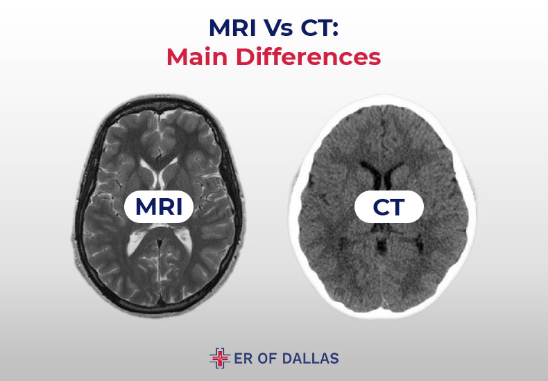
- CT Scans excel in visualizing dense structures like bones, acute hemorrhages, and air-filled cavities. It’s rapid, making it invaluable for emergencies where speed is paramount to diagnose life-threatening conditions.
- MRI Scans offer superior contrast resolution for soft tissues, allowing for clearer differentiation between different types of tissues and subtle abnormalities within them. This makes it ideal for evaluating conditions affecting the brain, spinal cord, joints, and soft tissue tumors.
Safety and Patient Considerations
- Radiation Exposure: CT scans involve ionizing radiation, which can be a concern, especially for children or patients requiring multiple follow-up scans. MRI does not use ionizing radiation, making it safer from this perspective.
- Patient Comfort: CT scans are quicker and typically involve a more open gantry, reducing claustrophobia. MRI scans are longer, louder, and often require patients to lie in a confined space, which can be challenging for some.
- Metallic Implants: The strong magnetic field of an MRI is a significant contraindication for patients with certain metallic implants or foreign bodies, whereas CT is generally safe for these individuals.
Cost and Accessibility
Generally, CT scans are more widely available and less expensive than MRI scans. This is partly due to the more complex technology, longer scan times, and specialized infrastructure required for MRI facilities. In many emergency settings, CT is the first-line imaging modality due to its speed and accessibility.
Specific Clinical Scenarios
- Head Trauma/Stroke: A CT scan is usually the first choice to quickly rule out a life-threatening hemorrhage. If no bleeding is found but stroke symptoms persist, an MRI may be performed for more detailed evaluation of brain tissue damage (ischemic stroke).
- Spinal Injuries: CT is excellent for assessing bone fractures or alignment issues in the spine. MRI is superior for evaluating the spinal cord itself, looking for damage, compression, or nerve root impingement.
- Knee Injuries: For suspected fractures, a CT scan might be useful. However, for soft tissue injuries like ligament tears (ACL, MCL) or meniscus damage, an MRI is the definitive imaging method.
- Cancer Diagnosis: Both can be used. CT is often used for staging large tumors and assessing metastasis to lungs or bones. MRI is particularly valuable for detailed evaluation of tumors in the brain, liver, pelvis, and musculoskeletal system due to its superior soft tissue contrast.
The Future of Medical Imaging: Evolution and Integration
Both CT and MRI technologies are continually evolving, driven by advancements in computing, physics, and artificial intelligence. The future promises even more precise, faster, and safer diagnostic capabilities.
Advancements in CT Technology
Modern CT scanners are becoming incredibly sophisticated. Innovations include:
- Lower Radiation Doses: Techniques like iterative reconstruction and dose modulation significantly reduce radiation exposure while maintaining diagnostic image quality.
- Faster Scans: Multi-detector CT (MDCT) scanners with hundreds of detector rows allow for incredibly fast data acquisition, enabling whole-body scans in seconds and reducing motion artifacts.
- Spectral CT (Dual-Energy CT): Uses two different X-ray energy levels to acquire more information about tissue composition, improving material characterization (e.g., distinguishing uric acid stones from calcium stones).
- AI Integration: Artificial intelligence is being deployed for image reconstruction, noise reduction, automated lesion detection, and quantitative analysis, enhancing diagnostic accuracy and efficiency.
Innovations in MRI Technology
MRI is also experiencing rapid advancements:
- Stronger Magnets and Faster Sequences: Higher field strength magnets (e.g., 3T, 7T) provide increased signal-to-noise ratio, leading to higher resolution images and faster scan times.
- Improved Coil Technology: Advanced receiver coils enhance signal detection and coverage, further improving image quality.
- Functional and Advanced Imaging: Techniques like fMRI map brain activity, Diffusion Tensor Imaging (DTI) visualizes nerve fiber pathways, and quantitative MRI allows for measuring tissue properties (e.g., fat fraction in the liver).
- More Open and Patient-Friendly Designs: Efforts are being made to create more spacious or “open” MRI scanners to alleviate claustrophobia, without compromising image quality.
- AI for Image Acquisition and Analysis: AI algorithms are optimizing scan parameters, reducing artifacts, and assisting radiologists in interpreting complex MRI data, potentially leading to earlier and more accurate diagnoses.
Hybrid Imaging and AI Integration
One of the most exciting developments is the rise of hybrid imaging systems, such as PET/CT and PET/MRI. These combine the functional information of Positron Emission Tomography (PET) with the anatomical detail of CT or MRI in a single scan. PET/CT is widely used in oncology to stage cancers and monitor treatment response, while PET/MRI offers the superior soft tissue contrast of MRI for even more precise localization of metabolic activity.
Furthermore, artificial intelligence is poised to revolutionize both CT and MRI. AI-powered software can assist in detecting subtle abnormalities, segmenting organs and lesions, predicting disease progression, and even personalizing imaging protocols. The integration of AI promises to make these technologies even more powerful, efficient, and accessible, ultimately benefiting patient care.
Conclusion
MRI and CT scans are indispensable diagnostic tools, each representing a triumph of technological ingenuity. While both provide invaluable insights into the human body, they are not interchangeable. CT scans, leveraging X-ray technology, excel in speed and visualizing bones, acute trauma, and hemorrhage, making them critical in emergency settings. Conversely, MRI scans, employing powerful magnetic fields and radio waves, offer unparalleled soft tissue contrast and anatomical detail, making them the preferred choice for neurological, musculoskeletal, and many oncological conditions.
The choice between an MRI and a CT scan hinges on the specific clinical question, the suspected pathology, and individual patient considerations. Often, they serve complementary roles, providing different pieces of the diagnostic puzzle. As technology continues to advance, with innovations in speed, image quality, radiation dose reduction, and the transformative power of artificial intelligence, both CT and MRI will remain at the forefront of medical diagnostics, continuing to enhance our understanding of human health and disease. Their ongoing evolution underscores the dynamic and vital role of technology in modern medicine.
aViewFromTheCave is a participant in the Amazon Services LLC Associates Program, an affiliate advertising program designed to provide a means for sites to earn advertising fees by advertising and linking to Amazon.com. Amazon, the Amazon logo, AmazonSupply, and the AmazonSupply logo are trademarks of Amazon.com, Inc. or its affiliates. As an Amazon Associate we earn affiliate commissions from qualifying purchases.