
The human skull is a marvel of biological engineering, a protective casing for the brain, and a framework for facial structures. Among its intricate components, the frontal bone stands out—literally—as the primary bone forming the forehead, part of the eye sockets (orbits), and the anterior cranial fossa. While its basic anatomical classification has been established for centuries, modern technology has profoundly reshaped our understanding, diagnosis, and treatment of this crucial skeletal element. No longer confined to purely anatomical descriptions, the “type” of bone the frontal bone represents is increasingly defined by its interaction with cutting-edge technological advancements, from high-resolution imaging to AI-driven diagnostics and personalized reconstructive solutions.
This article delves into the nature of the frontal bone, not just as a biological structure, but through the lens of the technological innovations that are revolutionizing its study and care. We explore how technology helps us fully appreciate its intricate structure, diagnose its pathologies with unprecedented precision, and develop innovative solutions for its repair and regeneration, ultimately redefining what it means to understand “what type of bone” the frontal bone truly is in the 21st century.
Defining the Frontal Bone: Its Anatomical and Functional Significance in the Digital Age
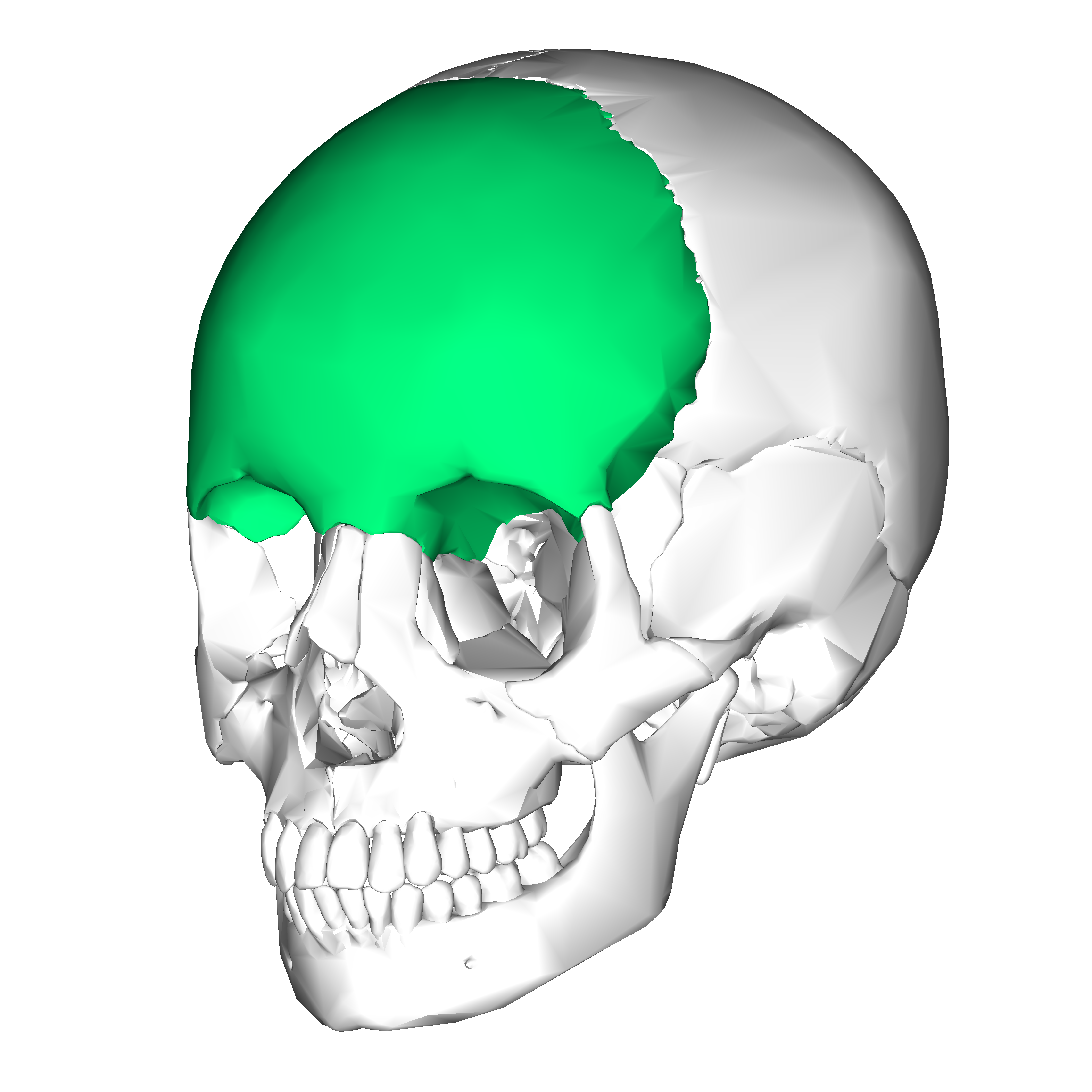
At its core, the frontal bone is classified as a flat bone, characterized by its broad, plate-like structure that provides extensive protection for underlying soft tissues and offers broad surfaces for muscle attachment. It is one of the eight cranial bones that form the neurocranium, the part of the skull that encloses the brain. Understanding its precise anatomical features and functional roles is paramount, and technology has significantly enhanced our ability to visualize and analyze these aspects.
The Cranial Vault’s Cornerstone: Digital Anatomy and Simulation
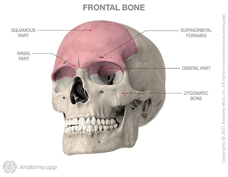
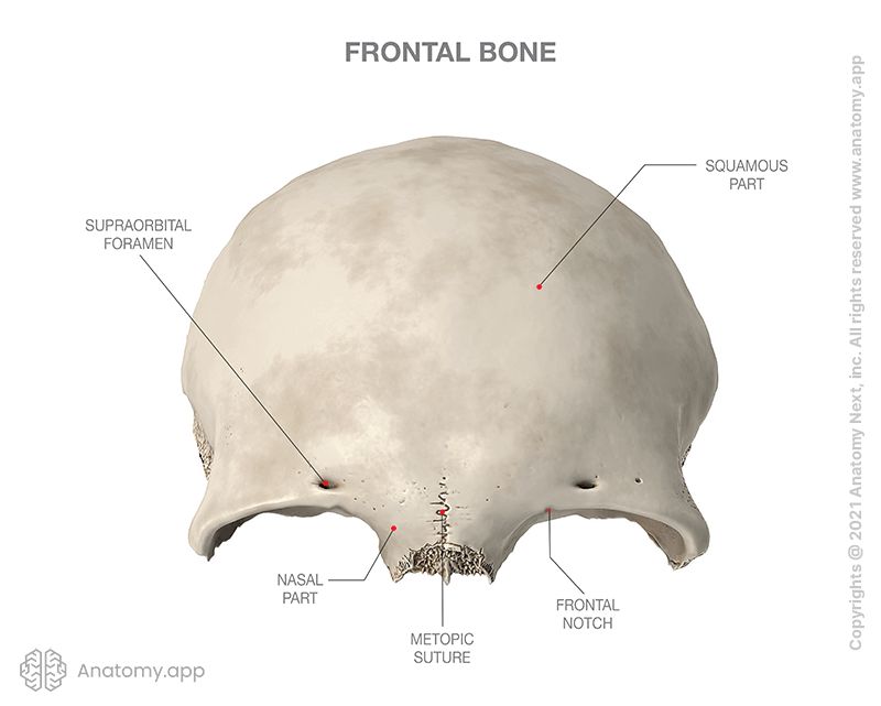
The frontal bone forms the squama frontalis, the large, convex forehead region; the orbital plates, which contribute to the roofs of the eye sockets; and the nasal part, which articulates with the nasal bones. Its internal surface features impressions for the brain’s gyri and sulci, and grooves for meningeal arteries, providing a detailed map of its complex relationship with the brain. Traditionally, this understanding was gained through cadaver dissection and two-dimensional schematics.
Today, advanced digital anatomy platforms and 3D simulation software have transformed this learning. Medical students and surgeons can interact with high-fidelity 3D models of the frontal bone, rotating them, peeling back layers, and even performing virtual dissections. These tools, often powered by sophisticated rendering engines, allow for an unparalleled appreciation of the bone’s precise curvature, its varying thickness, and its numerous foramina and fissures. Furthermore, biomechanical simulation software can model how the frontal bone responds to various forces, shedding light on its resistance to trauma and its role in distributing impact energy, offering insights far beyond static anatomical descriptions.
Protective Role and Beyond: Functional Analysis with Biofeedback
The primary function of the frontal bone is to protect the delicate frontal lobes of the brain from external trauma. Its robust yet somewhat flexible structure, coupled with its articulation with other cranial bones via sutures, creates a resilient protective shell. Beyond protection, it also contributes to the aesthetic contour of the face and supports the structures of the forehead, eyebrows, and upper eyelids.
Modern biofeedback systems and sensor technologies are beginning to allow for real-time analysis of the frontal bone’s functional performance. While still in nascent stages for direct bone monitoring, the integration of wearable sensors in sports and military contexts can track impact forces, providing data that helps engineers design better protective gear. This data, combined with advanced material science, informs the development of helmets and other headgear specifically optimized to mitigate frontal bone injuries, moving beyond empirical design to data-driven protective solutions. The “type” of bone, in this context, becomes defined by its dynamic interaction with external forces, measurable and analyzable through technological means.
Revolutionizing Diagnosis and Imaging with Advanced Tech
Accurate diagnosis of frontal bone pathologies—ranging from fractures and tumors to developmental anomalies—is critical for effective treatment. For centuries, diagnosis relied on palpation, X-rays, and physical examination. However, the advent of sophisticated imaging technologies and, more recently, artificial intelligence, has ushered in an era of unprecedented diagnostic precision, offering clinicians incredibly detailed insights into the frontal bone’s condition.
High-Resolution Imaging: CT, MRI, and Beyond
The evolution of medical imaging has been pivotal in understanding the frontal bone. Standard X-rays provide a basic outline, but their two-dimensional nature limits the appreciation of complex fractures or subtle lesions.
-
Computed Tomography (CT) scans are now the gold standard for visualizing the frontal bone. Modern multi-detector CT scanners can generate incredibly thin slices (sub-millimeter resolution) of the bone, producing highly detailed 3D reconstructions. These images allow clinicians to precisely map fracture lines, identify bone fragments, assess the involvement of the frontal sinuses, and detect subtle signs of osteomyelitis or tumors. Software tools can even generate virtual endoscopic views of the frontal sinuses, aiding surgical planning.
-
Magnetic Resonance Imaging (MRI), while less effective than CT for direct bone visualization due to its focus on soft tissues, is crucial for assessing the impact of frontal bone pathology on the brain and surrounding soft tissues. It can identify brain contusions, hemorrhages, and nerve damage that might accompany frontal bone trauma, or evaluate the extent of tumor invasion into intracranial structures.
-
Cone-Beam CT (CBCT) offers lower radiation doses and higher spatial resolution for specific craniofacial applications, particularly useful in dental and maxillofacial surgery, where precise visualization of the frontal bone’s orbital and nasal components is required.
These technologies provide not just images, but detailed volumetric data that can be manipulated and analyzed in various ways, giving clinicians an unparalleled view of “what type of bone” they are dealing with in terms of its structural integrity and relationship to vital organs.
AI-Powered Diagnostics and Predictive Analytics
The sheer volume and complexity of data generated by modern imaging techniques can be overwhelming for human interpretation. This is where artificial intelligence (AI) is transforming diagnosis. AI algorithms, particularly those leveraging deep learning, are being trained on vast datasets of CT and MRI scans to:
-
Automate Fracture Detection: AI can rapidly identify subtle hairline fractures or complex comminuted fractures in the frontal bone that might be missed by the human eye, especially in high-volume emergency settings.
-
Tumor Segmentation and Classification: AI can accurately segment tumors affecting the frontal bone, measure their size, and even help classify their type (e.g., benign osteoma vs. malignant sarcoma) based on imaging characteristics, aiding in treatment planning.
-
Predictive Analytics for Healing: Machine learning models are being developed to predict fracture healing rates, potential complications, and patient outcomes based on imaging features, patient demographics, and clinical data. This allows for personalized treatment strategies and proactive intervention.
By augmenting human expertise, AI not only speeds up diagnosis but also enhances its accuracy and consistency, providing a more comprehensive and nuanced understanding of the frontal bone’s pathological “type.”
Innovations in Frontal Bone Treatment and Reconstruction
When the frontal bone is damaged by trauma, disease, or developmental defects, advanced technology becomes indispensable for its reconstruction and restoration. The goal is to restore both function (protection of the brain, integrity of the orbital and nasal cavities) and aesthetics. Traditional methods involved using bone grafts from other parts of the patient’s body (autogenous grafts) or synthetic materials, often requiring significant surgical skill to shape them manually. Today, technology has ushered in an era of precision, customization, and minimally invasive approaches.
3D Printing and Custom Prosthetics
One of the most revolutionary advancements in frontal bone reconstruction is the use of 3D printing for patient-specific implants.
-
Personalized Design: Utilizing high-resolution CT scans, surgeons can create a precise 3D model of the patient’s skull and the defect. Specialized software is then used to design an implant that perfectly matches the missing bone segment, restoring the natural contour and volume of the forehead.
-
Additive Manufacturing: This digital design is then sent to a 3D printer that fabricates the custom implant layer by layer. Common materials include biocompatible polymers (e.g., PEEK), titanium, or even resorbable materials that encourage natural bone growth.
-
Benefits: Custom 3D-printed implants offer several advantages: they fit perfectly, reducing operating time and the need for intraoperative shaping; they achieve superior aesthetic results; and they minimize the risk of complications associated with donor site morbidity from autogenous grafts. The “type” of frontal bone reconstruction is no longer generic but uniquely tailored to each individual, thanks to this technology.
Robotic-Assisted Surgery and Minimally Invasive Techniques
While not yet as common in cranial surgery as in other fields, robotic-assisted surgery is gradually making inroads, particularly for complex, delicate procedures involving the frontal bone.
-
Enhanced Precision and Stability: Surgical robots can provide superhuman precision and tremor filtration, which is invaluable in navigating around critical structures like nerves, blood vessels, and the brain. This allows for more precise drilling, cutting, and placement of implants.
-
Minimally Invasive Approaches: Robotic systems can facilitate minimally invasive techniques, such as endoscopic surgery for frontal sinus pathologies. Smaller incisions lead to less pain, faster recovery times, and reduced scarring. For instance, endoscopic endonasal approaches, aided by navigation systems and specialized micro-instruments, can address issues in the posterior frontal sinus without external incisions, preserving the aesthetic integrity of the forehead.
-
Surgical Navigation Systems: Even without full robotics, advanced surgical navigation systems (similar to GPS for the operating room) are routinely used. These systems track the position of surgical instruments in real-time on a patient’s CT scan, providing surgeons with a “heads-up display” that enhances accuracy during tumor removal, fracture reduction, or implant placement within the frontal bone.
These technological tools not only improve surgical outcomes but also push the boundaries of what is surgically possible, allowing for the repair and reconstruction of even the most complex frontal bone defects with unparalleled safety and efficacy.
The Future of Frontal Bone Health: Wearables, Monitoring, and Personalized Medicine
The trajectory of technological innovation suggests an even more integrated future for frontal bone health. Beyond current diagnostic and treatment modalities, emerging technologies promise to usher in an era of proactive monitoring, advanced regeneration, and truly personalized care.
Real-time Monitoring and Biofeedback
The concept of continuous health monitoring is rapidly expanding beyond vital signs. Future wearables and smart implants could provide real-time data on frontal bone health.
-
Integrated Sensors: Imagine cranial implants embedded with tiny sensors that monitor pressure, detect early signs of infection, or even measure the progress of bone healing post-surgery. These sensors could wirelessly transmit data to healthcare providers, allowing for immediate intervention if issues arise.
-
Concussion Monitoring: For athletes or individuals in high-risk professions, advanced helmets with integrated impact sensors and brain activity monitors could provide immediate feedback on the severity of head trauma, including potential frontal bone injuries, guiding decisions on immediate care and return-to-play protocols. This moves beyond post-injury diagnosis to real-time risk assessment and proactive management of potential frontal bone damage.
Gene Editing and Regenerative Technologies
The ultimate frontier in frontal bone health lies in regenerative medicine, where technology aims not just to replace but to regrow the bone itself.
-
Tissue Engineering Scaffolds: Combining 3D printing with advanced biomaterials, researchers are developing scaffolds that mimic the natural extracellular matrix of bone. These scaffolds, laden with growth factors or stem cells, can be implanted into frontal bone defects, providing a template for new bone tissue to form and eventually resorbing as native bone replaces them.
-
Gene Editing (CRISPR-Cas9): While still largely experimental for bone regeneration, gene editing technologies like CRISPR-Cas9 hold the promise of correcting genetic predispositions to bone fragility or enhancing the bone’s natural healing capabilities. For instance, genes involved in osteogenesis could be targeted to stimulate more robust bone formation in cases of severe frontal bone loss.
-
Patient-Specific Stem Cells: Harvesting a patient’s own mesenchymal stem cells and culturing them in bioreactors, then implanting them with or without scaffolds, offers another avenue for personalized frontal bone regeneration, minimizing immune rejection and maximizing healing potential.
These future technologies promise to fundamentally change “what type of bone” the frontal bone is viewed as—from a static anatomical structure to a dynamic, regenerative component, amenable to sophisticated biological engineering.
Conclusion
The frontal bone, a seemingly simple flat bone forming our forehead, is anything but simple when viewed through the lens of modern technology. From its precise anatomical delineation and functional analysis through digital simulations to its intricate diagnosis via high-resolution imaging and AI, and its precise reconstruction using 3D printing and robotic assistance, technology has redefined our interaction with this vital cranial component. The “type” of bone it is has expanded beyond its histological classification to encompass its digital representation, its dynamic mechanical properties, its pathological manifestations as interpreted by algorithms, and its potential for regenerative repair. As technological advancements continue their rapid pace, our understanding and mastery of the frontal bone will only deepen, promising a future where its health and integrity can be maintained and restored with unprecedented precision and personalization.
aViewFromTheCave is a participant in the Amazon Services LLC Associates Program, an affiliate advertising program designed to provide a means for sites to earn advertising fees by advertising and linking to Amazon.com. Amazon, the Amazon logo, AmazonSupply, and the AmazonSupply logo are trademarks of Amazon.com, Inc. or its affiliates. As an Amazon Associate we earn affiliate commissions from qualifying purchases.