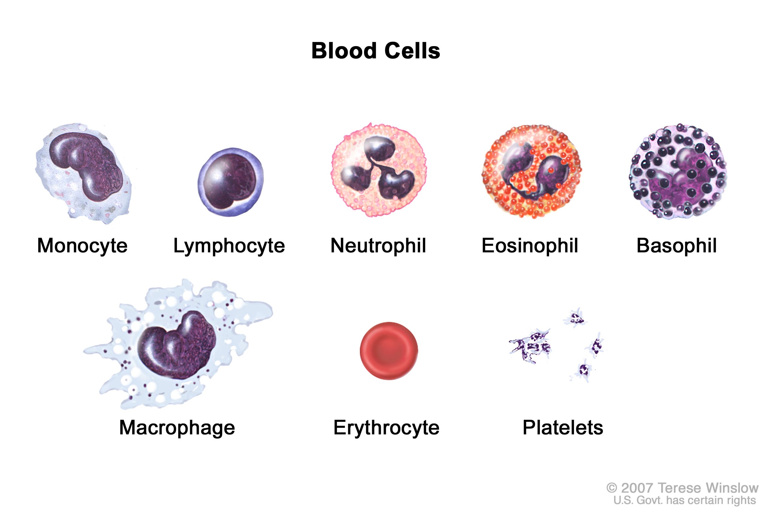
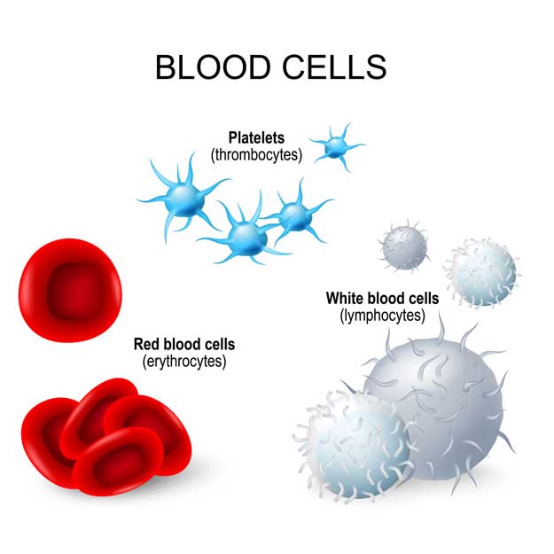
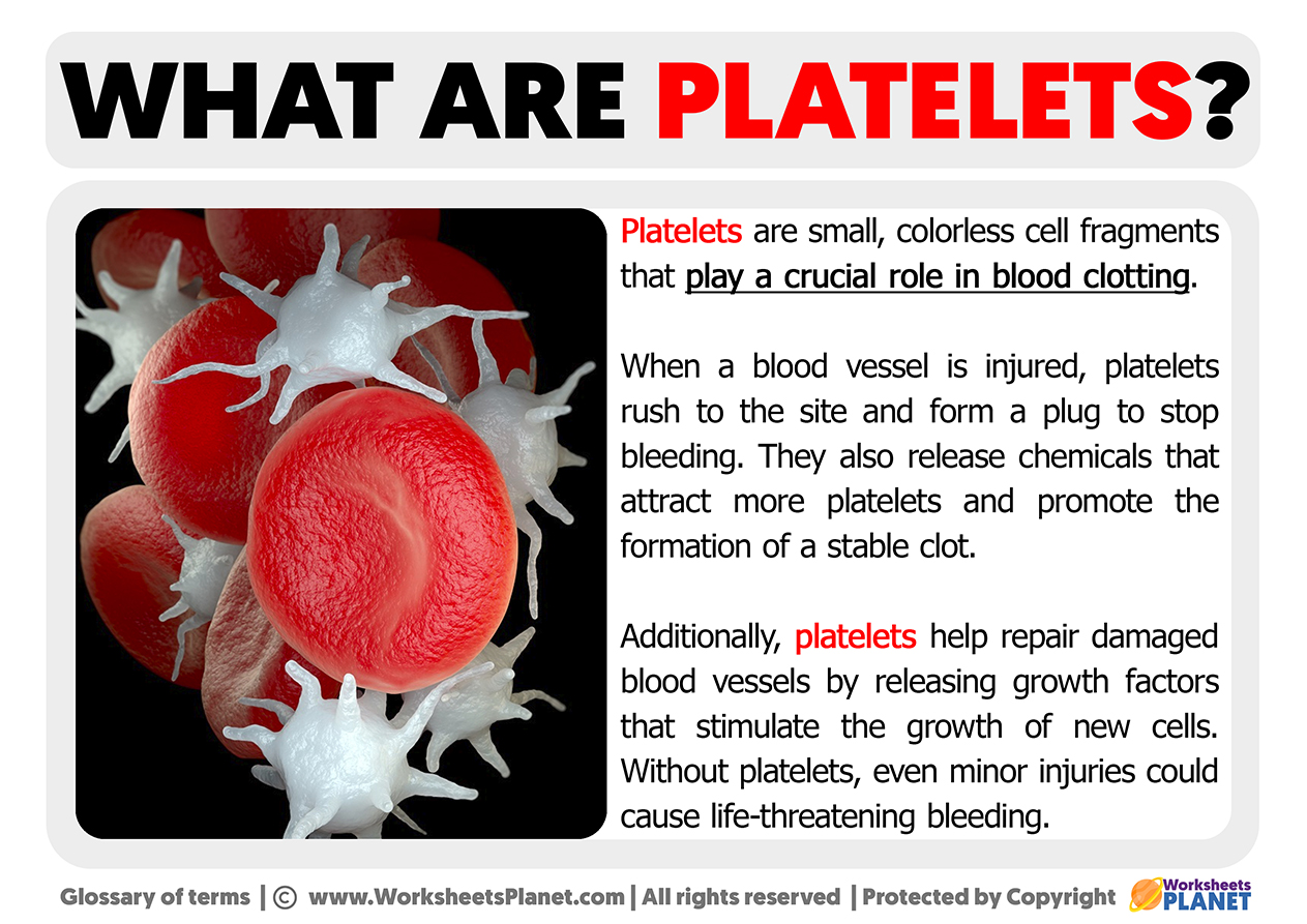
Platelets, often referred to as thrombocytes, are tiny, anucleated cell fragments circulating in our bloodstream, playing a critical role in hemostasis – the process of stopping bleeding. While their fundamental biological function has been understood for decades, the lens through which we now study and leverage their capabilities is increasingly shaped by sophisticated technological advancements. From advanced imaging techniques that visualize their intricate dance to cutting-edge diagnostic tools that assess their function, technology is revolutionizing our understanding and application of platelet biology. This article will delve into the multifaceted roles of platelets, examining them through the prism of modern technology, exploring how these innovations are transforming diagnostics, therapeutics, and our fundamental comprehension of blood health.
The Microscopic Mechanics: Visualizing Platelet Function with Advanced Imaging
The intricate choreography of platelet activation and aggregation, crucial for wound healing and blood clot formation, is a process that remained largely a black box for centuries. However, advancements in microscopy and imaging technologies have granted us unprecedented views into this microscopic world, revealing the dynamic nature of platelets and their complex interactions. These technologies not only deepen our scientific understanding but also underpin the development of more precise diagnostic tools.
Electron Microscopy: Unveiling the Granular Details
Transmission Electron Microscopy (TEM) and Scanning Electron Microscopy (SEM) have been instrumental in providing high-resolution images of platelet ultrastructure. TEM allows us to peer into the internal architecture of platelets, revealing the granular content – alpha granules and dense granules – which are packed with a diverse array of signaling molecules, growth factors, and proteins essential for hemostasis and inflammation. SEM, on the other hand, offers detailed surface topography, showcasing the characteristic shape changes platelets undergo upon activation and their remarkable ability to adhere to damaged blood vessels. These techniques, while requiring highly specialized equipment and expertise, were foundational in identifying platelet morphology and its relation to function. The ability to visualize these granular releases in real-time or in specific activation states has been a cornerstone in understanding the signaling cascades that platelets orchestrate.
Advanced Light Microscopy and Fluorescence Techniques: Dynamic Observation
While electron microscopy provides static, high-resolution snapshots, modern light microscopy techniques offer dynamic insights into platelet behavior in living systems. Confocal microscopy and super-resolution microscopy have revolutionized our ability to observe platelet aggregation, adhesion, and degranulation in live cell imaging. By using fluorescently labeled antibodies or genetically encoded fluorescent proteins, researchers can track individual platelets and their interactions with other blood components and the vessel wall. Techniques like Förster Resonance Energy Transfer (FRET) allow us to monitor molecular interactions within and between platelets in real-time, providing a deeper understanding of signaling pathways. Furthermore, microfluidic devices coupled with advanced imaging platforms allow for the creation of controlled environments that mimic the physiological shear forces experienced by blood, enabling researchers to study platelet function under conditions closer to their natural state. This has been particularly valuable in studying platelet adhesion and aggregation in response to specific stimuli or in the presence of disease-associated factors.
Quantitative Phase Imaging and Digital Holography: Label-Free Analysis
Emerging imaging technologies like Quantitative Phase Imaging (QPI) and Digital Holography offer non-invasive, label-free methods for analyzing platelet behavior. These techniques measure the refractive index and scattering properties of cells, providing quantitative information about cell morphology, volume, and internal structure without the need for fluorescent dyes or chemical fixation. QPI can capture dynamic changes in platelet shape, volume, and internal complexity during activation, offering a continuous and quantitative assessment of their functional state. This has significant implications for high-throughput screening and point-of-care diagnostics, where rapid and objective assessment of platelet function is crucial. The ability to analyze large numbers of platelets without labeling reduces potential artifacts and simplifies experimental procedures.
Technological Diagnostics: Assessing Platelet Function and Health
The diagnostic landscape for platelet disorders has been dramatically transformed by technological innovation. Beyond simple platelet counts, modern diagnostics employ a range of sophisticated techniques to assess platelet function, identify specific deficiencies or abnormalities, and guide therapeutic interventions. These technologies are vital for diagnosing conditions ranging from bleeding disorders to thrombotic events.
Flow Cytometry: Multiparametric Platelet Analysis
Flow cytometry stands as a cornerstone of modern platelet diagnostics. This technology allows for the rapid, multi-parametric analysis of individual cells in a fluid stream. For platelets, flow cytometry enables the simultaneous measurement of cell surface markers (e.g., CD41, CD42b, CD61), intracellular proteins, and the extent of activation (e.g., P-selectin, activated GPIIb/IIIa). By using fluorescently conjugated antibodies, researchers and clinicians can identify different platelet populations, assess their response to various agonists (like ADP, collagen, or thrombin), and quantify the expression of key functional molecules. This multiparametric approach provides a more comprehensive picture of platelet function than traditional methods and is essential for diagnosing a wide spectrum of inherited and acquired platelet disorders, such as Glanzmann thrombasthenia and Bernard-Soulier syndrome. The automation and high throughput of flow cytometry make it indispensable in clinical settings for rapid diagnosis and monitoring.
Platelet Aggregometry: Quantifying Clotting Potential
Platelet aggregometry, a technique that measures the aggregation of platelets in response to various agonists, has been a standard diagnostic tool for decades. However, technological refinements have significantly enhanced its precision and applicability. Modern automated aggregometers utilize optical or impedance-based methods to continuously monitor the increase in light transmission or electrical resistance as platelets aggregate in a stirred suspension. The resulting aggregation curves provide quantitative data on the rate and extent of platelet aggregation, allowing for the characterization of platelet responsiveness. Further technological integration allows for the testing of platelets against a panel of agonists, providing a detailed functional profile. This is crucial for identifying patients with impaired platelet function who are at risk of bleeding or those with hyperactive platelets prone to clotting.
Thromboelastography (TEG) and Rotational Thromboelastometry (ROTEM): Whole Blood Hemostasis Assessment
TEG and ROTEM represent advanced technologies that provide a global assessment of hemostasis, including the contribution of platelets, coagulation factors, and fibrinolysis, within whole blood. These point-of-care devices measure the viscoelastic properties of a forming clot over time, generating a tracing that reflects the kinetics and strength of clot formation and lysis. By incorporating specific activators and inhibitors, TEG and ROTEM can specifically evaluate the role of platelets in clot formation. This technology offers a dynamic and integrated view of hemostatic function, which is particularly valuable in critical care settings for guiding blood product transfusions and managing complex bleeding or thrombotic disorders. Their ability to mimic in vivo conditions more closely than traditional aggregometry makes them powerful tools for real-time hemostatic management.
Next-Generation Sequencing (NGS) and Genetic Testing: Identifying Underlying Predispositions
While not directly measuring platelet function, advancements in genetic technologies, particularly Next-Generation Sequencing (NGS), are revolutionizing the identification of inherited platelet disorders. By analyzing an individual’s genome or exome, NGS can pinpoint genetic mutations in genes known to be involved in platelet development, signaling, or function. This is crucial for diagnosing rare inherited thrombocytopenias and platelet function defects where the underlying genetic cause was previously elusive. This technological capability allows for precise genetic diagnosis, facilitating genetic counseling, family screening, and potentially informing personalized therapeutic strategies.
Computational Modeling and AI: Predicting and Optimizing Platelet-Related Therapies
The sheer complexity of platelet biology and its involvement in a myriad of physiological and pathological processes presents a formidable challenge for traditional research methods. The advent of powerful computational tools, including bioinformatics and artificial intelligence (AI), is opening new frontiers in understanding platelet behavior, predicting disease risk, and optimizing therapeutic interventions.
Bioinformatics and Big Data Analysis: Unraveling Platelet Genomics and Proteomics
The massive datasets generated by high-throughput technologies like NGS and proteomics are being harnessed through bioinformatics to reveal intricate networks of gene and protein interactions within platelets. By analyzing these complex biological datasets, researchers can identify novel genes and pathways associated with platelet disorders, discover biomarkers for disease progression, and gain a deeper understanding of the molecular underpinnings of platelet function. Computational analysis can identify subtle patterns and correlations that might be missed by manual inspection, leading to new hypotheses and avenues of research. This is crucial for understanding the heterogeneity of platelet responses in different individuals and disease states.
Machine Learning and AI in Platelet Research and Diagnostics
Machine learning (ML) and artificial intelligence (AI) are increasingly being applied to platelet research and diagnostics. ML algorithms can be trained on large datasets of clinical and laboratory information to identify predictive models for various conditions. For instance, AI can be used to analyze flow cytometry data to automatically identify abnormal platelet populations or predict the risk of thrombotic events based on a patient’s platelet signature. In drug discovery, AI can accelerate the identification of novel antiplatelet agents by predicting their efficacy and potential side effects. Furthermore, AI-powered image analysis can automate the interpretation of microscopic images of platelets, improving efficiency and consistency in research and diagnostics. The ability of AI to learn from complex data and identify subtle patterns is transforming how we approach platelet biology and its clinical implications.
Computational Fluid Dynamics (CFD) and Microfluidics: Simulating Blood Flow and Platelet Interactions
Computational Fluid Dynamics (CFD) coupled with microfluidics offers powerful tools for simulating blood flow and platelet behavior under various physiological and pathological conditions. CFD models can recreate the complex shear forces and flow patterns within blood vessels, allowing researchers to study how these factors influence platelet adhesion, activation, and aggregation. Microfluidic devices, designed to mimic the microvasculature, provide a controlled environment for experimental validation of CFD predictions and for conducting high-throughput experiments on platelet function under precisely controlled flow conditions. This integrated approach is invaluable for understanding the biomechanics of thrombosis and for designing new devices and therapies that target platelet behavior in specific vascular environments. The ability to simulate and experimentally validate these complex interactions at the micro-scale provides unprecedented insights into in vivo processes.
In conclusion, the humble platelet, once understood primarily for its role in sealing wounds, is now recognized as a dynamic cellular player whose intricate functions are being illuminated and leveraged by a rapidly evolving technological landscape. From the nanoscale visualization of their internal machinery to the macro-level predictive power of AI, technology is not merely an aid but a fundamental driver in our comprehension and clinical application of platelet biology. As these technologies continue to advance, our ability to diagnose, treat, and potentially even prevent a vast array of cardiovascular and hematological diseases will undoubtedly reach new heights, underscoring the indispensable role of innovation in unlocking the full potential of these vital blood components.
aViewFromTheCave is a participant in the Amazon Services LLC Associates Program, an affiliate advertising program designed to provide a means for sites to earn advertising fees by advertising and linking to Amazon.com. Amazon, the Amazon logo, AmazonSupply, and the AmazonSupply logo are trademarks of Amazon.com, Inc. or its affiliates. As an Amazon Associate we earn affiliate commissions from qualifying purchases.