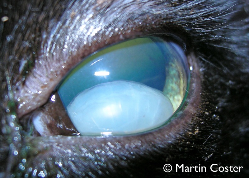
The delicate architecture of a dog’s eye, a marvel of biological engineering, is crucial for vision. Within this intricate system, the lens plays a pivotal role, focusing light onto the retina. However, for some canine companions, this vital structure can become dislodged, a condition known as luxation. While the underlying causes are often rooted in genetics and biology, the ability to accurately diagnose, understand, and manage lens luxation is increasingly reliant on sophisticated technological tools. This article delves into what makes a dog’s eye lens slip out of place, exploring the condition through the lens of veterinary technology.
The Technological Toolkit for Diagnosing Lens Luxation
Diagnosing lens luxation requires more than just a visual inspection. Modern veterinary practices employ a suite of advanced diagnostic technologies to pinpoint the issue, assess its severity, and understand the contributing factors. These tools allow for a precise and early identification, which is critical for effective treatment and preserving the dog’s vision.
Advanced Ophthalmic Examination Equipment
The cornerstone of any ophthalmic diagnosis, including lens luxation, is a thorough examination. While a manual examination is essential, technology significantly enhances its precision and scope.
Slit Lamp Biomicroscopy: Unveiling the Ocular Interior
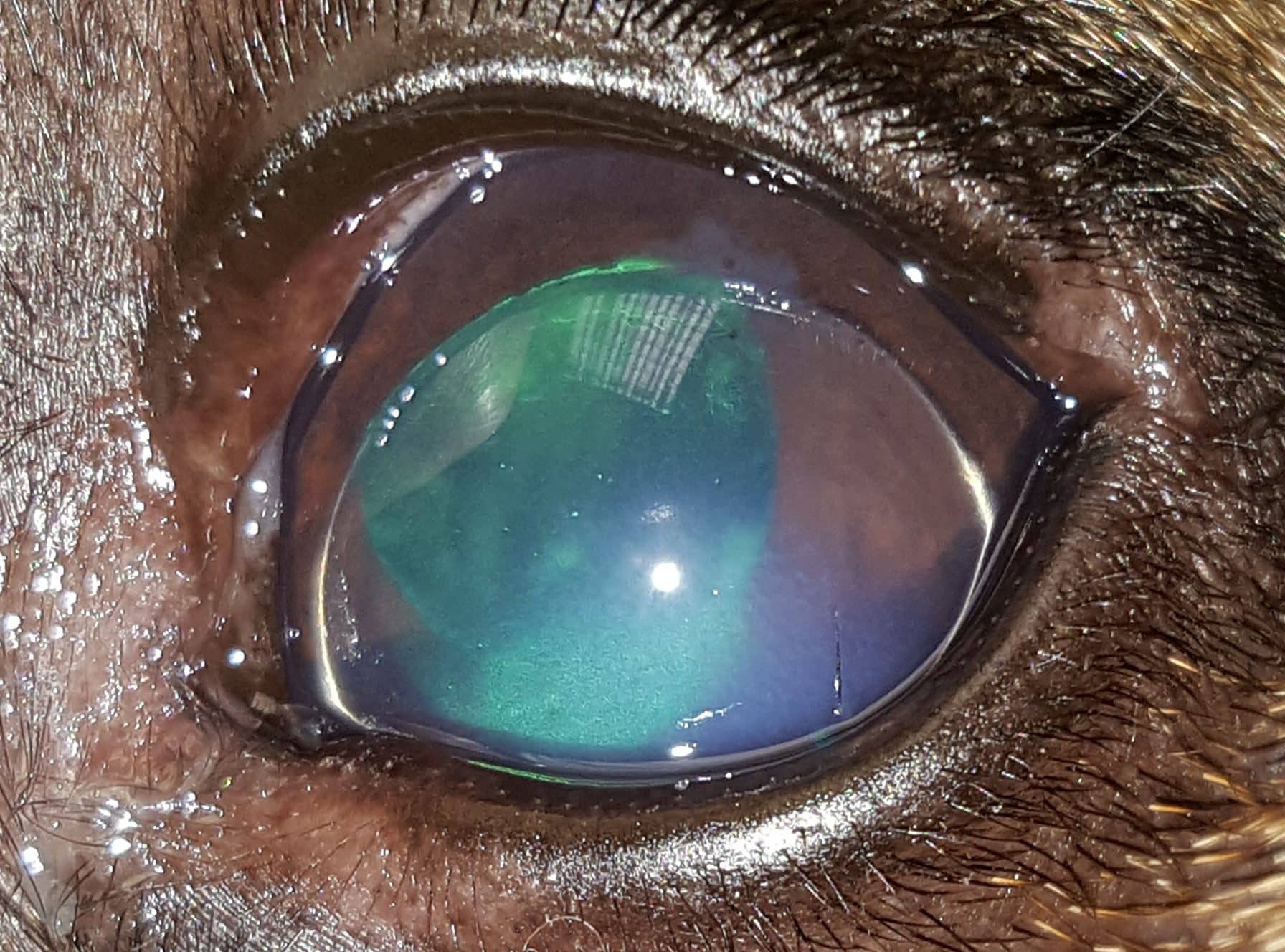
The slit lamp biomicroscope is an indispensable piece of technology in veterinary ophthalmology. This instrument combines a microscope with a specialized light source that can be narrowed into a thin beam. This “slit” allows veterinarians to illuminate and examine different layers of the eye in cross-section. For lens luxation, the slit lamp is crucial for:
- Visualizing Lens Position: The precise location of the lens can be meticulously observed. Displaced lenses will be immediately apparent, whether they have shifted anteriorly into the anterior chamber or posteriorly into the vitreous cavity.
- Assessing Associated Damage: Lens luxation rarely occurs in isolation. The slit lamp allows for the detection of secondary complications such as corneal edema (swelling), iritis (inflammation of the iris), and synechiae (adhesions between the iris and other structures). The intensity and configuration of the slit beam can highlight subtle changes in these tissues that might otherwise be missed.
- Measuring Anterior Chamber Depth: The depth of the anterior chamber is a critical parameter. A luxated lens can drastically alter this depth, and precise measurements can be obtained using calibrated reticles within the biomicroscope’s eyepiece. This quantitative data is vital for surgical planning.
- Documentation and Telemedicine: Many modern slit lamps are equipped with digital cameras or can be interfaced with cameras. This allows for high-resolution image capture, which is invaluable for documenting the condition, tracking its progression, and facilitating consultations with specialists through telemedicine platforms. These digital records form a crucial part of the patient’s technological diagnostic history.
Tonometry: Measuring Intraocular Pressure
Intraocular pressure (IOP) is a vital sign of ocular health. Conditions like lens luxation can significantly impact IOP, often leading to glaucoma, a condition characterized by elevated IOP that damages the optic nerve. Technological advancements in tonometry provide accurate IOP measurements.
- Applanation Tonometry (e.g., Tonopen): Handheld applanation tonometers are widely used. They measure IOP by briefly flattening (applanating) a small area of the cornea. The device then calculates the pressure based on the force required to achieve this flattening. This provides a rapid and relatively accurate assessment of IOP, helping to determine if glaucoma is a concurrent issue.
- Non-Contact Tonometry: While less common in routine veterinary practice, non-contact tonometers use a puff of air to flatten the cornea, offering a quick and less invasive way to screen for elevated IOP. This can be a useful initial screening tool, especially in anxious animals.
- Impact on Surgical Decisions: Accurate IOP readings are critical for determining the appropriate surgical intervention. If glaucoma is present, it may necessitate combined procedures or specific medical management alongside lens removal or repositioning.
Advanced Imaging Modalities: Beyond Visual Inspection
While visual examination is key, advanced imaging technologies offer a deeper, often quantitative, understanding of the ocular structures and the mechanics of lens luxation.
Ocular Ultrasound: Visualizing the Unseen
Ocular ultrasound (echography) is a non-invasive imaging technique that uses sound waves to create images of the eye’s internal structures. It is particularly valuable when direct visualization is compromised, such as in cases of corneal opacity or significant intraocular hemorrhage.
- Detecting Vitreal Changes: In cases of lens luxation, the vitreous humor (the gel-like substance filling the back of the eye) can be altered. Ultrasound can detect liquefaction, syneresis (shrinkage and separation of vitreous fibers), or the presence of inflammatory cells within the vitreous, which can predispose the lens to luxation.
- Assessing the Suspensory Ligaments: While direct visualization of the zonular fibers (the suspensory ligaments that hold the lens in place) can be challenging, ultrasound may indirectly reveal signs of their compromise, such as irregularity or thinning of the ciliary body where they attach.
- Evaluating Secondary Pathologies: Ultrasound can effectively image the retina and choroid, helping to identify detachment, tumors, or other abnormalities that might be associated with or exacerbated by lens luxation. This helps in a comprehensive assessment of ocular health.
- Pre-Surgical Planning: For procedures like phacoemulsification (lens removal), ultrasound can provide valuable information about the overall condition of the eye, guiding the surgical approach.
Optical Coherence Tomography (OCT): High-Resolution Cross-Sectional Imaging
Optical Coherence Tomography (OCT) is a relatively newer but increasingly important technological advancement in veterinary ophthalmology. It uses light waves to produce high-resolution, cross-sectional images of the eye’s tissues, akin to a biological MRI but at a much finer resolution.
- Quantifying Corneal Thickness: In cases of corneal edema secondary to lens luxation (e.g., due to the lens rubbing against the cornea), OCT can precisely measure corneal thickness, aiding in the assessment of severity and monitoring treatment response.
- Assessing Retinal Integrity: OCT provides detailed imaging of the retinal layers. This is crucial for evaluating any potential damage to the retina caused by the displaced lens or secondary glaucoma, helping to predict visual prognosis.
- Evaluating Anterior Segment Structures: OCT can visualize the anterior chamber angle, the iris, and the ciliary body with remarkable detail. This can aid in identifying structural abnormalities that might predispose to lens instability or provide information about the status of the zonular apparatus, albeit indirectly.
- Prognostic Value: By providing detailed structural information, OCT can offer a more refined prognosis for vision retention, guiding owner expectations and treatment decisions.
Genetic Predisposition and Technological Detection
While a direct “lens slip” is a mechanical event, the underlying predisposition is often genetic. Technological advancements are now playing a role in identifying these predispositions.
DNA Testing and Genetic Markers
Certain breeds are predisposed to inherited conditions that weaken the suspensory ligaments, making lens luxation more likely. While direct DNA tests for lens luxation might not be universally available for all breeds, the field of veterinary genomics is rapidly evolving.
- Genome-Wide Association Studies (GWAS): Researchers are increasingly using GWAS to identify genetic variations associated with common canine diseases, including ocular conditions. These studies involve analyzing the complete genomes of large cohorts of affected and unaffected dogs. By identifying specific genetic markers that are more prevalent in dogs with lens luxation, breeders and owners can be alerted to higher-risk individuals.
- Predictive Genetic Screening: As more genetic markers are identified, the development of predictive genetic tests becomes feasible. These tests can analyze a dog’s DNA to determine its likelihood of developing lens luxation, allowing for early intervention or informed breeding decisions. This represents a proactive technological approach to managing inherited diseases.
- Data Analysis and Machine Learning: The vast datasets generated by genomic research require sophisticated data analysis techniques, including machine learning algorithms. These algorithms can identify complex patterns and interactions between genes that might contribute to lens luxation, leading to a more nuanced understanding of its genetic underpinnings.
Advanced Imaging for Early Detection of Predisposition
Even before clinical signs of luxation appear, subtle structural changes may be present that predispose a dog to the condition. Technological imaging plays a role in detecting these early indicators.
High-Resolution Ultrasound for Zonular Assessment
While not always routine, specialized high-resolution ultrasound protocols can be employed to assess the integrity of the zonular fibers and the ciliary body. Subtle signs of weakness or laxity might be detectable by experienced sonographers, providing an early warning of potential instability. This is a frontier where technological refinement in ultrasound probes and software can improve diagnostic capabilities.
Scheimpflug Imaging for Corneal and Anterior Segment Analysis
Scheimpflug imaging is a non-contact method that uses a rotating camera to capture cross-sectional images of the anterior segment of the eye, including the cornea and iris.
- Corneal Thickness and Curvature: It provides precise measurements of corneal thickness at various points, which can be relevant in some forms of lens instability.
- Anterior Chamber Analysis: Scheimpflug imaging allows for detailed analysis of the anterior chamber, including its depth and the configuration of the iridocorneal angle. While its primary use is often in glaucoma diagnostics, the detailed anatomical mapping can contribute to understanding the overall biomechanical environment of the lens.
The Role of Computational Modeling and Simulation
While still in its nascent stages for veterinary ophthalmology, the application of computational modeling and simulation holds immense potential in understanding and predicting conditions like lens luxation.
Biomechanical Simulations of the Eye
By inputting data obtained from advanced imaging technologies (like OCT and ultrasound), sophisticated computer models can simulate the biomechanical forces acting on the ocular structures.
- Stress and Strain Analysis: These models can analyze how stress and strain are distributed within the suspensory ligaments and surrounding ocular tissues under various physiological conditions. This could help identify specific areas of weakness or predict where failure is most likely to occur, leading to luxation.
- Predicting Luxation Events: By simulating different scenarios of ocular movement or external forces, researchers could potentially predict the likelihood and trajectory of lens luxation in predisposed individuals. This predictive power would be revolutionary for preventative strategies.
Artificial Intelligence (AI) in Diagnostic Support
AI is increasingly being integrated into medical diagnostics, and veterinary ophthalmology is no exception.
- Image Analysis and Pattern Recognition: AI algorithms can be trained on vast datasets of ocular images from slit lamp examinations, ultrasound, and OCT scans. These algorithms can then assist veterinarians in identifying subtle abnormalities that might indicate early signs of lens instability or associated pathologies. For example, AI could be programmed to recognize patterns in vitreous changes or ciliary body morphology indicative of increased risk.
- Predictive Analytics: Leveraging genetic data and diagnostic imaging, AI could be employed to build predictive models that estimate a dog’s individual risk for developing lens luxation. This would allow for highly personalized preventative care strategies.
- Aid in Surgical Planning: AI could potentially analyze pre-operative imaging data to assist surgeons in planning the most effective approach for lens removal or management, optimizing outcomes.
![]()
Conclusion: Technology as the Key to Understanding and Managing Lens Luxation
The question of “what makes a dog’s eye lens slip out of place” is a complex interplay of genetics and biomechanics. However, the ability to definitively answer this question for individual cases, to predict risk, and to effectively manage the condition hinges critically on technological advancements. From the high-resolution imaging capabilities of slit lamps and OCT to the genetic insights gleaned from DNA analysis and the future potential of AI and computational modeling, technology is not just assisting in diagnosis; it is fundamentally transforming our understanding and management of lens luxation in veterinary medicine. By embracing and advancing these technological tools, we can continue to improve the vision and quality of life for our canine companions.
aViewFromTheCave is a participant in the Amazon Services LLC Associates Program, an affiliate advertising program designed to provide a means for sites to earn advertising fees by advertising and linking to Amazon.com. Amazon, the Amazon logo, AmazonSupply, and the AmazonSupply logo are trademarks of Amazon.com, Inc. or its affiliates. As an Amazon Associate we earn affiliate commissions from qualifying purchases.