
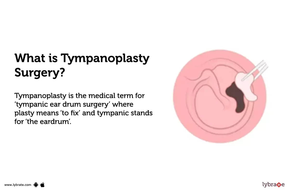
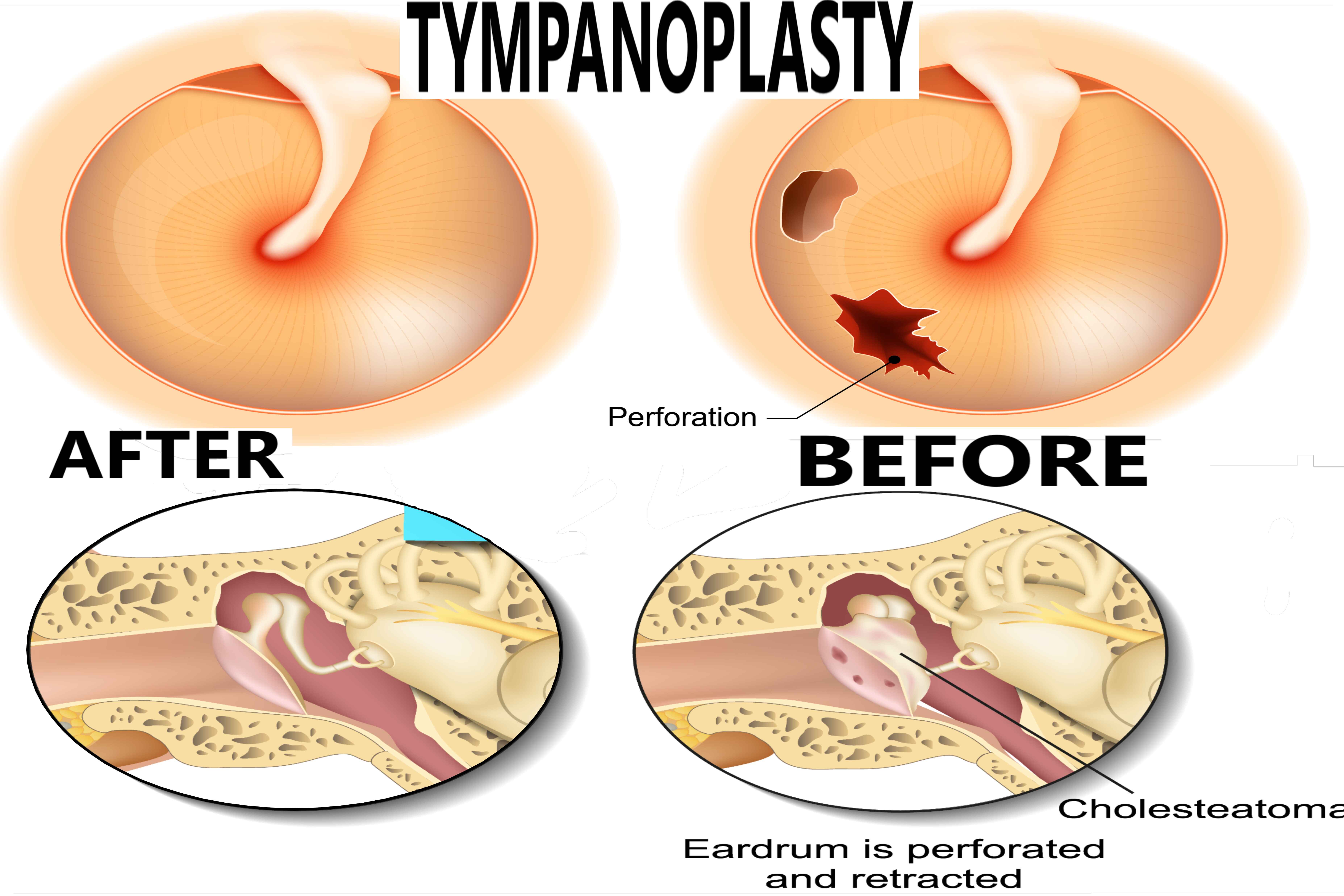
Tympanoplasty, at its core, is a surgical procedure designed to repair or reconstruct the eardrum (tympanic membrane) and/or the small bones (ossicles) of the middle ear. Historically, this delicate operation relied heavily on the surgeon’s skill and rudimentary tools. However, in the 21st century, understanding “what is tympanoplasty” requires a deep dive into the sophisticated technologies that have transformed it from an intricate manual task into a highly precise, often minimally invasive, and technologically driven specialty. This exploration delves into how advanced diagnostics, innovative surgical instruments, novel materials, and digital integration collectively elevate tympanoplasty, making it a testament to medical technological progress.
The Core Procedure: A Symphony of Precision and Imaging Technology
Modern tympanoplasty is less about brute force and more about meticulous precision, enabled by an array of diagnostic and surgical technologies that guide every step of the process.
Diagnosing the Need: Advanced Imaging and Audiometry
Before a single incision is made, technology plays a critical role in accurately diagnosing the need for tympanoplasty and mapping out the surgical strategy.
-
Audiometry and Tympanometry: These are foundational diagnostic tools. Audiometry, an electronic test, uses specialized computer-controlled equipment to measure hearing thresholds across various frequencies, precisely identifying the degree and type of hearing loss. Tympanometry, another electronically driven test, assesses the function of the middle ear by measuring eardrum movement in response to changes in air pressure. These technologies provide crucial data on the acoustic performance of the ear, helping pinpoint issues related to eardrum perforation or ossicular chain dysfunction.
-
High-Resolution Computed Tomography (CT) Scans: For more complex cases, especially those involving the ossicular chain or suspicion of cholesteatoma (a skin cyst in the middle ear), high-resolution CT scans are indispensable. These advanced imaging technologies generate detailed cross-sectional images of the temporal bone and middle ear structures, providing surgeons with a three-dimensional understanding of the pathology, bone erosion, and anatomical variations. The digital processing of these images allows for precise pre-operative planning and identification of potential surgical challenges.
-
Magnetic Resonance Imaging (MRI): While less common for routine tympanoplasty, MRI may be utilized in specific scenarios, such as differentiating soft tissue lesions, evaluating inflammatory processes, or assessing complications like intracranial extensions. The sophisticated magnetic fields and radio waves of MRI technology offer superior soft tissue contrast compared to CT, providing complementary information when indicated.
Surgical Techniques and Micro-Instrumentation
The execution of tympanoplasty is a masterclass in micro-engineering and precision. The surgical field, often no larger than a fingernail, necessitates tools far beyond typical surgical instruments.
-
Operating Microscopes: The cornerstone of traditional tympanoplasty, the operating microscope, provides magnified, stereoscopic vision (typically 6x to 25x magnification) of the middle ear structures. These highly advanced optical systems are equipped with powerful illumination, often fiber-optic, to illuminate the deep, narrow surgical field. Many modern microscopes also integrate digital cameras for recording procedures, allowing for detailed documentation, teaching, and real-time display for assistants.
-
Endoscopic Systems: A significant technological leap has been the advent of endoscopic tympanoplasty. Rigid endoscopes, typically 2.7 mm or 3 mm in diameter with various angled views (0, 30, 45 degrees), are inserted directly into the ear canal. Connected to a high-definition camera and monitor, these systems provide a wide, panoramic view of the middle ear that can often exceed the limited straight-line view of a microscope. The high-resolution digital image projected onto a large screen allows for enhanced visualization, particularly of hidden areas, and facilitates a less invasive approach.
-
Microsurgical Tools: The instruments used in tympanoplasty are miniaturized versions of standard surgical tools, designed with unparalleled precision. These include fine picks, hooks, elevators, scissors, and forceps, all crafted from high-grade metals to ensure durability and sterility. Their ergonomic design minimizes surgeon fatigue and maximizes control in the confined space of the middle ear. Advanced manufacturing techniques are crucial for producing these incredibly delicate yet robust instruments.
Grafting Materials and Bioengineering
The materials used to repair the eardrum or reconstruct the ossicular chain are a testament to advancements in biomaterials and tissue engineering.
-
Autologous Grafts: The most common graft materials are harvested from the patient’s own body (autologous), reducing rejection risk. These include fascia (from behind the ear muscle), perichondrium (from cartilage), or tragal cartilage. While naturally occurring, the preparation and shaping of these grafts often involve microscopic precision and the use of specialized cutting and shaping tools.
-
Synthetic Grafts and Prostheses: For ossicular chain reconstruction (ossiculoplasty), synthetic prostheses made from biocompatible materials like titanium, hydroxyapatite, or polyethylene are frequently employed. These prostheses are precisely engineered with specific dimensions and designs to mimic the natural ossicles, restoring sound conduction. The development of these advanced biomaterials has drastically improved long-term functional outcomes and biocompatibility, minimizing adverse reactions within the delicate middle ear environment. Research into biodegradable scaffolds and tissue engineering is also paving the way for future regenerative solutions.
Innovations in Tympanoplasty: Pushing the Boundaries of Surgical Tech
The field of tympanoplasty is in constant evolution, driven by technological breakthroughs that enhance precision, minimize invasiveness, and improve patient outcomes.
Endoscopic Tympanoplasty: A Minimally Invasive Revolution
The rise of endoscopic tympanoplasty represents a paradigm shift. Unlike the traditional microscopic approach that often requires a post-auricular incision and removal of bone (canalplasty) for access, the endoscope allows for direct visualization through the ear canal.
-
Technological Advantages: Endoscopic systems offer a panoramic, wide-angle view, often with superior illumination and magnification on a high-definition monitor. This “keyhole surgery” approach leads to smaller incisions (or no external incision at all), reduced post-operative pain, faster recovery times, and often preserved external ear anatomy. The technology of these sophisticated endoscopes, with their precise optics, integrated light sources, and robust camera systems, has made this minimally invasive technique a viable and often preferred option.
-
Ergonomics and Training: The two-dimensional view of the endoscope requires a different skill set than the 3D microscopic view. However, advancements in training simulators, often utilizing virtual reality, are helping surgeons adapt to this newer technology, offering a safe environment to master hand-eye coordination and surgical maneuvers.
Laser-Assisted Techniques and Precision Energy Devices
Lasers have found their niche in otologic surgery, offering unparalleled precision in tissue manipulation.
-
CO2 Lasers: In tympanoplasty, CO2 lasers can be used to precisely remove scar tissue, vaporize small lesions, or reshape cartilage grafts with minimal bleeding and collateral damage. The laser’s ability to ablate tissue with a high degree of control reduces trauma to surrounding delicate structures.
-
Other Energy Devices: Beyond lasers, various precision energy devices, such as radiofrequency ablation tools, are used for hemostasis (controlling bleeding) and precise tissue cutting. These technologies allow for a cleaner surgical field and improved visualization, critical for microscopic and endoscopic procedures within the middle ear.
3D Printing and Custom Implants
Additive manufacturing, or 3D printing, is beginning to revolutionize the fabrication of custom medical devices.
-
Custom Ossicular Prostheses: For complex ossicular chain defects, 3D printing allows for the creation of patient-specific prostheses that perfectly match the individual anatomy. This “tailor-made” approach, leveraging high-resolution CT data to design the implant, promises improved fit, stability, and auditory outcomes compared to off-the-shelf options. Biocompatible materials, including titanium and advanced polymers, are used in these custom fabrications.
-
Bio-Scaffolds for Regeneration: Looking ahead, 3D printing could create scaffolds for regenerative medicine, potentially supporting the growth of new eardrum tissue using the patient’s own cells, moving beyond simple repair to true biological reconstruction. This represents a significant shift from material replacement to biological regeneration, entirely enabled by advanced manufacturing technology.

The Role of Digital Integration and AI in Pre- and Post-Operative Care
Beyond the operating room, digital technologies and artificial intelligence are increasingly augmenting tympanoplasty care, improving diagnostics, planning, and patient management.
AI in Diagnostic Support and Surgical Planning
Artificial intelligence and machine learning algorithms are emerging as powerful tools in otology.
-
Enhanced Diagnostics: AI can analyze vast amounts of imaging data (CT, MRI) with greater speed and accuracy than the human eye, identifying subtle pathologies, quantifying disease extent, and even predicting the likelihood of certain complications. For example, AI could assist in precisely measuring the size and location of eardrum perforations or detecting early signs of cholesteatoma.
-
Predictive Analytics for Surgical Outcomes: By analyzing patient data, surgical parameters, and post-operative results, AI models can potentially predict surgical success rates or identify patients at higher risk for complications, allowing surgeons to refine their approach or better counsel patients.
-
Automated Measurement and Segmentation: AI-powered software can automatically segment and measure anatomical structures from imaging studies, providing surgeons with precise metrics for surgical planning, such as the exact dimensions required for a graft or prosthesis.
Telemedicine and Remote Monitoring
Digital communication platforms have transformed accessibility and follow-up care.
-
Pre-Operative Consultations: Telemedicine allows patients, particularly those in remote areas, to have initial consultations with specialists, review imaging, and discuss treatment plans without the need for extensive travel. This technology leverages secure video conferencing and digital medical record systems.
-
Post-Operative Follow-up and Monitoring: Remote monitoring tools, including wearable sensors or app-based questionnaires, can track patient recovery, identify early signs of infection or complications, and facilitate timely intervention. Secure online portals allow patients to communicate directly with their care team, upload images of their healing ear, and receive personalized advice.
Virtual Reality (VR) and Augmented Reality (AR) in Surgical Training
The complexity of middle ear surgery makes high-fidelity training essential, and VR/AR technologies are providing innovative solutions.
-
VR Surgical Simulators: VR platforms offer immersive, interactive environments where aspiring otologic surgeons can practice tympanoplasty procedures. These simulators provide haptic feedback, mimicking the tactile sensations of drilling bone or manipulating delicate tissues, allowing repeated practice without patient risk. The metrics provided by VR simulators help track progress and identify areas for improvement.
-
AR for Intraoperative Guidance: In the future, augmented reality could overlay patient-specific anatomical data and surgical plans directly onto the surgeon’s view during the actual procedure, providing real-time guidance and enhancing precision, especially for complex cases with challenging anatomy.
Future Horizons: AI, Robotics, and Regenerative Medicine
The trajectory of tympanoplasty continues upwards, with cutting-edge technologies promising even greater precision, less invasiveness, and potentially true biological regeneration.
Robotic-Assisted Microsurgery
Robotics are poised to enter the delicate world of otologic surgery.
-
Enhanced Precision and Tremor Reduction: Robotic systems offer unparalleled precision, capable of movements far steadier and more controlled than even the most skilled human hand. This can significantly reduce tremor, particularly crucial in the microscopic environment of the middle ear where millimeters make all the difference.
-
Minimally Invasive Approaches: Future robotic systems could enable even smaller incisions or entirely endoscopic, single-port approaches, accessing the middle ear through natural orifices with minimal disruption to surrounding tissues.
-
Remote Surgery: While still largely conceptual for such delicate procedures, robotic telepresence could eventually allow expert surgeons to perform tympanoplasties from remote locations, extending specialized care globally.
Advanced Regenerative Strategies
The ultimate goal for many medical conditions is regeneration, and tympanoplasty is no exception.
-
Stem Cell Research: Research is actively exploring the use of stem cells, often in conjunction with biocompatible scaffolds, to regenerate damaged eardrum tissue or even entire ossicles. This moves beyond merely patching a hole or inserting a prosthesis to truly restoring the native biological structure and function.
-
Bio-fabrication: Advanced bio-fabrication techniques, including bioprinting, aim to create functional tissues and organs. Applying this to the middle ear could lead to customized, living grafts that integrate seamlessly with the patient’s body, offering a more durable and physiologically sound repair.
Integrated Digital Operating Rooms
The operating room of the future will be a highly integrated digital ecosystem.
-
Seamless Data Flow: Diagnostic images, real-time physiological data, surgical planning models, and instrument telemetry will all be displayed and analyzed concurrently, providing the surgical team with a comprehensive and dynamic view of the patient and procedure.
-
Intelligent Navigation and Feedback: AI-driven systems could provide real-time feedback on surgical maneuvers, warn of potential risks, or even suggest optimal pathways based on anatomical mapping and pre-operative planning.
Conclusion
Tympanoplasty is a remarkable illustration of how medical science and technological innovation converge to restore vital sensory function. From the sophisticated imaging technologies that enable precise diagnosis to the meticulously engineered micro-instruments, endoscopic systems, and biocompatible materials used in reconstruction, every aspect of modern tympanoplasty is deeply rooted in technological advancement. As we look to the future, the integration of AI, robotics, and advanced regenerative medicine promises to continue pushing the boundaries, making tympanoplasty an ever more precise, minimally invasive, and effective procedure. Understanding “what is tympanoplasty” today is intrinsically linked to appreciating the continuous march of technological progress in medical science, forever shaping how we heal and restore the miracle of hearing.
aViewFromTheCave is a participant in the Amazon Services LLC Associates Program, an affiliate advertising program designed to provide a means for sites to earn advertising fees by advertising and linking to Amazon.com. Amazon, the Amazon logo, AmazonSupply, and the AmazonSupply logo are trademarks of Amazon.com, Inc. or its affiliates. As an Amazon Associate we earn affiliate commissions from qualifying purchases.