
The human body is a marvel of intricate design, a complex system where every component plays a crucial role. Among its many landmarks, the sternal angle, also known as the Angle of Louis, stands out as a critical anatomical reference point. Far from being just a static feature, its accurate identification and measurement are paramount in numerous medical, diagnostic, and even rehabilitative contexts. In an era defined by rapid technological advancement, our understanding and interaction with the sternal angle have been revolutionized, moving beyond manual palpation to sophisticated imaging, AI-driven analysis, and real-time biometric monitoring. This article explores the sternal angle through a technological lens, examining how innovative tools and platforms enhance our ability to visualize, analyze, and leverage this vital anatomical junction.
The Anatomical Blueprint: Why the Sternal Angle Matters to Technology
Before delving into the technological applications, it’s essential to understand what the sternal angle is and why its precise identification is so significant. This foundational knowledge allows us to appreciate the transformative impact technology has had on its study.
Defining the Sternal Angle (Angle of Louis)
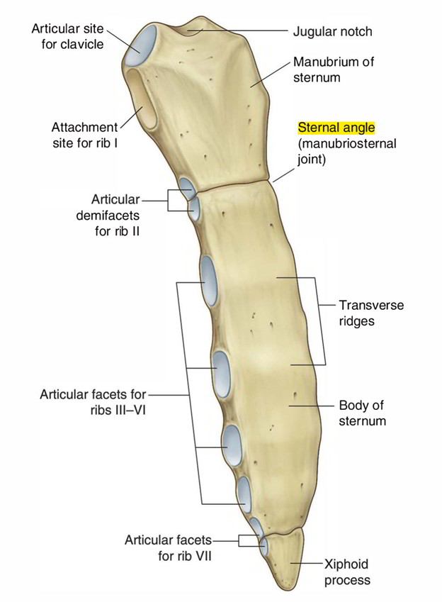
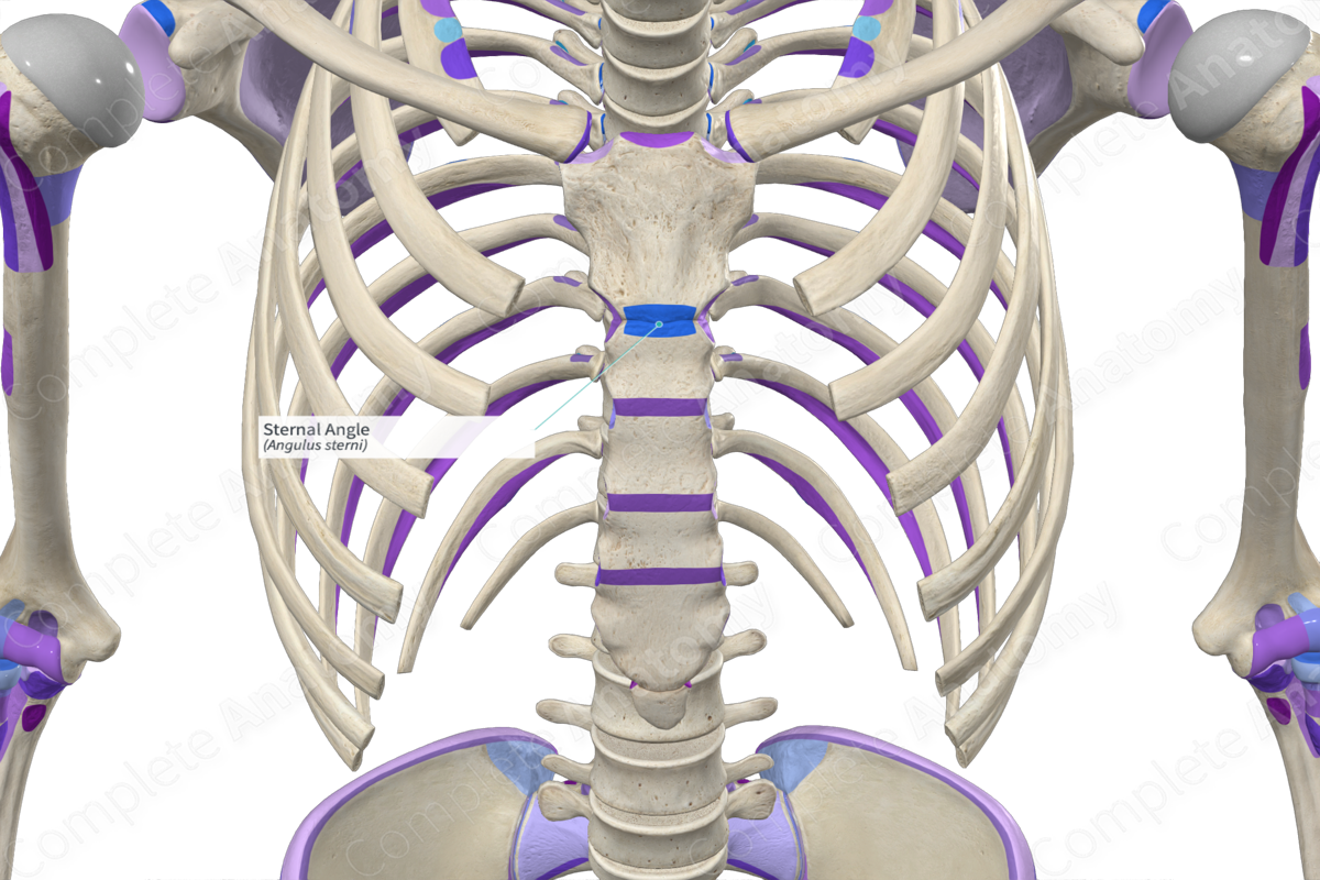
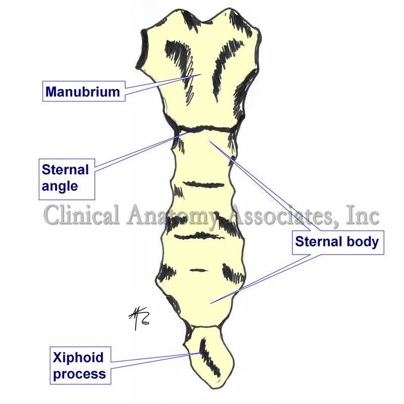
The sternal angle is the palpable, horizontal ridge located at the junction between the manubrium (the upper part of the sternum or breastbone) and the body of the sternum. It marks a slight angulation that is typically convex anteriorly. This angle corresponds to several other critical anatomical landmarks: it is at the level of the second costal cartilage, which articulates with the second rib; it aligns with the intervertebral disc between the T4 and T5 thoracic vertebrae; and it often indicates the level of the aortic arch, the bifurcation of the trachea into the main bronchi, and the upper border of the atria of the heart. For clinicians, it’s a consistent and easily identifiable landmark, making it invaluable for anatomical orientation.
Clinical and Biomechanical Relevance
The sternal angle’s significance extends far beyond mere anatomical reference. Clinically, it serves as a critical starting point for counting ribs, assessing chest wall deformities, and pinpointing the location of intrathoracic structures during physical examinations. Deviations or abnormalities in the sternal angle can be indicative of various conditions, from congenital anomalies to post-traumatic changes or even the progression of certain diseases affecting the chest wall and spine.
From a biomechanical perspective, the sternal angle plays a role in the mechanics of breathing and chest wall expansion. The articulation of the second ribs at this point means that its position and mobility influence the overall movement of the rib cage during respiration. Understanding its dynamics is crucial for diagnosing restrictive lung diseases, assessing the impact of musculoskeletal disorders on respiratory function, and designing effective rehabilitative interventions. Given these multifaceted roles, the demand for precise, repeatable, and non-invasive methods to assess the sternal angle has spurred significant technological innovation.
Advanced Imaging: Visualizing the Sternal Angle with Precision
The evolution of medical imaging has dramatically transformed our ability to “see” inside the human body, providing unparalleled detail and allowing for highly accurate measurements of complex anatomical structures like the sternal angle.
From X-rays to 3D Reconstruction
Traditional plain film X-rays offered a foundational view of the sternum, allowing clinicians to identify gross abnormalities. However, their two-dimensional nature often made precise angular measurements challenging due to superimposition of structures and projection distortions. The advent of Computed Tomography (CT) scans marked a significant leap forward. CT provides cross-sectional images, eliminating superimposition and offering clearer visualization of bone and soft tissue structures around the sternal angle. More importantly, advancements in CT technology, particularly multi-detector CT (MDCT), enable rapid acquisition of volumetric data, which can then be used for sophisticated 3D reconstructions.
Magnetic Resonance Imaging (MRI), while less ideal for direct bone visualization, excels in soft tissue differentiation and can provide complementary information, especially in cases where soft tissue pathology around the sternum is suspected. For measuring the sternal angle itself, specialized techniques like kinematic MRI can even capture the dynamic movement of the chest wall, offering insights into how the angle changes during respiration or movement, a capability unattainable with static imaging.
Digital Measurement and Analysis Tools
The raw data from CT and MRI scans is invaluable, but its true power is unlocked by advanced digital measurement and analysis software. These tools allow radiologists and researchers to perform highly precise measurements of the sternal angle in three dimensions. Specialized software platforms can:
- Render 3D models: Recreate the sternum and surrounding rib cage in a virtual 3D space, allowing for viewing from any angle.
- Define anatomical planes: Precisely establish reference planes and axes based on bony landmarks.
- Calculate angles and distances: Automatically or manually measure the angle formed by the manubrium and sternal body, as well as distances to other structures.
- Segment structures: Isolate the sternum from other tissues, facilitating focused analysis.
These digital tools not only enhance accuracy and reproducibility compared to manual methods but also enable comparative studies, track changes over time, and facilitate pre-operative planning for corrective surgeries, all with unprecedented detail and efficiency.
Artificial Intelligence and Machine Learning: Automating Sternal Angle Insights
The sheer volume and complexity of medical imaging data make it an ideal candidate for analysis by artificial intelligence (AI) and machine learning (ML). These technologies are increasingly being leveraged to automate, accelerate, and enhance the interpretation of sternal angle data.
Automated Detection and Anomaly Identification
AI algorithms, particularly deep learning models trained on vast datasets of medical images, can be programmed to automatically identify the sternal angle within CT or MRI scans. This capability is revolutionary for several reasons:
- Speed and Efficiency: What might take a human expert several minutes to meticulously identify and measure can be done by an AI in seconds.
- Consistency: AI reduces inter-observer variability, ensuring highly consistent measurements across different scans and different clinicians.
- Automated Anomaly Detection: Beyond just identifying the angle, advanced AI can be trained to recognize subtle deviations from a “normal” range. This could include detecting unusually acute or obtuse angles, or structural irregularities that might indicate congenital defects, fractures, or early signs of disease like pectus excavatum or carinatum. Such early and automated detection can flag cases for further human review, potentially leading to earlier diagnoses and interventions.
Predictive Analytics for Chest Wall Health
Building on automated detection, machine learning models can process sternal angle data in conjunction with other clinical parameters (patient demographics, symptoms, genetic markers, etc.) to develop powerful predictive analytics tools. These tools can:
- Predict Disease Progression: For conditions affecting the chest wall, ML models might predict how a deformity (e.g., pectus excavatum) is likely to progress over time, or how a surgical correction might alter chest mechanics.
- Assess Treatment Outcomes: By analyzing pre- and post-intervention imaging, AI can help evaluate the success of surgical corrections or other treatments, correlating changes in the sternal angle with functional improvements.
- Identify Risk Factors: Large-scale analysis of sternal angle data across diverse populations can uncover subtle correlations between sternal angle morphology and increased risk for certain respiratory or cardiovascular conditions, contributing to proactive healthcare strategies. This integration of AI allows for a more comprehensive and forward-looking approach to chest wall health.
Wearable Technology and Biometric Sensors: Real-time Sternal Angle Monitoring
The application of technology isn’t confined to radiology suites. Wearable devices and embedded biometric sensors are bringing the assessment of anatomical landmarks like the sternal angle into everyday life, albeit often indirectly.
Posture Correction and Ergonomic Design
While no wearable directly “measures” the sternal angle in isolation, many advanced posture monitoring systems use sensors placed on the chest or upper back to track overall torso alignment. These devices often incorporate gyroscopes, accelerometers, and magnetometers to create a real-time model of a user’s posture. The sternal angle, as a key component of chest wall geometry, plays an indirect but vital role in these models.
- Reference Point: The area around the sternal angle serves as a general reference for sensors attempting to determine if the chest is slumped, excessively arched, or symmetrically aligned.
- Feedback Mechanisms: When a user deviates from an ideal posture, the wearable can provide haptic (vibration) or auditory feedback, encouraging correction. For instance, prolonged slouching, which would alter the relative orientation of the manubrium and sternal body, could trigger an alert.
- Ergonomic Optimization: Data collected over time can inform ergonomic adjustments in workspaces or guide the design of supportive furniture and apparel that promote healthier postural alignment.
Respiratory Kinematics and Movement Analysis
The sternal angle’s role in respiratory mechanics makes it a valuable anatomical point for devices aimed at monitoring breathing.
- Strain Gauges and Smart Fabrics: Wearable vests or smart fabrics embedded with strain gauges or other textile sensors can detect minute changes in chest circumference and shape during breathing. The expansion and contraction of the chest wall, including movements around the sternal angle, are captured by these sensors.
- Asthma and COPD Management: For individuals with respiratory conditions like asthma or COPD, real-time monitoring of chest wall expansion patterns (which are influenced by sternal angle mobility) can provide valuable data on breathing efficiency, early signs of distress, or the effectiveness of bronchodilator use.
- Fitness and Performance: Athletes and fitness enthusiasts use wearables to optimize breathing techniques. By analyzing chest wall motion, these devices can help improve diaphragmatic breathing, monitor respiratory rate, and even detect patterns indicative of fatigue or overexertion, all of which are influenced by the dynamics of the sternal angle region.
The Future of Sternal Angle Applications: Robotics and Personalized Medicine
Looking ahead, the integration of advanced technologies with anatomical understanding promises even more sophisticated applications for the sternal angle. Robotics and personalized medicine stand at the forefront of these innovations.
Robotic-Assisted Procedures and Surgical Planning
Robotic surgery platforms, such as the Da Vinci system, are renowned for their precision and ability to perform complex procedures through tiny incisions. The sternal angle, as a gateway to the mediastinum and a reference for intrathoracic organs, plays a crucial role in surgical planning.
- Precise Navigation: Surgeons can utilize 3D anatomical models derived from CT/MRI data, including the precise location of the sternal angle, to plan robotic port placement and instrument trajectories with sub-millimeter accuracy. This minimizes tissue damage and improves surgical outcomes.
- Virtual Reality (VR) and Augmented Reality (AR) Training: Surgical trainees can practice complex procedures on virtual patients, where anatomical landmarks like the sternal angle are rendered with photorealistic detail. AR can overlay digital anatomical data onto the patient during actual surgery, providing real-time guidance and enhancing the surgeon’s spatial awareness of structures relative to the sternal angle.
- Minimally Invasive Interventions: For conditions like pectus excavatum, robotic-assisted techniques could potentially offer less invasive methods for sternal elevation or stabilization, guided by precise measurements and real-time feedback referencing the sternal angle.
Personalized Biomechanical Modeling
The concept of “digital twins”—virtual replicas of a patient’s anatomy and physiology—is gaining traction in personalized medicine. The sternal angle will be an integral feature in these complex models.
- Patient-Specific Simulations: Engineers and clinicians can build highly detailed biomechanical models of individual patients, incorporating their unique sternal angle morphology and its relationship to the rib cage, spine, and internal organs.
- Treatment Simulation: These digital twins allow for virtual simulations of various treatments, such as the impact of different surgical approaches for pectus deformities, the effect of spinal fusion on chest wall mechanics, or the response to specific respiratory therapies. This can help predict outcomes and optimize interventions tailored to the individual.
- Drug Delivery and Device Design: Future applications might involve simulating drug dispersion within the chest cavity or designing custom medical devices (e.g., sternal implants) that perfectly match a patient’s specific sternal angle and surrounding anatomy, ensuring optimal fit and function.
Conclusion
The sternal angle, a seemingly modest anatomical landmark, underscores the profound synergy between human biology and technological innovation. From advanced imaging and AI-driven diagnostics to wearable sensors and futuristic robotic interventions, technology has not only elucidated the sternal angle’s critical importance but also transformed how we measure, monitor, and clinically apply this knowledge. As technological frontiers continue to expand, our capacity to understand, manage, and even enhance human health by leveraging precise anatomical insights will only grow, promising a future where personalized and highly effective care is the norm. The sternal angle serves as a compelling example of how foundational anatomical knowledge, when augmented by cutting-edge technology, can lead to revolutionary advancements in medicine and beyond.
aViewFromTheCave is a participant in the Amazon Services LLC Associates Program, an affiliate advertising program designed to provide a means for sites to earn advertising fees by advertising and linking to Amazon.com. Amazon, the Amazon logo, AmazonSupply, and the AmazonSupply logo are trademarks of Amazon.com, Inc. or its affiliates. As an Amazon Associate we earn affiliate commissions from qualifying purchases.