
The human body is an astonishingly complex system, built from trillions of cells working in harmony. Each cell performs specific functions, adhering to a strict regimen of growth, division, and death. This delicate balance is what defines health. However, sometimes, this intricate system goes awry. A single cell can break free from these natural controls, embarking on a path of relentless, unregulated growth, ultimately leading to cancer. Understanding the fundamental differences between these renegade cancer cells and their healthy counterparts is paramount, and it is here that technology plays an indispensable, transformative role.
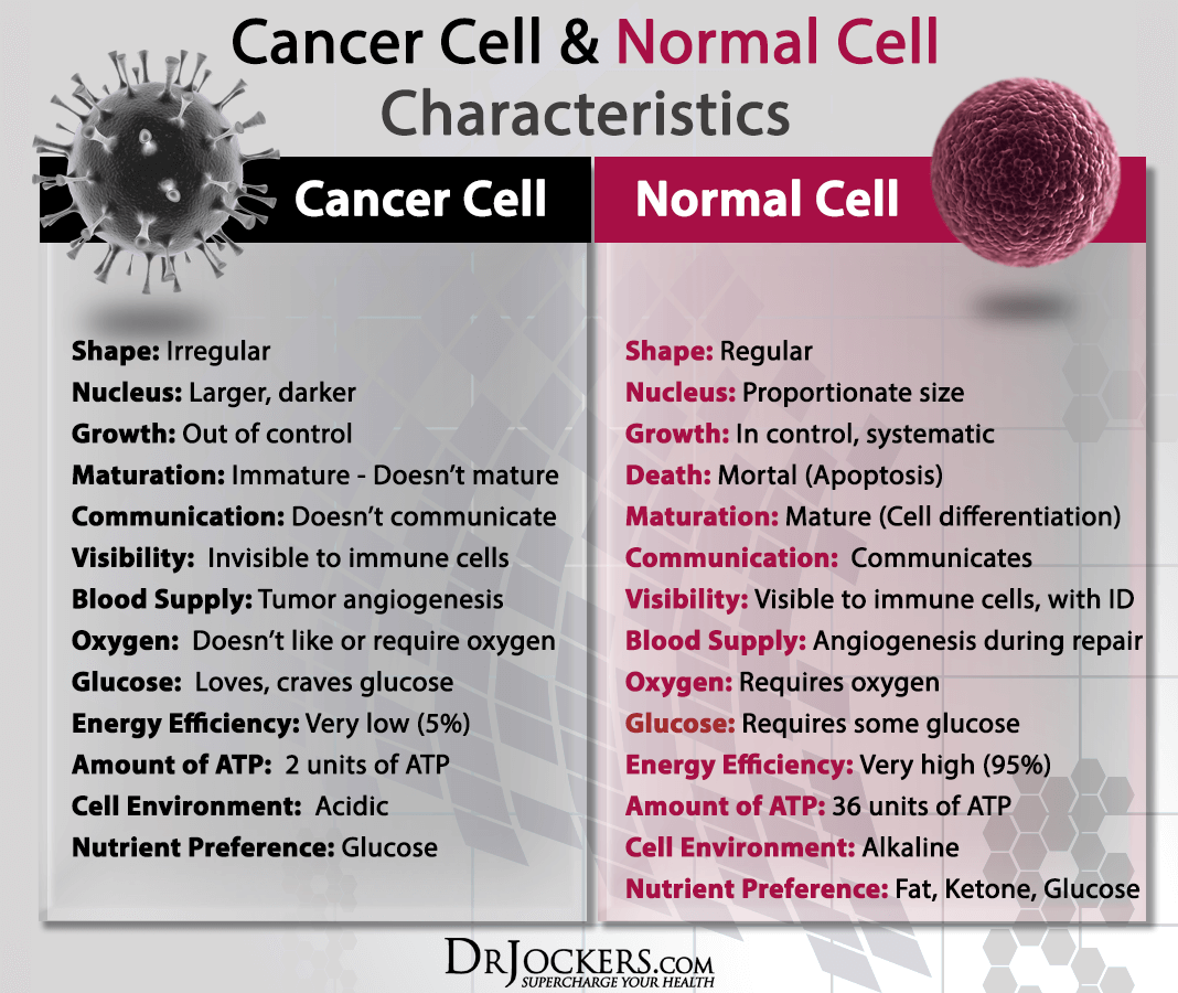
From pioneering microscopy techniques to cutting-edge AI-driven diagnostics and gene-editing tools, technology has not only illuminated these critical distinctions but also empowered us to detect, track, and combat cancer with unprecedented precision. This article delves into the core differences between cancer and normal cells, viewed through the lens of the technological innovations that make this understanding possible and drive the future of oncology.
The Fundamental Divide: Cellular Characteristics Unveiled by Technology
At its heart, cancer is a disease of cellular malfunction. While a casual glance under a conventional microscope might reveal some morphological differences, it’s the deeper molecular and functional distinctions, brought to light by advanced technological tools, that truly define the threat.
Uncontrolled Proliferation: The Engine of Disease
Normal cells grow and divide in a controlled manner, responding to intricate signaling pathways that dictate when to multiply and when to stop. This process is tightly regulated by cell cycle checkpoints. Cancer cells, however, largely ignore these signals. They possess an almost limitless replicative potential, dividing relentlessly without regard for normal boundaries.
Technological Insight: Traditional histology and immunohistochemistry, enhanced by digital pathology and image analysis software, allow pathologists to quantify mitotic activity and assess cellular density, clearly indicating uncontrolled growth. Beyond morphology, flow cytometry uses lasers and detectors to analyze thousands of cells per second, measuring DNA content and identifying cells stuck in various phases of the cell cycle, a hallmark of dysregulated proliferation. Furthermore, real-time PCR and next-generation sequencing (NGS) technologies can detect overexpression of genes that promote cell division (oncogenes) or mutations in genes that suppress it (tumor suppressor genes) – the very genetic drivers of this uncontrolled growth.
Loss of Apoptosis: Immortal Cells
Programmed cell death, or apoptosis, is a vital process by which damaged or unnecessary cells are eliminated. It’s the body’s natural quality control mechanism. Cancer cells, remarkably, often develop mechanisms to evade apoptosis, essentially becoming “immortal.” This evasion allows them to persist and accumulate, even when they are damaged or abnormal.
Technological Insight: Investigating the evasion of apoptosis requires sophisticated molecular techniques. Western blotting and immunofluorescence microscopy are critical for detecting the presence or absence of key apoptotic proteins (like caspases, Bcl-2 family proteins, p53). Gene expression profiling using microarrays or RNA sequencing (RNA-seq) can identify altered transcriptional landscapes where anti-apoptotic genes are upregulated and pro-apoptotic genes are downregulated. These technologies provide a detailed molecular blueprint of how cancer cells cheat death.
Technological Advancements in Identifying Cellular Differences
The journey from observing a lump to understanding its cellular pathology has been revolutionized by technology. We no longer just see; we analyze, quantify, and predict.
Genomic Sequencing and Proteomics: Unveiling Molecular Signatures
Perhaps the most profound technological leap in understanding cancer comes from our ability to read the genetic code and analyze the protein machinery of cells.
Genomic Sequencing (NGS): NGS platforms can rapidly sequence entire genomes, exomes, or specific gene panels, revealing somatic mutations, chromosomal rearrangements, and copy number variations that drive cancer. Technologies like single-cell sequencing take this a step further, allowing researchers to analyze the genetic landscape of individual cells within a tumor, uncovering heterogeneity that was previously invisible. This precision allows for the identification of actionable mutations that can be targeted by specific drugs.
Proteomics: While genomics tells us what can happen, proteomics tells us what is happening at the protein level. Mass spectrometry (MS)-based proteomics allows for the identification and quantification of thousands of proteins from a cellular sample, revealing altered signaling pathways, abnormal protein modifications, and the presence of biomarkers unique to cancer cells. Integrating genomic and proteomic data, often using bioinformatics software and AI algorithms, provides a holistic view of the cellular machinery gone awry.
Advanced Imaging and Microscopy: Visualizing the Micro-World
Beyond traditional light microscopy, a suite of advanced imaging technologies offers unprecedented views into cellular architecture and function.
Confocal and Multiphoton Microscopy: These advanced optical microscopes enable high-resolution, three-dimensional imaging of living cells and tissues, reducing out-of-focus blur and allowing for visualization of sub-cellular structures and dynamic processes. This helps researchers observe morphological changes, cellular interactions, and the movement of organelles in real-time, differentiating between normal and cancerous phenotypes.

Electron Microscopy (EM): For ultra-structural detail, EM offers resolutions far beyond light microscopy, revealing intricate details of cellular organelles, membrane structures, and viral particles within cells. Differences in mitochondrial structure, nuclear envelope integrity, and cell-junction formations, subtle in cancer cells, become apparent.
Live-Cell Imaging: Coupled with genetically encoded fluorescent reporters and advanced image analysis software, live-cell imaging allows scientists to track cellular processes—like cell division, migration, and apoptosis—in real-time, providing dynamic insights into how cancer cells behave differently from normal cells in a living context.
AI and Machine Learning: Pattern Recognition in Pathology
The sheer volume and complexity of data generated by modern diagnostic tools would be overwhelming without computational assistance. Artificial intelligence and machine learning are revolutionizing how we identify and interpret cellular differences.
AI in Digital Pathology: AI algorithms are now being trained on vast datasets of digitized pathology slides to identify microscopic patterns indicative of cancer with remarkable accuracy. They can classify tumor types, grade malignancy, detect micrometastases, and even predict patient outcomes, often surpassing human capabilities in speed and consistency. This technology assists pathologists by highlighting suspicious regions, reducing diagnostic errors, and speeding up turnaround times.
Radiomics and Imaging Biomarkers: AI is also transforming medical imaging (MRI, CT, PET scans). Radiomics involves extracting numerous quantitative features from medical images that are not discernible to the human eye. Machine learning models then analyze these features to differentiate between benign and malignant lesions, assess tumor aggressiveness, and monitor treatment response, providing non-invasive insights into cellular behavior within living tissues.
The Tech Frontier: From Diagnosis to Targeted Therapies
The precise identification of cancer cell differences isn’t just an academic exercise; it’s the bedrock for developing highly effective, personalized treatments. Technology continues to push the boundaries from early detection to therapeutic intervention.
Liquid Biopsies and Early Detection Technologies
One of the holy grails in oncology is early detection. Liquid biopsies, a revolutionary technology, analyze blood or other bodily fluids for circulating tumor DNA (ctDNA), circulating tumor cells (CTCs), or tumor-derived exosomes. These components carry genetic and molecular signatures of the tumor, often before a solid mass is detectable by conventional imaging.
Technological Edge: NGS, advanced PCR techniques (like digital PCR), and highly sensitive microfluidic devices are critical for isolating and analyzing these minute quantities of tumor material. This non-invasive technology holds immense promise for screening high-risk individuals, monitoring treatment efficacy, and detecting recurrence, drastically improving patient prognoses by catching cancer at its most treatable stages.
CRISPR and Gene Editing: Precision Intervention
The ability to precisely edit genes represents an extraordinary technological leap. CRISPR-Cas9 (Clustered Regularly Interspaced Short Palindromic Repeats) technology allows scientists to make targeted changes to the DNA sequence with unprecedented accuracy.
Therapeutic Potential: In the context of cancer, CRISPR can be used to correct cancer-driving mutations, disable oncogenes, or enhance the immune system’s ability to recognize and destroy cancer cells (e.g., in CAR-T cell therapy development). While still largely in experimental stages for direct human therapy, it offers a glimpse into a future where the genetic instructions that make a cell cancerous can be precisely rewritten, offering a level of targeted intervention unimaginable just decades ago.
Digital Security and Ethical Considerations in Cancer Data
As technology becomes increasingly intertwined with cancer research and patient care, the accompanying challenges around data management, privacy, and ethics become critical, firmly placing these discussions within the “Tech” niche.
Protecting Patient Data in Genomic Research
The vast amounts of sensitive genetic and health data generated through sequencing and electronic health records present significant cybersecurity challenges. Ensuring the privacy and security of this information is paramount to maintaining patient trust and preventing misuse. Advanced encryption techniques, blockchain technology for data provenance, and robust access control systems are vital for protecting this highly personal information.
The Promise and Peril of AI in Healthcare
AI’s integration into diagnostics and personalized medicine promises immense benefits but also raises ethical questions. Biases in training data can lead to disparities in care, and the “black box” nature of some AI models can make it difficult to understand their decision-making processes. Developing transparent, explainable AI (XAI) and establishing clear regulatory frameworks are crucial to harness AI’s potential responsibly and ensure equitable, ethical application in oncology.

Conclusion
The differences between cancer cells and normal cells are not merely biological curiosities; they are the battleground where health and disease contend. The relentless pursuit of understanding these distinctions has been propelled by an extraordinary surge in technological innovation. From the microscopic lens that first hinted at cellular anomalies to the AI algorithms that now sift through vast genomic datasets, technology is the engine driving progress in oncology. It allows us to pinpoint the genetic aberrations, visualize the abnormal structures, predict the cellular behaviors, and ultimately, develop targeted therapies that distinguish and destroy cancer cells while sparing healthy ones. As we continue to push the boundaries of computing, imaging, and bioengineering, the hope for a future where cancer is not just treatable, but often preventable or even curable, moves ever closer to reality, firmly rooted in our technological prowess.
aViewFromTheCave is a participant in the Amazon Services LLC Associates Program, an affiliate advertising program designed to provide a means for sites to earn advertising fees by advertising and linking to Amazon.com. Amazon, the Amazon logo, AmazonSupply, and the AmazonSupply logo are trademarks of Amazon.com, Inc. or its affiliates. As an Amazon Associate we earn affiliate commissions from qualifying purchases.