
The dicrotic notch, a subtle but significant feature in the arterial blood pressure waveform, represents a crucial physiological event. While often discussed in medical contexts, its detection, analysis, and interpretation have become increasingly intertwined with advancements in technology. Far from being a mere blip on a graph, understanding this notch in the digital age involves sophisticated sensors, advanced signal processing, artificial intelligence, and a growing ecosystem of interconnected health monitoring devices. From basic physiological principles to its role in cutting-edge diagnostics, the dicrotic notch provides a compelling example of how technological innovation is revolutionizing our understanding and management of human health data.

The Physiological Basis and its Digital Representation
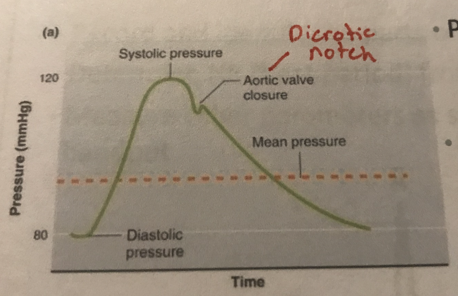
At its core, the dicrotic notch is a transient dip in the descending limb of the arterial pressure waveform, observed just after the peak systolic pressure. Physiologically, this dip marks the closure of the aortic valve following ventricular ejection. As the left ventricle relaxes, blood briefly flows backward towards the heart, causing the aortic valve to snap shut. This sudden closure creates a small, transient increase in pressure (known as the dicrotic wave or incisura) followed immediately by the “notch” as the pressure equilibrates. The subsequent pressure decline reflects the continuous flow of blood into the peripheral vasculature.
Digitally representing this phenomenon requires specialized sensors capable of capturing minute pressure changes. Transducers, whether invasive (like an arterial line) or non-invasive (like those used in oscillometric blood pressure monitors or certain photoplethysmography sensors), convert mechanical pressure waves into electrical signals. These analog signals are then digitized through analog-to-digital converters (ADCs), transforming the continuous waveform into discrete data points. The fidelity of this digital representation is critical; high sampling rates and resolution are necessary to accurately capture the rapid fluctuations that define the dicrotic notch. Without precise digital capture, subsequent analysis by software and algorithms would be compromised, potentially leading to misinterpretation or missed diagnostic clues. The resulting time-series data, comprising thousands of data points per second, forms the raw material for technological processing and analysis.
Technological Measurement and Monitoring
The ability to accurately measure and monitor the dicrotic notch has evolved significantly with technological advancements. These technologies range from highly precise clinical instruments to burgeoning consumer-grade wearables, each offering different levels of fidelity and application.
Non-Invasive Wearables and Peripheral Monitoring
The rise of wearable technology has sparked interest in non-invasive methods to assess cardiovascular health. While direct, high-fidelity arterial pressure waveforms are challenging to obtain from wearables, many devices employ photoplethysmography (PPG). PPG sensors, commonly found in smartwatches and fitness trackers, measure volumetric changes in blood flow in the capillaries near the skin surface. These optical signals produce a pulse waveform that, while not identical to a direct arterial pressure waveform, shares similar characteristics, including an inflection point often correlated with the dicrotic notch.
Advanced algorithms are employed to analyze these PPG waveforms, attempting to identify the dicrotic notch or its equivalent and derive parameters like pulse transit time (PTT) or arterial stiffness indicators. While still facing limitations in accuracy compared to clinical-grade devices, the continuous, non-invasive nature of wearables offers unprecedented opportunities for long-term trend analysis, early detection of deviations from baseline, and personalized health insights. The technological challenge lies in improving sensor sensitivity, compensating for motion artifacts, and developing robust algorithms that can reliably extract meaningful physiological markers from inherently noisy data.
Clinical and Diagnostic Devices
In clinical settings, the dicrotic notch is a cornerstone for continuous hemodynamic monitoring. Invasive arterial lines provide the gold standard, directly measuring intra-arterial pressure and generating high-resolution waveforms that clearly display the notch. These systems integrate advanced signal processing to filter noise, automatically identify key waveform features, and display real-time physiological parameters.
Beyond invasive methods, non-invasive oscillometric blood pressure devices also utilize principles of waveform analysis. While they typically provide mean, systolic, and diastolic pressures, some advanced models capture and analyze the full waveform, processing the oscillations to infer arterial characteristics. Continuous non-invasive blood pressure monitors, often utilizing finger cuffs or applanation tonometry, are also becoming more sophisticated, striving to provide continuous waveform data that includes discernible dicrotic notches. These devices require sophisticated sensor technology, often employing piezoelectric or optical elements, coupled with embedded microprocessors for real-time signal acquisition and initial processing. The data collected by these devices is frequently integrated into hospital information systems or specialized patient monitoring platforms, enabling comprehensive data visualization and historical tracking.
Remote Patient Monitoring (RPM) Systems
The integration of dicrotic notch data, alongside other physiological metrics, into Remote Patient Monitoring (RPM) systems represents a significant technological leap. RPM platforms collect data from various patient-worn or in-home devices and transmit it securely to healthcare providers. For cardiovascular monitoring, this includes devices capable of capturing waveforms, even if indirectly. The value here lies in longitudinal data collection, allowing clinicians to observe trends over weeks or months, rather than just snapshot measurements.
These systems leverage cloud computing for data storage and processing, secure communication protocols (e.g., encrypted Wi-Fi, Bluetooth LE, cellular), and user-friendly dashboards for clinicians. The ability to track parameters related to the dicrotic notch over time can help in managing chronic conditions like hypertension or heart failure, identifying early signs of deterioration, and optimizing medication regimens without requiring frequent in-person visits. AI and machine learning are increasingly integrated into RPM platforms to automatically flag anomalous waveform patterns or significant changes in notch characteristics, providing alerts to care teams.
AI and Advanced Analytics for Dicrotic Notch Data
The sheer volume and complexity of physiological waveform data necessitate sophisticated analytical tools, with Artificial Intelligence (AI) and machine learning leading the charge. These technologies unlock deeper insights from the dicrotic notch, moving beyond simple identification to predictive modeling and diagnostic support.

Pattern Recognition and Anomaly Detection
AI algorithms, particularly deep learning models like Convolutional Neural Networks (CNNs) and Recurrent Neural Networks (RNNs), are exceptionally adept at pattern recognition within time-series data. When trained on vast datasets of arterial waveforms, these models can reliably identify the dicrotic notch, even amidst noise or artifacts. Beyond mere identification, AI can characterize the notch’s morphology: its depth, width, sharpness, and its precise timing relative to other waveform features.
This capability is vital for anomaly detection. A “blunted” or absent dicrotic notch, for example, can be indicative of certain cardiovascular pathologies such as aortic regurgitation or a reduced peripheral vascular resistance. Conversely, a very prominent notch might suggest other conditions. AI models can learn the spectrum of normal notch variations and flag any deviations that fall outside established parameters. This automated anomaly detection can assist clinicians by drawing attention to potentially concerning changes that might otherwise be subtle or missed in a manual review of continuous data streams.
Predictive Modeling and Diagnostic Support
One of the most promising applications of AI in analyzing dicrotic notch data lies in predictive modeling. By correlating specific characteristics of the dicrotic notch (identified by AI) with patient outcomes or disease progression, machine learning algorithms can build predictive models. For instance, changes in notch morphology over time, combined with other physiological data points (e.g., heart rate, blood pressure, patient demographics), could be used to predict the risk of future cardiovascular events, such as heart attacks or strokes, or to forecast the worsening of conditions like congestive heart failure.
AI also offers significant potential for diagnostic support. While the dicrotic notch is one piece of the diagnostic puzzle, its characteristics, when analyzed alongside a comprehensive suite of patient data, can contribute to more accurate and earlier diagnoses. AI-powered systems can provide clinicians with probabilistic assessments, suggesting potential conditions based on waveform analysis, thereby augmenting human expertise and reducing diagnostic delays. This moves beyond simply identifying the notch to understanding its implications within a broader clinical context.
Data Visualization and Interpretation Tools
Even with advanced AI analytics, the output needs to be interpretable by human users. Technological solutions focus on creating intuitive data visualization tools that transform raw waveform data and AI insights into actionable information. Interactive dashboards, specialized medical software, and mobile applications present complex physiological data in clear, graphical formats.
These tools allow clinicians to zoom in on specific segments of a waveform, compare current readings to historical data, and overlay different physiological parameters. For the dicrotic notch, this might include visualizing its depth profile, timing variations, or classifying its morphology. Explanations from AI models, often termed “explainable AI” (XAI), can also be integrated, providing insights into why the AI flagged a particular notch or made a specific prediction, thereby fostering trust and aiding clinical decision-making.
Future Innovations and Challenges
The intersection of the dicrotic notch and technology is a dynamic field, brimming with potential yet facing significant hurdles.
Miniaturization and Integration
Future innovations will likely focus on even greater miniaturization and seamless integration of sensors capable of capturing high-fidelity waveforms into everyday objects. Imagine smart clothing, patches, or even contact lenses that continuously monitor cardiovascular parameters, including subtle features like the dicrotic notch. This continuous, unobtrusive monitoring could provide unprecedented longitudinal data, moving healthcare from episodic to proactive. The challenge lies in maintaining accuracy and reliability in highly variable real-world conditions while ensuring user comfort and power efficiency.
Data Privacy and Security
As more sensitive physiological data, including detailed waveform characteristics, are collected and transmitted, the issues of data privacy and security become paramount. Robust encryption, secure cloud infrastructure, and strict adherence to regulatory frameworks like HIPAA (Health Insurance Portability and Accountability Act) and GDPR (General Data Protection Regulation) are crucial. Developing decentralized data storage and processing models (e.g., blockchain-based solutions) could also play a role in empowering individuals with greater control over their health data while ensuring its integrity and privacy.
Regulatory Hurdles and Validation
For any technology analyzing the dicrotic notch to be adopted in clinical practice, rigorous regulatory approval is essential. This requires extensive clinical validation to prove accuracy, reliability, and clinical utility. The path for medical devices and AI algorithms, especially those making diagnostic or prognostic claims, is long and demanding. Future advancements will need to navigate these regulatory landscapes effectively, demonstrating consistent performance across diverse patient populations and clinical scenarios.
Democratizing Health Insights
Ultimately, technological progress in understanding the dicrotic notch has the potential to democratize health insights. By making sophisticated physiological monitoring more accessible and interpretable, individuals can gain a deeper understanding of their own cardiovascular health. This empowers preventative care, facilitates earlier intervention, and fosters a more personalized approach to medicine. The ongoing challenge is to ensure equitable access to these technologies and to educate both healthcare providers and the public on their capabilities and limitations.
aViewFromTheCave is a participant in the Amazon Services LLC Associates Program, an affiliate advertising program designed to provide a means for sites to earn advertising fees by advertising and linking to Amazon.com. Amazon, the Amazon logo, AmazonSupply, and the AmazonSupply logo are trademarks of Amazon.com, Inc. or its affiliates. As an Amazon Associate we earn affiliate commissions from qualifying purchases.