
The human body is an intricate symphony of systems, each performing vital functions to sustain life. Among these, the airway—the pathway for air to travel from our nose and mouth to our lungs—is paramount. When this critical conduit experiences a narrowing, particularly in the region just below the vocal cords, a condition known as subglottic stenosis arises. While not as widely discussed as some other medical conditions, understanding subglottic stenosis is crucial, as it can significantly impact breathing and overall quality of life for those affected.
This article aims to demystify subglottic stenosis, exploring its causes, symptoms, diagnostic approaches, and treatment options. Drawing parallels to the innovation and problem-solving inherent in the worlds of technology, branding, and finance, we’ll examine how this condition presents a unique challenge that requires careful analysis, strategic management, and often, technological or procedural advancements for effective resolution.

Understanding the Anatomy: The Subglottic Region
Before delving into the stenosis itself, it’s essential to grasp the anatomy of the larynx, or voice box. The larynx sits at the top of the trachea (windpipe) and houses the vocal cords. The glottis is the space between the vocal cords, responsible for sound production. The subglottis is the area immediately below the glottis and above the first tracheal ring. This narrow section of the airway is critical for airflow to the lungs.
The subglottic region is characterized by its relatively small diameter, especially in infants and children, making it susceptible to narrowing from various causes. A healthy subglottic space allows for unobstructed breathing, essential for everything from simple respiration to vocalization. When this space becomes constricted, the effort required to breathe increases, leading to a range of symptoms.
Causes of Subglottic Stenosis: A Multifaceted Origin
Subglottic stenosis isn’t a single entity with a singular cause. Its origins are diverse, often stemming from factors that damage or irritate the delicate lining of the airway. Understanding these causes is the first step in effective diagnosis and management.
Congenital vs. Acquired Causes
One primary distinction in the causes of subglottic stenosis is between congenital (present at birth) and acquired (developing after birth) origins.
Congenital Subglottic Stenosis
Congenital subglottic stenosis is present from birth, though symptoms may not manifest immediately. It is often due to abnormalities in the cartilage of the larynx or a developmental issue during fetal formation. While the exact mechanisms are not always fully understood, factors such as:
- Abnormal Cartilage Development: The C-shaped cartilage rings that support the trachea can be malformed, leading to a naturally narrower subglottis from birth.
- Vascular Abnormalities: Sometimes, blood vessels near the developing larynx can compress the airway, contributing to narrowing.
- Genetic Predispositions: While not strongly linked to specific genetic disorders, some families may have a higher incidence, suggesting a possible genetic component.
Congenital cases are more common in infants and young children, and the degree of narrowing can vary significantly. Early identification is crucial as it can impact feeding, growth, and overall development.
Acquired Subglottic Stenosis
Acquired subglottic stenosis is far more common and arises from injury or inflammation to the subglottic area after birth. The most frequent culprit is prolonged intubation, a medical procedure where a breathing tube is inserted into the trachea.
-
Prolonged Endotracheal Intubation: This is arguably the leading cause of acquired subglottic stenosis. The presence of a breathing tube, particularly if it’s too large, inflated too much, or left in place for an extended period, can cause pressure on the tracheal lining. This pressure can lead to:
- Inflammation: The tube irritates the delicate mucosa.
- Ischemia: Reduced blood flow to the compressed areas.
- Granulation Tissue Formation: The body’s response to injury is to form scar tissue, which can thicken and narrow the airway.
- Cartilage Weakening or Collapse: In severe cases, the cartilage rings can be damaged, leading to instability and airway collapse.
-
Trauma: Direct injury to the neck, such as from a blunt force or a penetrating wound, can damage the larynx and subglottis, leading to stenosis.
-
Infections: Severe infections of the upper airway, like epiglottitis or croup, can cause significant inflammation and swelling that, if recurrent or severe, might lead to scarring and narrowing.
-
Gastroesophageal Reflux Disease (GERD): Chronic exposure of the airway to stomach acid through reflux can cause irritation and inflammation, potentially contributing to subglottic narrowing, especially in infants.
-
Surgical Procedures: While less common, certain surgeries on the neck or airway can inadvertently lead to scarring and stenosis in the subglottic region.
The distinction between congenital and acquired causes guides the diagnostic and treatment strategies. For acquired stenosis, the focus is often on mitigating the damaging factors and managing the resulting scar tissue.
Identifying Subglottic Stenosis: Symptoms and Diagnosis
The symptoms of subglottic stenosis can be subtle at first, often mistaken for other respiratory issues, especially in infants. As the narrowing progresses, the signs become more pronounced, reflecting the increasing difficulty of air passage.
Common Symptoms
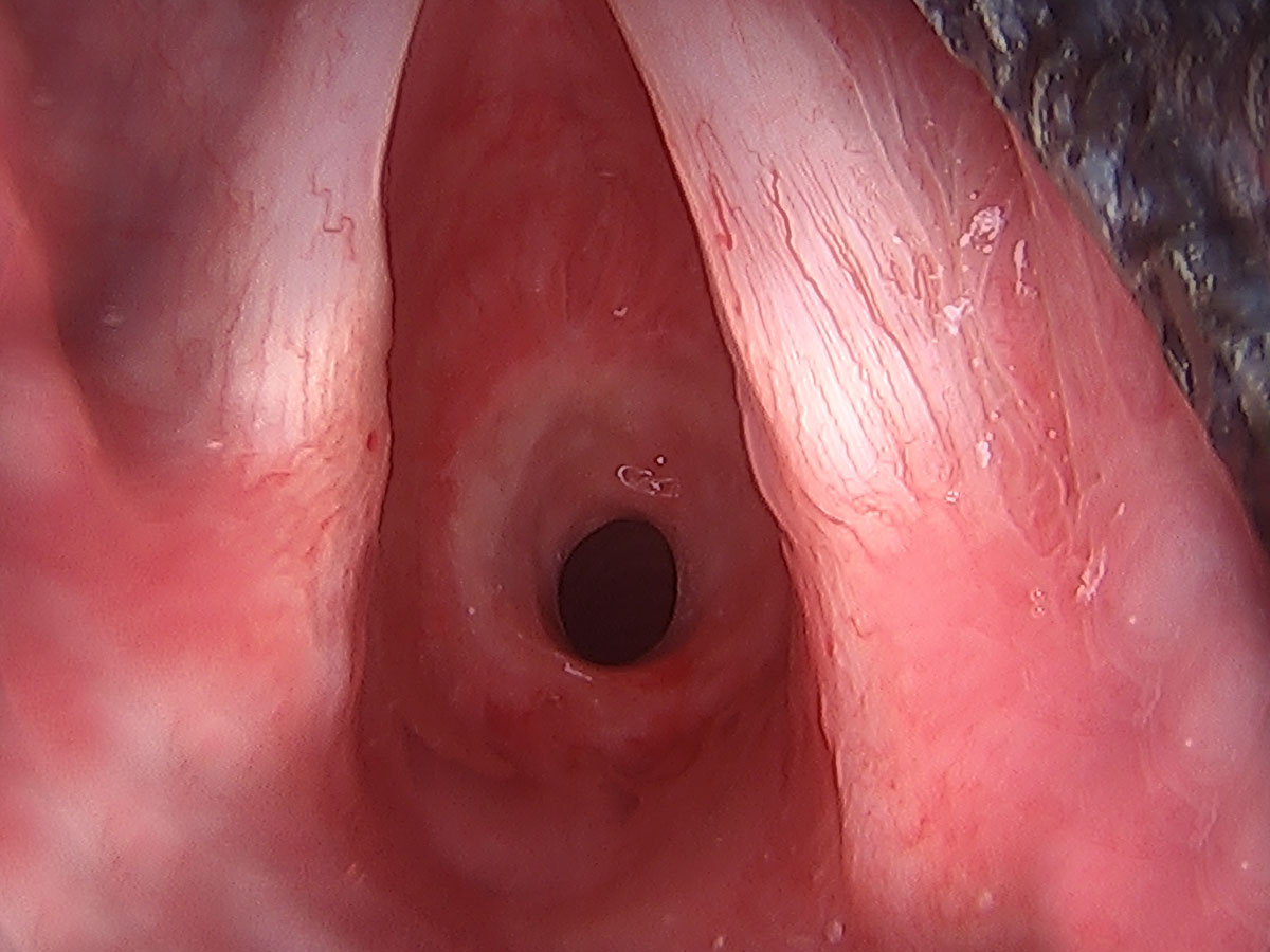
The hallmark symptom is stridor, a high-pitched, musical whistling sound that occurs during inhalation. This sound arises from turbulent airflow through the narrowed segment. Other symptoms can include:
- Noisy Breathing: Beyond stridor, any breathing that sounds labored or unusual can be a red flag.
- Shortness of Breath: Especially with exertion, but in severe cases, even at rest.
- Difficulty Breathing (Dyspnea): A feeling of not getting enough air.
- Recurrent or Persistent Croup-like Cough: A barking cough that doesn’t resolve.
- Feeding Difficulties and Poor Weight Gain: In infants, the effort of breathing can interfere with their ability to feed effectively, leading to failure to thrive.
- Apnea: Episodes of paused breathing, particularly in severe cases.
- Hoarseness or Weak Cry: If the vocal cords are affected by the stenosis or the swelling associated with it.
- Retractions: The skin between the ribs or above the collarbone pulls inward with each breath, indicating increased effort.

The severity of these symptoms is directly related to the degree of airway narrowing. A mild stenosis might only cause stridor with exertion, while a severe stenosis can be life-threatening, requiring immediate medical intervention.
Diagnostic Pathways: Pinpointing the Problem
Diagnosing subglottic stenosis requires a systematic approach, often involving visual inspection and imaging.
-
Physical Examination and History: A thorough medical history, noting any risk factors like prematurity, intubation history, or recurrent respiratory infections, is crucial. The doctor will listen to the breathing pattern for characteristic sounds like stridor.
-
Laryngoscopy: This is the gold standard for visualizing the subglottic area. A flexible or rigid scope with a camera is inserted through the nose or mouth to directly view the larynx and subglottis. This allows the doctor to assess the location, length, and severity of the narrowing, as well as the condition of the airway lining (e.g., presence of granulation tissue).
- Flexible Laryngoscopy: Performed with local anesthetic, this is often done in an outpatient setting and is less invasive.
- Rigid Laryngoscopy: Performed under general anesthesia, it allows for a more detailed and comprehensive examination, often including biopsy or intervention if needed.
-
Imaging Studies: While laryngoscopy is primary, imaging can provide complementary information.
- X-rays: A plain radiograph of the neck may show narrowing of the subglottis, but it’s not always definitive.
- CT Scan: A computed tomography scan can provide detailed cross-sectional images of the airway, helping to define the extent of the stenosis and assess surrounding structures.
- MRI Scan: Magnetic resonance imaging can offer excellent soft tissue visualization and is particularly useful for assessing vascular structures or masses that might be contributing to the narrowing.
The diagnostic process aims to confirm the presence of stenosis, determine its cause and severity, and rule out other conditions that might mimic its symptoms.
Managing Subglottic Stenosis: Treatment Strategies and Innovations
The management of subglottic stenosis is highly individualized, depending on the severity of the narrowing, the patient’s age, overall health, and the underlying cause. The primary goals are to restore adequate airflow, prevent further damage, and improve the patient’s quality of life.
Non-Surgical Approaches
For mild cases or when there’s active inflammation, non-surgical interventions may be attempted.
- Observation and Monitoring: In very mild cases, particularly in infants where the airway may grow, close monitoring may be sufficient.
- Steroid Therapy: Topical or systemic steroids can help reduce inflammation and potentially shrink granulation tissue.
- Dilations: In some instances, endoscopic dilations can be performed to gradually widen the narrowed segment. This often involves using balloons or dilators inserted through a scope. However, this approach can sometimes lead to further scarring and is more effective for certain types of stenosis.
- Treatment of Underlying Conditions: If GERD is a contributing factor, aggressive management of reflux is essential.
Surgical Interventions
When stenosis is moderate to severe, surgical intervention is often necessary.
-
Endoscopic Surgery: This minimally invasive approach involves using specialized instruments passed through a laryngoscope.
- Laser Surgery: A CO2 laser can be used to precisely ablate scar tissue or granulation tissue.
- Microdebrider Use: A powered instrument that can shave away scar tissue.
- Balloon Dilation: As mentioned, this can also be performed endoscopically.
The advantage of endoscopic surgery is less trauma and faster recovery, but it’s generally best suited for less complex or shorter stenoses.
-
Open Surgical Reconstruction: For more severe or complex stenoses, open surgery is required.
- Cricotracheal Resection (CTR): This is the most common and definitive surgical treatment for significant subglottic stenosis. It involves surgically removing the stenotic segment of the trachea and then rejoining the healthy ends. This is a major surgery with a long recovery period.
- Laryngotracheal Reconstruction (LTR): This procedure involves augmenting the narrowed airway by grafting cartilage (often from the rib) or other tissues to expand the subglottic space. LTR is more complex and may be used when a simple resection isn’t feasible or desirable.
The success of surgery depends on many factors, including the surgeon’s expertise, the patient’s healing response, and the prevention of post-operative complications like infection or recurrence of stenosis.
The Role of Technology and Innovation
The management of subglottic stenosis is increasingly benefiting from technological advancements, mirroring the progress seen in other fields.
- Advanced Imaging and Simulation: 3D modeling of the airway based on CT or MRI scans can allow surgeons to meticulously plan complex reconstructions, visualize the exact extent of the stenosis, and even simulate surgical approaches before operating. This is akin to using sophisticated software to test and refine designs in tech or model financial scenarios.
- Minimally Invasive Surgical Tools: The development of finer, more maneuverable endoscopic instruments and lasers allows for more precise interventions with less collateral damage to healthy tissue. This echoes the trend in consumer tech towards smaller, more powerful devices.
- Biomaterials and Tissue Engineering: While still in its nascent stages for airway reconstruction, research into biodegradable scaffolds and engineered tissues holds promise for future LTR procedures, potentially offering better integration and reduced risk of rejection. This aligns with the drive for innovation in materials science across industries.
- Artificial Intelligence (AI) in Diagnosis: AI algorithms are being developed to analyze medical images and identify subtle signs of airway compromise, potentially aiding in earlier diagnosis, especially in challenging cases. This parallels the application of AI in identifying patterns in market data or optimizing software performance.
Long-Term Management and Rehabilitation
Post-treatment, individuals with subglottic stenosis often require ongoing care. This can include:
- Speech and Swallowing Therapy: The airway is intimately connected to voice production and swallowing. Rehabilitation may be needed to regain normal function.
- Pulmonary Rehabilitation: Helping patients improve their breathing capacity and manage any residual breathing difficulties.
- Regular Follow-ups: Close monitoring is essential to detect any signs of restenosis or complications.
Conclusion: A Lifelong Airway Journey
Subglottic stenosis, while a challenging condition, is increasingly manageable thanks to advances in medical understanding, diagnostic capabilities, and surgical techniques. It serves as a powerful reminder of the delicate nature of our respiratory system and the profound impact airway compromise can have on life.
Just as in the realms of technology, where innovative solutions tackle complex problems, and in branding, where strategic thinking builds resilience and impact, the approach to subglottic stenosis requires a blend of precision, foresight, and continuous adaptation. From the meticulous planning of a surgical reconstruction to the ongoing rehabilitation of a patient, every step is a testament to the power of dedicated expertise and the relentless pursuit of improving lives. Understanding what subglottic stenosis is—its causes, symptoms, and treatments—empowers patients, families, and healthcare professionals to navigate this unseen airway challenge with greater confidence and hope.
aViewFromTheCave is a participant in the Amazon Services LLC Associates Program, an affiliate advertising program designed to provide a means for sites to earn advertising fees by advertising and linking to Amazon.com. Amazon, the Amazon logo, AmazonSupply, and the AmazonSupply logo are trademarks of Amazon.com, Inc. or its affiliates. As an Amazon Associate we earn affiliate commissions from qualifying purchases.