
The Magnetic Resonance Imaging (MRI) machine stands as one of the most sophisticated pieces of technology ever developed for clinical use. Unlike X-ray or CT scanners, which rely on ionizing radiation, the MRI machine is a masterpiece of quantum physics, high-performance computing, and precision engineering. It utilizes the fundamental properties of hydrogen atoms within the human body to create high-resolution, three-dimensional maps of internal structures. In the modern tech landscape, the MRI machine is no longer just a passive diagnostic tool; it has become a hub for artificial intelligence (AI), edge computing, and advanced materials science.
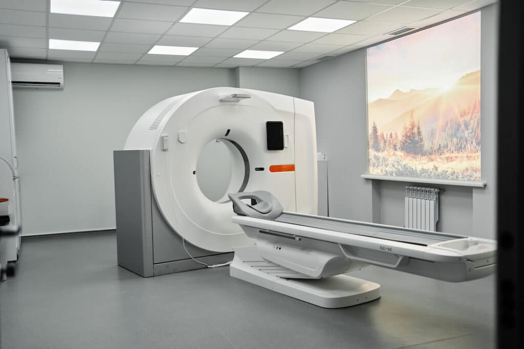
To understand what an MRI machine is, one must look beyond the imposing “donut” shape and explore the complex interplay of superconducting magnets, radiofrequency (RF) systems, and the software algorithms that translate raw electromagnetic signals into visible diagnostic data.
The Physics and Engineering Behind Magnetic Resonance Imaging
At its core, an MRI machine is a massive, high-precision electromagnetic sensor. The hardware is designed to manipulate the magnetic spin of protons—specifically hydrogen nuclei—which are abundant in the water and fat molecules of the human body.
The Role of Superconducting Magnets
The most critical component of an MRI machine is the main magnet. Most modern clinical systems utilize a superconducting magnet, typically cooled by liquid helium to temperatures near absolute zero (-269°C or 4 Kelvin). At these temperatures, the wire coils lose all electrical resistance, allowing a massive current to flow indefinitely. This creates a powerful, static magnetic field, measured in Teslas (T). While a typical refrigerator magnet is about 0.005T, a standard clinical MRI operates at 1.5T or 3.0T. This field aligns the “spin” of protons in the patient’s body, creating a state of equilibrium that the machine can then manipulate.
Gradient Coils and Spatial Localization
Inside the main magnet bore are three sets of gradient coils. These are secondary electromagnets that can be turned on and off rapidly to create deliberate, controlled variations in the magnetic field along the X, Y, and Z axes. By slightly altering the magnetic strength at specific points in space, the system can “tune” the resonance frequency of protons in a specific slice of the body. This spatial encoding is what allows the computer to determine exactly where a signal is coming from, enabling the construction of a 3D image rather than a flat 2D projection.
Radiofrequency (RF) Systems and Signal Acquisition
Once the protons are aligned by the main magnet and localized by the gradients, the machine emits a pulse of radiofrequency energy. This pulse is tuned to the Larmor frequency—the specific frequency at which the protons are “precessing.” The pulse tips the protons out of alignment. When the RF pulse is turned off, the protons relax back to their original state, emitting their own faint RF signal in the process. Sensitive receiver coils, often placed directly over the body part being scanned, capture these signals. This raw data, known as K-space, is then processed using a complex mathematical operation called a Fourier Transform to generate the final image.
Evolution of MRI Hardware: From Low-Field to Ultra-High-Field Systems
The technology of MRI has evolved significantly since the first human scan in 1977. Today, the industry is bifurcating into two distinct technological directions: ultra-high-field systems for extreme precision and portable, low-field systems for decentralized access.
The Standard 1.5T and 3.0T Platforms
For decades, the 1.5T scanner has been the workhorse of the industry, offering a balance between cost, image quality, and patient safety (as higher fields can cause more significant interference with metallic implants). However, the tech trend has shifted toward 3.0T systems as the new standard for neurology and musculoskeletal imaging. The higher field strength provides a superior signal-to-noise ratio, allowing for thinner slices, faster scan times, and the ability to visualize smaller anatomical structures with unprecedented clarity.
The Frontier of 7T and Beyond
The cutting edge of MRI technology is found in 7T (Tesla) and even 11.7T systems. These ultra-high-field (UHF) magnets are primarily used in research but are increasingly finding clinical applications in mapping the fine architecture of the brain’s cortex. At 7T, researchers can observe the metabolic processes of the brain in real-time and identify biomarkers for neurodegenerative diseases like Alzheimer’s and Parkinson’s years before symptoms appear. The engineering challenge for these machines is immense, requiring specialized RF coils and sophisticated software to manage the “dielectric effect,” where the wavelength of the RF pulse becomes smaller than the body part being imaged, causing uneven lighting in the scan.
Sustainable Innovations: Helium-Free Cooling Systems
One of the most significant recent tech shifts in MRI hardware is the development of “helium-free” or “low-helium” cooling. Traditional MRIs require approximately 1,500 to 2,000 liters of liquid helium, a finite and increasingly expensive resource. New “sealed” magnet technologies, such as Philips’ BlueSeal or GE’s Freelium-ready designs, use only a few liters of helium that is permanently sealed within the unit. This reduces the weight of the machine, eliminates the need for expensive vent pipes, and makes the technology much more sustainable and easier to install in remote locations.
The Role of Artificial Intelligence and Software in Modern MRI
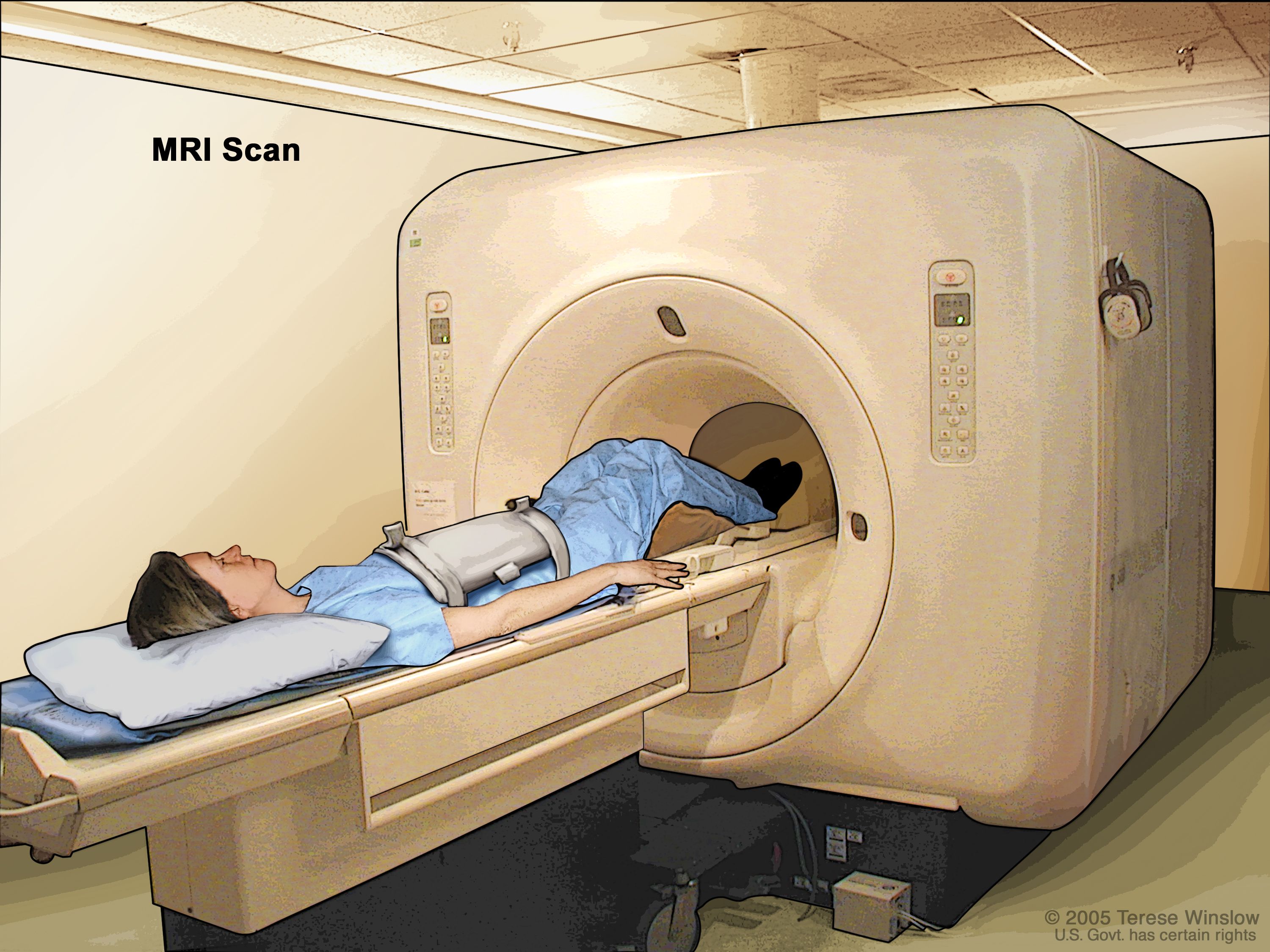
While the magnets provide the raw power, the “brain” of the modern MRI machine is its software. We have entered the era of the “Software-Defined MRI,” where AI and machine learning are revolutionizing how data is captured and interpreted.
Deep Learning for Image Reconstruction
Historically, the biggest drawback of MRI has been the scan time; a comprehensive exam could take 45 minutes or more. Deep learning algorithms are now being used to perform “reconstruction from undersampled data.” By training neural networks on millions of high-quality scans, AI can “fill in the gaps” of a scan that was performed in half the time. This doesn’t just speed up the process; it reduces the artifacts caused by patient movement, making the technology more accessible to children or patients with claustrophobia.
Automated Protocoling and Patient Positioning
The efficiency of an MRI suite is often limited by the manual setup required for each patient. Modern tech integrations include “smart touch” interfaces and 3D camera-based positioning systems. These cameras use AI to recognize the patient’s anatomy and automatically suggest the optimal table height and coil placement. Furthermore, “Auto-protocoling” software analyzes the doctor’s order and automatically selects the correct pulse sequences, reducing human error and ensuring consistency across different technicians.
Predictive Maintenance through IoT
MRI machines are now essentially Internet of Things (IoT) devices. They are equipped with hundreds of sensors that monitor cryogenic levels, gradient temperatures, and vacuum integrity. This data is streamed back to the manufacturer’s cloud, where predictive analytics models identify potential hardware failures before they occur. This “zero-downtime” tech strategy is vital for high-volume hospitals where a single day of an MRI being offline can result in significant financial loss and patient backlogs.
Specialized MRI Technologies and Their Industrial Applications
MRI technology is not a monolith; specialized variants have been developed to study specific physiological functions, moving the technology into the realm of functional and molecular mapping.
Functional MRI (fMRI) and Mapping the Brain
Functional MRI is a tech breakthrough that allows scientists to see the brain in action. It measures the BOLD (Blood Oxygen Level Dependent) signal, identifying which areas of the brain are consuming more oxygen during specific tasks. This has massive implications for the tech industry, particularly in neuro-ergonomics and Brain-Computer Interface (BCI) research. By understanding how the brain responds to different digital stimuli, developers can design more intuitive user interfaces.
Diffusion Tensor Imaging (DTI)
DTI is a specialized software application of MRI that maps the diffusion of water molecules along white matter tracts in the brain. This “tractography” provides a 3D wiring diagram of the human mind. In the tech world, these datasets are used to inform the development of more human-like neural networks in AI research, as engineers look to the biological “hardware” of the brain to design more efficient silicon-based intelligence.
Intraoperative MRI (iMRI) in High-Tech Surgery
The integration of MRI into the operating room is a feat of engineering. These systems involve magnets that are either mobile (on a ceiling track) or have a patient table that can slide into an adjacent room. This allows neurosurgeons to take real-time scans during a procedure to ensure a tumor has been completely removed. The challenge here is “MR-compatibility”—developing surgical tools, anesthesia machines, and monitoring equipment that contain no ferrous metals and can function within a high-intensity magnetic field.
Data Security and Interoperability in the MRI Ecosystem
As MRI machines become more connected, they become part of the broader digital health infrastructure, bringing challenges in data management and cybersecurity.
The DICOM Standard and Data Portability
MRI images are stored in the DICOM (Digital Imaging and Communications in Medicine) format. This is a robust standard that includes both the pixel data and a “header” containing metadata about the patient and the machine settings. Modern MRI tech focuses on “vendor-neutral archiving,” allowing these massive datasets (often several gigabytes per patient) to be shared seamlessly between different hospital systems and cloud-based AI diagnostic tools.

Protecting Connected Medical Devices from Cyber Threats
Because modern MRI machines run on standard operating systems (like Windows or Linux) for their user interfaces and are connected to hospital networks, they are targets for cyberattacks. The tech community is currently focused on “Hardening” these devices. This includes the implementation of hardware-level encryption, multi-factor authentication for technicians, and the use of isolated VLANs (Virtual Local Area Networks) to ensure that a breach in a hospital’s administrative network cannot compromise the life-saving hardware of the MRI suite.
In conclusion, an MRI machine is a multidisciplinary tech marvel. It combines the extreme conditions of superconductivity and cryogenics with the cutting edge of AI-driven signal processing. As we look forward, the trend is toward making this technology smaller, faster, and smarter, transforming it from a massive, stationary installation into a versatile, data-driven engine of modern medicine.
aViewFromTheCave is a participant in the Amazon Services LLC Associates Program, an affiliate advertising program designed to provide a means for sites to earn advertising fees by advertising and linking to Amazon.com. Amazon, the Amazon logo, AmazonSupply, and the AmazonSupply logo are trademarks of Amazon.com, Inc. or its affiliates. As an Amazon Associate we earn affiliate commissions from qualifying purchases.