The human body, a marvel of biological engineering, is susceptible to a myriad of conditions, some of which manifest as subtle anomalies within its intricate vascular network. Among these is the cavernoma, a type of vascular malformation that, while not a common household term, represents a significant area of research and technological development within the realm of neurology and medical imaging. Understanding what a cavernoma is, from a technical perspective, involves delving into the advanced tools and methodologies that allow us to identify, monitor, and manage these delicate formations. This exploration will not focus on the clinical symptoms or direct patient care, but rather on the technological underpinnings that enable our current understanding and approach to cavernomas.
![]()
The Technological Foundation of Cavernoma Identification
The identification of cavernomas has been profoundly shaped by advancements in medical imaging technology. Without these sophisticated tools, these small, often asymptomatic lesions would remain largely undetected, leading to potential complications that might be preventable with early diagnosis. The precision and resolution offered by modern imaging modalities are crucial for differentiating cavernomas from other neurological abnormalities and for understanding their unique characteristics.
High-Resolution Magnetic Resonance Imaging (MRI)
Magnetic Resonance Imaging (MRI) stands as the cornerstone technology for detecting cavernomas. Unlike X-rays or CT scans, which utilize ionizing radiation, MRI employs powerful magnetic fields and radio waves to generate detailed cross-sectional images of the body’s internal structures. For cavernomas, the specific sequences and parameters used in MRI are critical.
Advanced MRI Sequences for Cavernoma Detection
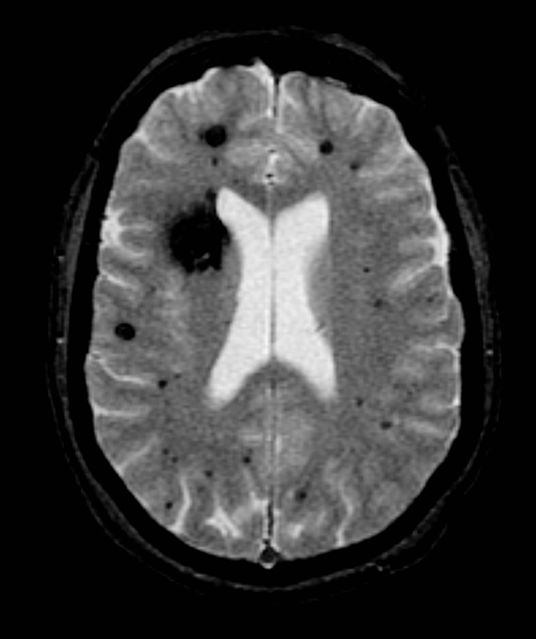
- Gradient Echo (GRE) and Susceptibility Weighted Imaging (SWI): These are particularly sensitive sequences for identifying cavernomas due to their paramagnetic properties. Cavernomas are characterized by abnormal, thin-walled blood vessels that are prone to microhemorrhages. The presence of hemosiderin, an iron-containing pigment resulting from the breakdown of red blood cells during bleeding, causes susceptibility artifacts on GRE and SWI sequences. These artifacts appear as characteristic dark signal spots, often referred to as “popcorn-like” or “mulberry-like” lesions, which are highly indicative of cavernomas. SWI, in particular, amplifies these susceptibility effects, offering superior visualization of small cavernomas and signs of previous bleeding.
- T2-weighted Imaging (T2W): While not as specific as GRE or SWI for detecting hemosiderin, T2-weighted images provide excellent anatomical detail of the brain parenchyma. They help to delineate the size and location of the cavernoma and assess any associated edema or gliosis (scarring) in the surrounding brain tissue.
- Contrast-Enhanced MRI: Although cavernomas typically do not show significant enhancement after the administration of gadolinium contrast agents (unlike some other vascular malformations like arteriovenous malformations), contrast can sometimes be used to rule out other lesions or to further characterize the lesion’s vascularity in complex cases.
The ability of modern MRI scanners, operating at higher field strengths (e.g., 3 Tesla or more), to acquire images with increased signal-to-noise ratio and better spatial resolution has dramatically improved the detection rate of small cavernomas, even those measuring just a few millimeters in diameter. Furthermore, advancements in image processing software allow for 3D reconstructions and multiplanar viewing, providing clinicians and researchers with a comprehensive understanding of the lesion’s morphology and its relationship to surrounding neural structures.
Computed Tomography (CT) Scans: A Complementary Role
While MRI is the preferred modality for cavernoma diagnosis, Computed Tomography (CT) scans can play a complementary role, especially in emergent situations or when MRI is contraindicated. CT utilizes X-rays to create cross-sectional images.
CT Characteristics and Limitations
- Calcifications: Cavernomas can sometimes calcify, and these calcifications are readily visible on CT scans as hyperdense (bright) areas. However, calcification is not a universal feature and is less common than hemosiderin deposition.
- Hemorrhage Detection: In cases of acute hemorrhage, CT can be effective in identifying blood products, which appear hyperdense. However, differentiating acute hemorrhage from other hyperdense lesions can be challenging on CT alone, and MRI is generally superior for characterizing the precise nature of the lesion.
- Resolution Limitations: The spatial resolution of CT is generally lower than that of MRI, making it less sensitive for detecting small cavernomas or subtle signs of microhemorrhage. Therefore, a negative CT scan does not rule out the presence of a cavernoma if clinical suspicion remains high.
The integration of advanced CT techniques, such as dual-energy CT, is also being explored for its potential in better differentiating tissues and identifying subtle abnormalities, though its role in cavernoma diagnosis remains secondary to MRI.
Technological Advancements in Cavernoma Monitoring and Characterization
Beyond initial detection, technology plays a vital role in monitoring the evolution of cavernomas and characterizing their risk profile. This is particularly important as cavernomas carry a risk of hemorrhage, which can lead to neurological deficits.
Serial Imaging and Longitudinal Studies

The non-invasive nature of MRI allows for repeated imaging sessions, enabling the monitoring of cavernomas over time. Technological advancements in MRI hardware and software facilitate more efficient and comfortable scanning for patients undergoing serial evaluations.
Quantifying Change and Predicting Risk
- Volume Measurement and Tracking: Sophisticated image analysis software can now accurately segment and measure the volume of cavernomas and any associated hemosiderin rings or hemosiderin-laden macrophages. By comparing serial scans, clinicians can quantify any changes in size, appearance, or extent of hemosiderin deposition, which can be indicative of recent or ongoing bleeding events. This quantitative approach moves beyond subjective visual assessment, providing more objective data for risk stratification.
- Diffusion Tensor Imaging (DTI) and Tractography: While not a primary diagnostic tool for cavernomas themselves, DTI and tractography are advanced MRI techniques that can map the white matter tracts of the brain. This technology is crucial for understanding how a cavernoma, particularly one located near critical neural pathways, might be affecting neuronal connectivity and function. It allows for visualization of potential disruption or displacement of these tracts, aiding in surgical planning if intervention is considered.
- Functional MRI (fMRI): In cases where cavernomas are suspected of causing or are located near eloquent brain regions (areas responsible for specific functions like speech, motor control, or sensation), fMRI can be employed. This technique maps brain activity by detecting changes in blood flow. By identifying which areas of the brain are active during specific tasks, fMRI helps to delineate the functional boundaries of critical brain regions, informing surgical decisions to minimize the risk of post-operative neurological deficits.
Artificial Intelligence (AI) and Machine Learning in Cavernoma Analysis
The burgeoning field of artificial intelligence (AI) and machine learning (ML) is beginning to make inroads into the analysis of medical imaging, including for conditions like cavernomas. These technologies offer the potential for faster, more consistent, and potentially more accurate interpretations of complex imaging data.
Enhancing Diagnostic Efficiency and Predictive Capabilities
- Automated Lesion Detection and Segmentation: AI algorithms are being trained on large datasets of MRI scans to automatically identify and segment cavernomas. This can significantly reduce the time radiologists spend manually reviewing scans and can help flag subtle lesions that might otherwise be overlooked.
- Predictive Modeling for Hemorrhage Risk: ML models are being developed to analyze various imaging features (e.g., size, location, presence and extent of hemosiderin, associated blood products) and patient-specific factors to predict the likelihood of future hemorrhage. Such predictive models, if validated, could revolutionize the management of cavernomas by allowing for more personalized risk stratification and informed treatment decisions.
- Radiomics and Feature Extraction: Radiomics involves extracting a large number of quantitative features from medical images, many of which are imperceptible to the human eye. AI can be used to analyze these high-dimensional radiomic datasets to identify patterns associated with cavernoma behavior, such as propensity for bleeding or growth.
Technological Frontiers in Cavernoma Management
While the primary management of symptomatic or high-risk cavernomas often involves neurosurgical intervention, technological advancements are also influencing these therapeutic approaches and exploring less invasive alternatives.
Advanced Neurosurgical Techniques
For cavernomas requiring surgical removal, advancements in surgical technology have made these procedures safer and more precise.
Minimally Invasive and Image-Guided Surgery
- Neuronavigation Systems: These systems, akin to GPS for the brain, integrate pre-operative MRI and CT scans with intra-operative imaging and instrument tracking. They provide surgeons with real-time, three-dimensional guidance, allowing for precise localization of the cavernoma and meticulous dissection, minimizing damage to surrounding healthy brain tissue.
- Intraoperative MRI (iMRI): The integration of MRI scanners directly into the operating room allows for intraoperative imaging. This is invaluable for confirming complete tumor resection, assessing for any residual lesion, and identifying new anatomical information that may have emerged during surgery.
- Microscope Technology and Endoscopy: High-definition surgical microscopes offer magnified and illuminated views of the surgical field, crucial for delicate dissections around small vascular lesions. Endoscopic techniques, using minimally invasive instruments with cameras, are also being explored for accessing and removing cavernomas in certain locations, leading to smaller incisions and faster recovery times.
Investigating Novel Technological Interventions
While surgical resection remains a primary treatment for symptomatic cavernomas, ongoing research explores technological avenues for less invasive or even non-surgical management, particularly for specific types or locations of these lesions.
Radiation Therapy and Emerging Technologies
- Stereotactic Radiosurgery (SRS): For select cases, particularly cavernomas that are difficult to access surgically or when surgery carries significant risk, stereotactic radiosurgery (using focused beams of radiation) can be considered. While SRS is not typically curative for cavernomas (as they are vascular malformations, not tumors), it can potentially lead to vessel occlusion and stabilization over time, reducing the risk of future hemorrhage. Advanced techniques in SRS planning and delivery, guided by precise imaging, ensure that radiation is targeted effectively to the lesion while sparing surrounding healthy tissue.
- Focused Ultrasound Technologies: Emerging research is exploring the potential of focused ultrasound technologies, such as focused ultrasound surgery (FUS), for treating various neurological conditions. While still in its nascent stages for cavernomas, the principle of using focused acoustic energy to precisely target and ablate tissue or induce targeted effects holds promise for future, non-invasive treatment modalities.
In conclusion, the understanding and management of cavernomas are deeply intertwined with the continuous evolution of technology. From the high-resolution imaging that first reveals these complex vascular structures to the sophisticated AI algorithms that promise to enhance diagnostic accuracy and predictive capabilities, technology is at the forefront of our ability to address cavernomas. As imaging modalities become more refined, AI capabilities mature, and surgical techniques advance, the technological landscape surrounding cavernomas will undoubtedly continue to transform, offering greater hope for improved detection, more accurate prognostication, and ultimately, better patient outcomes.
aViewFromTheCave is a participant in the Amazon Services LLC Associates Program, an affiliate advertising program designed to provide a means for sites to earn advertising fees by advertising and linking to Amazon.com. Amazon, the Amazon logo, AmazonSupply, and the AmazonSupply logo are trademarks of Amazon.com, Inc. or its affiliates. As an Amazon Associate we earn affiliate commissions from qualifying purchases.