
An “infiltrate in the lung” is a term commonly used in medical imaging to describe any substance denser than air—such as fluid, pus, blood, or cells—that appears within the lung tissue (parenchyma) and is visible on an X-ray, CT scan, or other imaging modalities. Traditionally, identifying and characterizing these infiltrates has relied heavily on the trained eye and experience of radiologists and clinicians. However, in an era increasingly defined by data, digital innovation, and artificial intelligence, understanding what an infiltrate is transcends mere visual identification. It’s about sophisticated algorithms, advanced imaging hardware, integrated digital platforms, and the intelligent processing of vast datasets that are fundamentally redefining pulmonary diagnostics and patient care. This article explores the concept of lung infiltrates through a technological lens, examining how tech tools are revolutionizing their detection, analysis, and management.
The Enigma of Pulmonary Infiltrates: A Data Challenge
At its core, an infiltrate, when viewed from a technological perspective, represents a complex pattern within medical imaging data. It’s an anomaly, a deviation from the expected ‘normal’ lung tissue signature. The challenge lies not just in identifying its presence, but in rapidly and accurately interpreting its characteristics, extent, and potential implications—a task that generates immense amounts of data and demands increasingly sophisticated analysis.
Defining the “Infiltrate” through Imaging Data
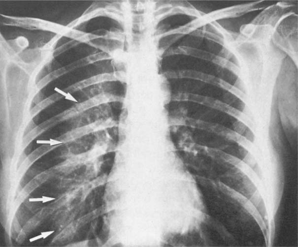
Medical imaging, whether it’s a standard chest X-ray, a computed tomography (CT) scan, or more advanced techniques, functions as a data acquisition system. Each pixel or voxel in these images holds specific intensity values that correspond to tissue density. An infiltrate manifests as regions of altered density, indicating a departure from healthy, air-filled lung tissue. For example, on a chest X-ray, an infiltrate might appear as an opaque white patch, signifying consolidation. A CT scan, offering far greater detail, can reveal the specific patterns—ground-glass opacities, reticular patterns, nodules, or masses—each providing granular data points about the nature of the infiltrate.
From a technological standpoint, defining an “infiltrate” involves segmenting these anomalous regions from the surrounding healthy tissue. This segmentation is a data processing task that converts raw image signals into quantifiable features. Software can measure the size, shape, density, and distribution of these infiltrates, generating a rich dataset that goes far beyond what a human eye can easily process. The more sophisticated the imaging, the richer the data, presenting both opportunities and challenges for analysis.
The Limitations of Traditional Interpretation
Historically, interpreting medical images has been a highly skilled, subjective process prone to human variability. Radiologists meticulously review films or digital images, drawing on years of experience to identify subtle changes. However, this traditional approach faces several inherent limitations, which technology is uniquely positioned to address:
-
Subjectivity and Variability: What one radiologist identifies as a subtle infiltrate, another might overlook, or interpret differently. This inter-observer variability can impact diagnostic accuracy and consistency.
-
Fatigue and Workload: Radiologists often face immense workloads, reviewing hundreds of images daily. Fatigue can lead to missed findings or reduced attention to detail, particularly with subtle or small infiltrates.
-
Complexity of Data: Modern imaging generates incredibly complex 3D datasets. Manually sifting through hundreds of CT slices to identify and characterize every abnormality is time-consuming and cognitively demanding.
-
Lack of Standardization: While guidelines exist, the descriptive language for infiltrates can vary, making it challenging to standardize reporting and compare findings across different institutions or over time.
These limitations underscore the need for technological solutions that can augment human capabilities, standardize interpretation, and extract deeper insights from imaging data, paving the way for more precise and consistent pulmonary diagnostics.
AI and Machine Learning: Revolutionizing Infiltrate Detection
The advent of artificial intelligence (AI) and machine learning (ML), particularly deep learning, has proven to be a transformative force in medical imaging, offering unprecedented capabilities for detecting and analyzing lung infiltrates. These technologies move beyond simple image processing to intelligent pattern recognition and predictive modeling.
Automated Image Analysis and Pattern Recognition
AI algorithms, trained on vast datasets of annotated medical images, can now automatically detect infiltrates with remarkable speed and accuracy. Deep convolutional neural networks (CNNs), a specific type of AI, are particularly adept at identifying complex visual patterns that characterize different types of infiltrates. For instance, an AI model can be trained to distinguish between bacterial pneumonia (often presenting as lobar consolidation), viral pneumonia (more diffuse ground-glass opacities), or pulmonary edema (fluid accumulation), based on subtle textural and spatial patterns that might be challenging for the human eye to discern consistently.
This automated analysis offers several advantages:
-
Speed: AI can process images in seconds, significantly reducing the time to diagnosis, especially crucial in emergency settings.
-
Consistency: Unlike humans, AI doesn’t experience fatigue, ensuring consistent performance regardless of workload.
-
Early Detection: AI can often identify subtle infiltrates that might be missed by the human eye, enabling earlier intervention.
-
Quantification: AI tools can precisely measure the volume and extent of infiltrates, providing objective metrics for tracking disease progression or response to treatment, a capability vital for research and personalized medicine.
Predictive Analytics for Early Intervention
Beyond mere detection, AI is increasingly being used for predictive analytics. By analyzing the characteristics of an infiltrate alongside patient clinical data (e.g., electronic health records, lab results, vital signs), AI models can predict the likelihood of certain outcomes, such as disease progression, response to specific treatments, or even the risk of developing complications.
For example, in the context of emerging infectious diseases, AI can analyze lung infiltrates and predict the severity of a patient’s condition, helping clinicians prioritize care and allocate resources. In chronic lung diseases, AI could predict exacerbations by identifying subtle changes in infiltrates over time, allowing for proactive interventions before a crisis develops. This shift from reactive diagnosis to proactive prediction represents a significant leap forward in patient management, enabled entirely by sophisticated data analytics and machine learning.
Overcoming Bias and Enhancing Accuracy
While AI models require careful validation to avoid perpetuating biases present in their training data, they offer the potential to reduce human cognitive biases in diagnosis. By providing objective assessments based on learned patterns, AI can act as a “second pair of eyes,” flagging potential abnormalities that a human might overlook due to confirmation bias or cognitive load. This enhances overall diagnostic accuracy and can lead to improved patient outcomes. Furthermore, AI systems can be continuously updated and refined with new data, ensuring they remain at the forefront of diagnostic precision.
Advanced Imaging Technologies: Peering Deeper

While AI processes the data, the quality and type of data available are determined by the imaging hardware. The evolution of advanced imaging technologies continues to provide richer, more detailed, and multi-dimensional datasets, enabling a more profound understanding of lung infiltrates than ever before.
High-Resolution CT and Beyond
The standard for detailed lung imaging, High-Resolution Computed Tomography (HRCT), has itself advanced significantly. Modern multi-detector CT (MDCT) scanners offer faster acquisition times, lower radiation doses, and exquisite anatomical detail, allowing for the visualization of even the smallest infiltrates and their precise structural characteristics. Beyond conventional CT, techniques like dual-energy CT (DECT) provide functional information, differentiating materials based on their atomic number. This can help distinguish between different types of infiltrates, such as iodine uptake in vascular lesions versus calcium deposits, adding a chemical dimension to the anatomical data.
Functional Imaging and Molecular Insights
The next frontier lies in moving beyond purely anatomical imaging to functional and molecular imaging. Technologies like Positron Emission Tomography (PET) scans, often combined with CT (PET/CT), can detect metabolic activity associated with inflammation, infection, or malignancy, offering insights into the biological nature of an infiltrate. For example, a PET scan can identify hypermetabolic activity in a lung nodule, suggesting malignancy even before significant morphological changes are evident. This functional data, when integrated with anatomical information, provides a more holistic view of the infiltrate’s biological signature.
The Role of Digital Pathology
While imaging provides macroscopic views, digital pathology offers microscopic insights. With whole-slide imaging technology, traditional glass slides of lung tissue biopsies are scanned at high resolution, creating vast digital files. These digital slides can then be analyzed by AI algorithms to identify cellular characteristics, tissue architecture, and molecular markers associated with specific types of infiltrates or diseases. Integrating this digital pathology data with imaging data (radiomics) and genomic data creates a multi-modal dataset that allows for a comprehensive, multi-scale understanding of pulmonary conditions, enabling highly precise diagnoses and personalized treatment plans.
Digital Health Platforms and Telemedicine: Bridging Gaps in Care
Technology extends beyond image acquisition and analysis into the infrastructure of healthcare delivery itself. Digital health platforms and telemedicine are crucial for disseminating imaging data, facilitating collaborative diagnostics, and empowering patients, thereby ensuring that insights gained from advanced tech are translated into improved patient care, regardless of geographical barriers.
Remote Monitoring and Data Integration
Digital health platforms serve as centralized hubs for patient data, integrating imaging reports, lab results, electronic health records (EHRs), and even data from wearable devices. This seamless integration ensures that all relevant information about a patient’s lung infiltrate is readily accessible to healthcare providers. Furthermore, for patients with chronic lung conditions, remote monitoring technologies (e.g., smart spirometers, pulse oximeters that connect to apps) can continuously track key physiological parameters. Any deviation can alert clinicians to potential changes in their condition, potentially indicating a developing infiltrate before symptoms become severe, thus enabling proactive management.
Collaborative Diagnostics Across Geographic Divides
Telemedicine and cloud-based image sharing platforms have revolutionized collaborative diagnostics. A radiologist in a rural hospital can securely transmit high-resolution CT scans to a sub-specialist in a major urban center for expert review, facilitating second opinions and access to specialized expertise. This is particularly vital for complex or rare lung infiltrates that require highly specialized interpretation. Secure digital pathways ensure that patient data remains protected while enabling efficient communication and shared decision-making among a geographically dispersed team of experts. This global connectivity ensures that patients, wherever they are, can benefit from the collective knowledge of the medical community.
Patient Engagement Through Health Tech
Beyond professional collaboration, technology also empowers patients. Digital health apps and secure online portals allow patients to access their own imaging reports, understand their diagnosis (e.g., the nature of their lung infiltrate), track their health metrics, and communicate directly with their care team. This transparency and engagement lead to better adherence to treatment plans and a more active role in managing their own health, turning complex medical information into actionable insights for the individual.
The Future Landscape: Personalized Medicine and Proactive Lung Health
The confluence of AI, advanced imaging, and digital platforms is rapidly accelerating the shift towards personalized medicine and a proactive approach to lung health. Understanding “what is an infiltrate in the lung” will increasingly mean understanding its unique digital fingerprint within an individual’s comprehensive health profile.
Data-Driven Treatment Strategies
In the future, decisions regarding treatment for lung infiltrates will be increasingly data-driven. AI algorithms, combining insights from imaging, genomics, proteomics, and clinical data, will predict the most effective therapies for specific patients, considering their unique biological makeup and the precise characteristics of their infiltrate. This precision medicine approach aims to optimize therapeutic outcomes while minimizing adverse effects, moving away from a ‘one-size-fits-all’ strategy. For instance, AI might identify a specific gene mutation associated with a certain type of lung infiltrate and suggest a targeted molecular therapy that has a high probability of success.

Wearable Tech for Continuous Monitoring
The ultimate goal is to move beyond reacting to symptoms and towards preventing or detecting disease at its earliest, most treatable stages. Wearable devices, equipped with advanced sensors, are poised to play a crucial role. While still in nascent stages for lung conditions, future wearables might monitor subtle changes in breathing patterns, cough characteristics, oxygen saturation, or even early inflammatory markers. These devices could continuously feed data into AI-powered platforms, which would then flag early indicators of an evolving lung infiltrate, prompting timely medical intervention, often before the patient even perceives any symptoms. This proactive approach has the potential to transform lung health management, making it more preventative and personalized than ever before.
In conclusion, “what is an infiltrate in the lung” is no longer just a medical definition; it’s a profound technological question. From the data acquisition of advanced scanners to the intelligent analysis by AI, and the seamless integration via digital health platforms, technology is not just aiding diagnosis but fundamentally reshaping our understanding of pulmonary conditions. The future of lung health lies in the continued integration and innovation of these tech tools, leading to earlier detection, more accurate diagnoses, personalized treatments, and ultimately, healthier lives.
aViewFromTheCave is a participant in the Amazon Services LLC Associates Program, an affiliate advertising program designed to provide a means for sites to earn advertising fees by advertising and linking to Amazon.com. Amazon, the Amazon logo, AmazonSupply, and the AmazonSupply logo are trademarks of Amazon.com, Inc. or its affiliates. As an Amazon Associate we earn affiliate commissions from qualifying purchases.