The phrase “stereotactic breast biopsy” might sound technical, even daunting, but at its core, it represents a significant advancement in medical technology, particularly within the realm of diagnostic imaging and minimally invasive procedures. In an era where precision and early detection are paramount in healthcare, understanding this procedure sheds light on how cutting-edge technology is revolutionizing patient care. For those interested in the intersection of Tech, the strategic application of Brand in healthcare, and the financial implications of advanced medical treatments, a stereotactic breast biopsy offers a compelling case study.
Understanding the Core of Stereotactic Breast Biopsy: Precision Imaging Meets Targeted Intervention
At its heart, a stereotactic breast biopsy is a sophisticated diagnostic tool. It leverages advanced imaging techniques to guide a needle biopsy with exceptional accuracy. Unlike traditional needle aspirations, which might rely on palpation or less precise imaging methods, stereotactic biopsy uses imaging (typically mammography) to create a three-dimensional map of the breast, pinpointing the exact location of a suspicious abnormality. This precise localization is crucial for obtaining the most representative tissue sample for pathological examination, leading to more accurate diagnoses.
The term “stereotactic” itself hints at the technological underpinnings. Derived from the Greek words “stereos” (solid) and “taktikos” (orderly), it refers to a method of accurately locating points in three-dimensional space. In the context of a breast biopsy, this means precisely identifying the coordinates of a lesion within the breast tissue. This spatial understanding is achieved through specialized imaging equipment that captures multiple views of the breast from different angles.
The process typically begins with a mammogram. However, instead of just two standard views, a stereotactic mammogram takes a series of images from various angles around the breast. Advanced computer software then processes these images, creating a 3D reconstruction that allows the radiologist to determine the exact location of any suspicious area, whether it’s a calcification cluster, a mass, or a distortion in the breast tissue. This level of detail is critical, especially for abnormalities that are too small to be felt or seen on a standard mammogram.
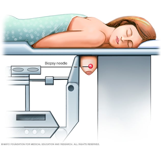
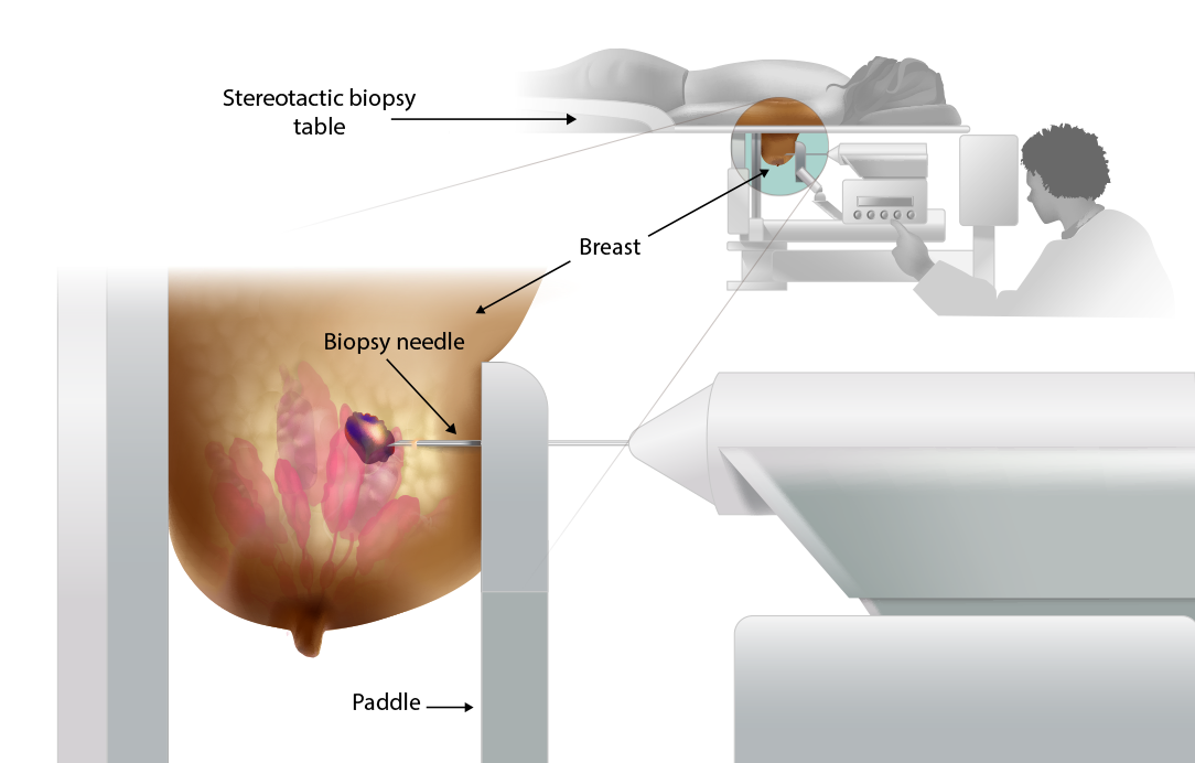
Once the target is precisely located, the patient is positioned on a specialized examination table. This table has an opening through which the breast is positioned. The patient typically lies face down with the breast gently compressed within an opening in the table. This compression is essential for immobilizing the breast and ensuring clear, stable imaging throughout the procedure. The stereotactic biopsy machine, which houses the mammography equipment and the biopsy needle system, is then precisely aligned with the identified abnormality.
The biopsy itself involves using a needle or a vacuum-assisted device to extract small samples of tissue from the suspicious area. The stereotactic guidance system ensures that the needle is inserted at the exact predetermined coordinates. The system continuously monitors the needle’s position, making minute adjustments as needed to maintain accuracy. Multiple tissue samples are usually collected to ensure a comprehensive analysis by the pathologist. The entire process is minimally invasive, requiring only a small incision, often no larger than a pencil eraser, through which the biopsy needle is inserted.
The benefits of this technological approach are manifold. It offers a higher rate of diagnostic accuracy compared to less image-guided methods. By precisely targeting the abnormality, it minimizes the risk of missing the lesion or obtaining an inadequate sample. This, in turn, can reduce the need for repeat biopsies or more invasive surgical procedures, ultimately saving time, reducing patient discomfort, and potentially lowering healthcare costs.
The Technological Backbone: Innovation Driving Diagnostic Accuracy
The development and refinement of stereotactic breast biopsy technology represent a triumph of medical engineering and computer science. At its core, the system relies on high-resolution imaging hardware and sophisticated software algorithms.
Advanced Imaging Modalities: The Eyes of the System
The primary imaging modality used in stereotactic breast biopsy is digital mammography. Unlike older film-based systems, digital mammography captures images electronically, allowing for enhanced image quality, easier manipulation of contrast and brightness, and quicker transmission to radiologists. For stereotactic procedures, specialized mammography units are equipped with robotic arms that can precisely angle the X-ray source and detector to capture multiple views.
Beyond conventional digital mammography, some advanced systems may incorporate other imaging technologies to further enhance lesion detection and localization. These can include:
- Digital Breast Tomosynthesis (DBT) or 3D Mammography: This technology takes multiple low-dose X-ray images from different angles and reconstructs them into a series of thin slices, effectively creating a 3D view of the breast. This significantly reduces the overlap of breast tissue, which can obscure small abnormalities in traditional 2D mammography, making it easier to identify and locate lesions for stereotactic biopsy.
- Ultrasound: While mammography is the primary guiding tool for calcifications and many masses, ultrasound can be particularly useful for identifying cystic lesions or solid masses that are better visualized with sound waves. In some cases, ultrasound may be used in conjunction with stereotactic biopsy to further refine the target location, especially for lesions that are visible on ultrasound but not clearly delineated on mammography.
- Magnetic Resonance Imaging (MRI): MRI is highly sensitive for detecting breast cancer and can identify lesions that may be missed by mammography or ultrasound. For MRI-detected lesions that are not visible on mammography or ultrasound, specialized MRI-guided biopsy techniques are employed, which, while not stereotactic in the mammographic sense, share the principle of highly precise image guidance.
The Role of Software and Computation: From Images to Coordinates
The “stereotactic” aspect of the biopsy is heavily dependent on advanced software. After the multiple mammographic images are acquired, specialized software analyzes them. This software performs several critical functions:
- Image Reconstruction: It reconstructs the 2D images into a 3D representation of the breast, allowing the radiologist to visualize the lesion in its spatial context.
- Localization Algorithms: These algorithms pinpoint the exact coordinates (X, Y, and Z axes) of the suspicious abnormality within the 3D model.
- Guidance and Navigation: The software interfaces with the biopsy device, translating the determined coordinates into precise instructions for the needle insertion path. It ensures that the needle moves accurately towards the target, compensating for any slight patient movement.
The integration of AI (Artificial Intelligence) into these systems is also a growing trend. AI algorithms can be trained to detect subtle patterns in mammograms that might indicate malignancy, potentially improving the sensitivity and specificity of lesion detection. Furthermore, AI can assist in automating parts of the localization process, making it faster and potentially more consistent.
Biopsy Devices: Minimally Invasive Tools for Sample Collection
The biopsy needle itself is a crucial piece of technology. Stereotactic biopsies commonly employ either:
- Core Needle Biopsy Devices: These devices use a spring-loaded mechanism to rapidly advance a hollow needle into the target tissue, extracting a cylindrical core sample.
- Vacuum-Assisted Biopsy (VAB) Devices: These are often preferred for their ability to collect larger tissue samples. A slightly larger needle with a vacuum port is used. The needle rotates and moves slightly, collecting tissue that is then suctioned into a collection chamber. The vacuum system can also be used to irrigate the site after the biopsy, helping to control any minor bleeding.
The design of these biopsy needles is engineered for minimal invasiveness, allowing for rapid insertion and retrieval with the least possible disruption to surrounding tissue.
Strategic Branding and Patient Trust in Advanced Healthcare Tech
In the competitive landscape of healthcare, the Brand of a medical facility or a specific diagnostic service plays a pivotal role in patient acquisition and retention. For a procedure like a stereotactic breast biopsy, the “brand” encompasses not just the name of the hospital or clinic, but the entire patient experience, the perceived quality of care, and the level of trust instilled.
Building Trust Through Transparency and Education

A strong Brand in healthcare is built on transparency, clear communication, and demonstrated expertise. For stereotactic breast biopsies, this means clearly articulating the benefits of the technology, demystifying the procedure, and reassuring patients about their safety and comfort.
- Educational Content: Websites and patient brochures should explain what a stereotactic breast biopsy is in accessible language, highlighting its accuracy and minimally invasive nature. This content, similar to this article, educates potential patients and addresses their anxieties.
- Physician Expertise: Emphasizing the qualifications and experience of the radiologists and technologists performing the biopsy is crucial. Testimonials from satisfied patients, when ethically permissible, can further bolster trust.
- Technological Prowess as a Differentiator: Highlighting the state-of-the-art technology used can position a healthcare provider as a leader in diagnostic imaging. This appeals to patients who are actively seeking the best possible care and are willing to research and choose providers based on their technological capabilities.
The Corporate Identity of Precision Medicine
The corporate identity of a healthcare institution offering stereotactic biopsies should project an image of precision, innovation, and compassionate care. This is reflected in:
- Facility Design: A modern, clean, and welcoming environment can contribute to a positive patient experience.
- Digital Presence: A professional and informative website, online appointment booking, and clear communication channels are essential.
- Marketing and Outreach: Targeted marketing campaigns that educate the public about breast health and available advanced diagnostic tools can be highly effective. This includes partnerships with community organizations and participation in health fairs.
Case Studies and Reputation Management
Positive case studies, anonymized for patient privacy, demonstrating successful diagnoses and outcomes achieved through stereotactic biopsies can serve as powerful Brand-building tools. Managing online reviews and responding promptly and empathetically to patient feedback is also critical for maintaining a strong reputation. A facility known for its excellence in stereotactic biopsies builds a reputation that attracts both patients and referring physicians.
Financial Considerations: Investing in Early Detection and Advanced Care
The implementation and utilization of advanced technologies like stereotactic breast biopsy have significant Money implications, both for healthcare providers and for patients. Understanding these financial aspects is crucial for informed decision-making.
The Investment in Advanced Medical Technology
For healthcare institutions, investing in stereotactic biopsy equipment represents a substantial capital expenditure. This includes the cost of:
- High-Resolution Digital Mammography Units: These are significantly more expensive than standard mammography machines.
- Specialized Biopsy Devices: Including the vacuum-assisted systems.
- Software and IT Infrastructure: For image processing, storage, and network connectivity.
- Training and Ongoing Maintenance: Ensuring that medical staff are proficient in using the technology and that the equipment is regularly maintained.
This upfront investment is justified by the long-term benefits, including improved diagnostic accuracy, reduced need for repeat procedures, and the potential for earlier detection of cancer, which often leads to less aggressive and less costly treatment.
Reimbursement and Insurance Coverage
The cost of a stereotactic breast biopsy is typically covered by most health insurance plans, as it is considered a medically necessary diagnostic procedure. However, the specifics of coverage can vary depending on the insurance provider and the individual policy. Factors that influence reimbursement include:
- Medical Necessity: The procedure must be deemed medically necessary by a physician, usually based on abnormal findings on a screening mammogram or physical examination.
- Provider Network: Whether the healthcare facility and the performing physician are in-network with the patient’s insurance plan.
- Deductibles and Co-pays: Patients will likely be responsible for their deductible and co-payment amounts as outlined in their insurance policy.
It is crucial for patients to verify their insurance coverage with their provider and the healthcare facility before undergoing the procedure.
The Financial Value of Early Detection
From a personal finance perspective, the Money spent on a diagnostic procedure like a stereotactic breast biopsy, even if it incurs out-of-pocket costs, can be an investment in one’s long-term financial well-being. Early detection of breast cancer often leads to:
- Less Extensive Treatment: Early-stage cancers are typically treated with less aggressive and therefore less expensive therapies (e.g., lumpectomy instead of mastectomy, less extensive chemotherapy or radiation).
- Shorter Recovery Times: This means less time away from work, minimizing income loss.
- Improved Prognosis: The ultimate “return on investment” is the preservation of life and health, which far outweighs any financial cost.
Conversely, delaying diagnosis due to cost concerns can lead to more advanced cancer, requiring more complex and expensive treatments, longer periods of disability, and potentially a poorer outcome.
Financial Tools and Planning
For healthcare providers, managing the financial aspects of offering advanced diagnostic services involves careful financial planning and the use of financial tools. This includes:
- Cost-Benefit Analysis: Evaluating the financial return on investment for new technology.
- Revenue Cycle Management: Efficiently managing billing, coding, and claims processing to ensure timely reimbursement.
- Patient Financial Counseling: Offering transparent information about costs and payment options to patients.
For patients, understanding their insurance benefits, exploring payment plans if needed, and prioritizing preventative care are key financial strategies when it comes to their health.
In conclusion, a stereotactic breast biopsy is more than just a medical procedure; it’s a prime example of how technological innovation, strategic branding in healthcare, and careful financial consideration converge to improve patient outcomes. It underscores the importance of embracing advanced Tech for precise diagnostics, leveraging Brand building to foster trust and deliver exceptional patient experiences, and understanding the financial value of investing in one’s health through early detection.
aViewFromTheCave is a participant in the Amazon Services LLC Associates Program, an affiliate advertising program designed to provide a means for sites to earn advertising fees by advertising and linking to Amazon.com. Amazon, the Amazon logo, AmazonSupply, and the AmazonSupply logo are trademarks of Amazon.com, Inc. or its affiliates. As an Amazon Associate we earn affiliate commissions from qualifying purchases.