
In the rapidly evolving landscape of medical technology, few innovations have bridged the gap between internal visualization and high-resolution data acquisition as effectively as Endoscopic Ultrasound (EUS). While traditional imaging techniques like X-rays or standard ultrasounds provide a peripheral view of the human body, EUS represents a sophisticated fusion of two distinct technological domains: high-definition fiber optics and advanced acoustic engineering. To understand what an endoscopic ultrasound is from a tech-centric perspective is to appreciate the incredible miniaturization of hardware and the complex software algorithms that allow clinicians to see beyond the surface of internal tissues.
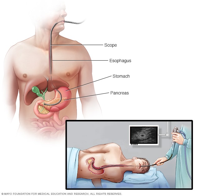
As we dive into the “Med-Tech” ecosystem, EUS stands out not just as a clinical tool, but as a masterpiece of instrumentation that leverages real-time signal processing and precision engineering. This article explores the technological architecture of EUS, the software driving its diagnostic accuracy, and the burgeoning role of Artificial Intelligence in its evolution.
The Convergence of Optics and Acoustics: The Hardware Behind EUS
At its core, an endoscopic ultrasound device is a hybrid piece of hardware. It combines the visual capabilities of a standard endoscope—a flexible tube equipped with a light source and camera—with an ultrasound transducer at the tip. This dual-sensor approach allows for a “macro” view of the gastrointestinal tract and a “micro” view of the surrounding organs and layers.
The Micro-Ultrasound Transducer
The defining piece of hardware in EUS is the miniaturized ultrasound transducer. Unlike the bulky probes used for external abdominal scans, the EUS transducer must be small enough to fit through narrow anatomical passages while remaining powerful enough to emit high-frequency sound waves. These transducers utilize piezoelectric crystals that convert electrical energy into mechanical vibrations (sound) and back again. The engineering challenge lies in maintaining a high signal-to-noise ratio within a confined form factor. By placing the transducer directly against the internal lining of an organ, the technology bypasses the interference of skin, fat, and air that often degrades the quality of external scans.
High-Definition Endoscopic Fiber Optics
While the ultrasound component looks “through” walls, the optical component provides the “navigation” system. Modern EUS devices employ high-definition (HD) CMOS sensors capable of transmitting 1080p or even 4K video feeds to a monitor. These sensors must operate under low-light conditions, supported by advanced LED fiber-optic bundles that provide cool, intense illumination. The synchronization between the optical video feed and the acoustic image is managed by a central processing unit, allowing the operator to toggle between visual and sonic data streams seamlessly.
Software Integration and Real-Time Data Processing
The hardware of an EUS device is only as effective as the software that interprets the raw data. When the ultrasound transducer receives returning echoes, it generates a massive amount of “noisy” electronic data. Transforming this into a legible, high-contrast image requires significant computational power and sophisticated digital signal processing (DSP).
Image Enhancement Algorithms
Digital image processing in EUS involves complex algorithms designed to sharpen edges, reduce “speckle” (a type of noise inherent in ultrasound), and enhance tissue differentiation. Advanced software packages now include “Elastography,” a feature that measures the stiffness of tissues in real-time. By calculating how much a tissue deforms under the pressure of the probe, the software color-codes the image—blue for hard tissues and red for soft. This digital mapping provides a layer of data that is invisible to the naked eye, highlighting potential anomalies through mechanical property analysis rather than just visual appearance.
Digital Interfacing and PACS Integration
Modern EUS systems are no longer standalone units; they are nodes within a hospital’s broader digital ecosystem. These devices are integrated with Picture Archiving and Communication Systems (PACS), utilizing the DICOM (Digital Imaging and Communications in Medicine) standard. This integration allows for the instantaneous sharing of high-resolution video and ultrasound captures across a network. Furthermore, contemporary EUS software supports “Picture-in-Picture” (PiP) and overlay capabilities, where a patient’s previous CT or MRI scans can be digitally superimposed over the live EUS feed to assist in spatial orientation and precision.
Minimally Invasive Innovation: The Engineering Feat
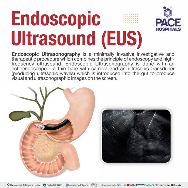
EUS is a prime example of the “minimally invasive” tech trend. The goal is to maximize data acquisition while minimizing the physical footprint of the intervention. This balance is achieved through the integration of mechanical tools within the digital probe.
Fine Needle Aspiration (FNA) and Biopsy Technology
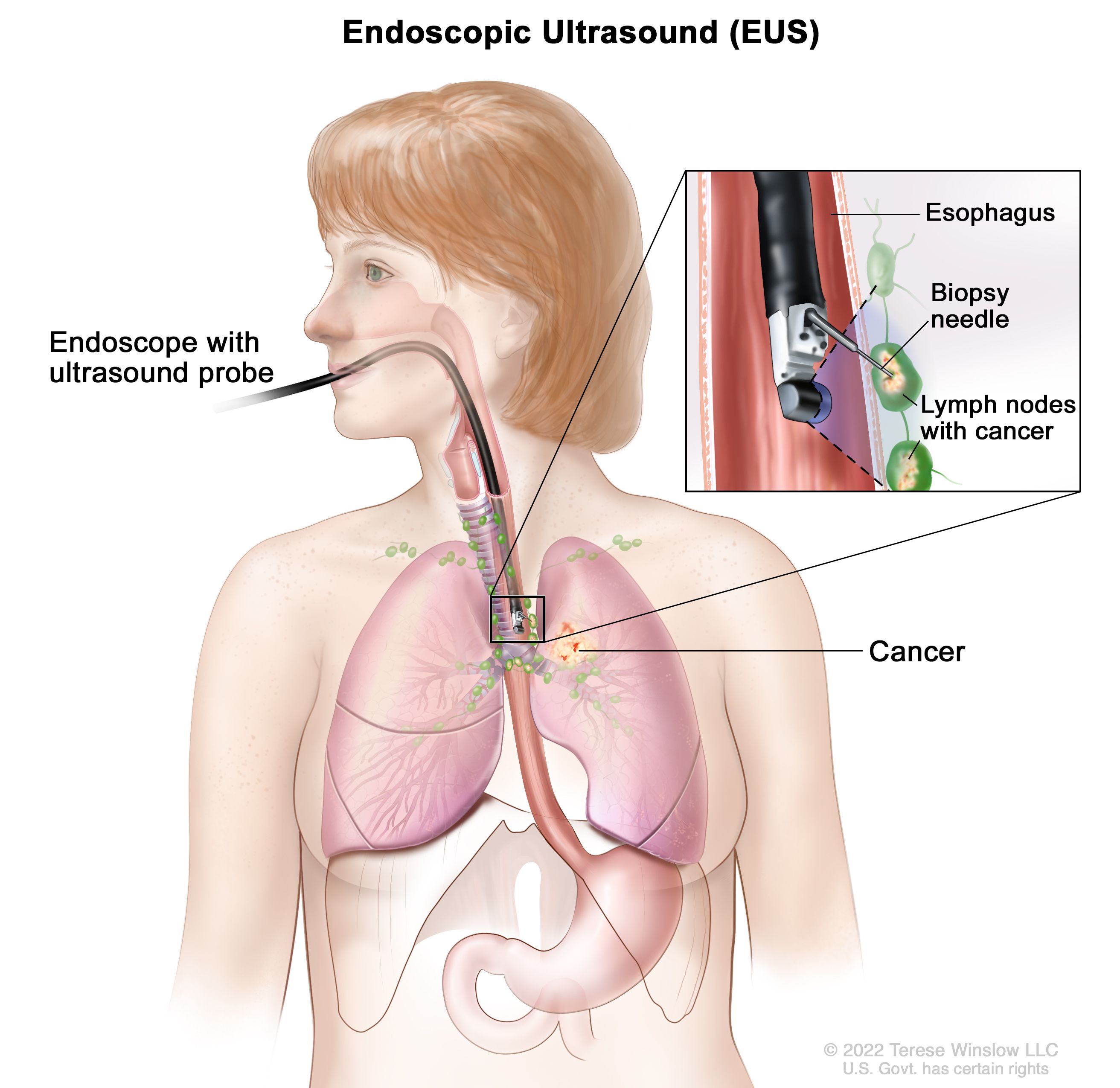
One of the most impressive technical features of EUS is the working channel. Despite the probe’s small diameter, it contains a channel for deploying mechanical instruments. Fine Needle Aspiration (FNA) needles are high-tech delivery systems that can be guided by the ultrasound feed in real-time. The engineering of these needles involves specialized coatings to make them more “echogenic” (visible on ultrasound). This allows the technologist to watch the needle enter a target area on the digital screen with sub-millimeter precision, effectively turning the diagnostic tool into a surgical delivery system.
Navigation and Spatial Awareness
Newer generations of EUS hardware are experimenting with electromagnetic tracking. By placing tiny sensors within the endoscope tip and using an external magnetic field generator, software can map the device’s exact 3D position within the patient’s body. This “GPS for the body” allows for a more comprehensive digital reconstruction of the internal anatomy, ensuring that no areas are missed during a scan and providing a digital “bread crumb” trail for follow-up procedures.
The Role of Artificial Intelligence in Modern EUS
Artificial Intelligence (AI) and Machine Learning (ML) are currently the most disruptive forces in medical technology, and EUS is a major beneficiary. The complexity of ultrasound images makes them an ideal candidate for computer vision enhancements.
AI-Assisted Lesion Detection
Because ultrasound images are often grainier and more difficult to interpret than CT scans, AI models are being trained on millions of previous EUS procedures to identify patterns associated with specific pathologies. These AI overlays act as a “second set of eyes,” highlighting suspicious regions of interest (ROI) in real-time. This reduces the cognitive load on the operator and minimizes the risk of human error in detecting subtle changes in tissue texture or vascular patterns.
Automated Measurement and Reporting
Software automation is also streamlining the administrative side of EUS. Previously, measuring the dimensions of a detected anomaly required manual input and calculations. Today’s AI-driven software can automatically detect the borders of a lesion, calculate its volume, and populate a digital report with precise metrics. This automation ensures consistency across different examinations and allows for more accurate longitudinal tracking of a patient’s condition over months or years.
Future Horizons: Wireless and Robotic Advancements
As we look toward the future of EUS, the trend is moving toward even greater autonomy and further miniaturization. The tech roadmap for EUS is heavily influenced by the broader trends in robotics and wireless communication.
Robotic-Assisted Endoscopy
One of the most exciting frontiers is the development of robotic-assisted EUS. Currently, the maneuverability of an endoscope is limited by the manual dexterity of the operator. Robotic systems, controlled via a console, offer 360-degree articulation and tremor-cancellation technology. This allows the ultrasound probe to reach deeper or more difficult-to-access areas with a level of stability that is physically impossible for a human hand. These robotic platforms also pave the way for remote procedures, where a specialist in one city could perform an EUS on a patient in another via high-speed, low-latency 5G networks.
The Miniaturization Trend: EUS-on-a-Chip
The long-term goal for many developers is “EUS-on-a-chip.” Currently, the transducer is a mechanical component, but research into CMUT (Capacitive Micromachined Ultrasonic Transducers) suggests a future where ultrasound can be generated by silicon chips. This would drastically reduce the cost of the hardware and allow for even thinner, more flexible probes. Eventually, we may see the convergence of EUS with “pill camera” technology, where a wireless capsule can emit ultrasound signals as it travels through the body, transmitting data to a wearable receiver.
In conclusion, an endoscopic ultrasound is far more than a simple medical exam; it is a high-tech interface between human biology and advanced digital engineering. Through the integration of high-definition optics, real-time acoustic signal processing, and the burgeoning power of AI, EUS continues to push the boundaries of what is possible in diagnostic technology. As the hardware becomes more refined and the software more intelligent, EUS will remain at the forefront of the technological revolution in healthcare.
aViewFromTheCave is a participant in the Amazon Services LLC Associates Program, an affiliate advertising program designed to provide a means for sites to earn advertising fees by advertising and linking to Amazon.com. Amazon, the Amazon logo, AmazonSupply, and the AmazonSupply logo are trademarks of Amazon.com, Inc. or its affiliates. As an Amazon Associate we earn affiliate commissions from qualifying purchases.