
The knee, a marvel of biological engineering, is crucial for mobility and bears significant stress daily. When pain, instability, or injury strikes this complex joint, understanding the underlying issue is paramount for effective treatment. While X-rays provide a snapshot of bone structure, and ultrasounds offer dynamic views of soft tissues, neither can match the comprehensive detail offered by Magnetic Resonance Imaging (MRI). An MRI scan of the knee provides an unparalleled view into the joint’s intricate anatomy, revealing a spectrum of conditions often invisible to other diagnostic methods. However, to truly appreciate the significance of knee MRI, it’s essential to look beyond its purely medical application and understand its profound connections to technology, brand strategy, and the economics of healthcare.

This article delves into the technological sophistication that makes MRI possible, explores how this diagnostic powerhouse shapes the brand identity of healthcare providers, and examines the financial implications for both institutions and individuals. We will dissect what specific knee conditions MRI vividly illustrates, all while keeping a keen eye on the digital innovations and strategic considerations that underpin this vital medical imaging tool.
The Technological Marvel Behind MRI for Knee Diagnostics
At its core, MRI is a triumph of modern engineering and software development, a sophisticated gadget that leverages fundamental physics to peer inside the human body without invasive procedures or ionizing radiation. For a joint as complex as the knee, this technological prowess is not just beneficial; it’s revolutionary, offering precision that impacts everything from diagnostic accuracy to treatment planning and patient outcomes.
How MRI Technology Works: A A Glimpse into the ‘Tech’
The magic of MRI lies in its interaction with the hydrogen atoms abundantly present in water molecules throughout the body. The patient is placed inside a powerful magnet that aligns these hydrogen protons. Then, a radiofrequency current is pulsed through the patient, briefly knocking these protons out of alignment. When the pulse is turned off, the protons relax back into alignment, releasing energy signals. These signals are detected by receiver coils in the MRI machine.
This is where the ‘Tech’ truly shines. Sophisticated software algorithms take these raw signals and process them into cross-sectional images, differentiating between various tissue types based on how quickly their hydrogen atoms realign. The clarity and detail are astounding – distinguishing between cartilage, ligaments, tendons, muscles, bone, and fluid with remarkable precision. Modern MRI systems integrate high-performance computing, advanced signal processing, and intricate networking solutions to capture, transmit, and render these complex datasets, reflecting the cutting edge of what’s possible in digital imaging and data management. Furthermore, the development of specialized “apps” within the MRI console allows for customized imaging sequences tailored to specific clinical questions, enhancing diagnostic utility for knee pathologies.
Advancements in MRI Software and Hardware for Enhanced Knee Imaging
The field of MRI technology is constantly evolving, driven by innovations in both hardware and software, echoing broader trends in high-tech industries.
- Higher Field Strengths: Modern MRI scanners operate at higher magnetic field strengths (e.g., 3T vs. 1.5T), which translates to improved signal-to-noise ratio and consequently, higher resolution images. This enhanced detail is critical for visualizing subtle injuries within the knee, such as small meniscal tears or early cartilage degradation.
- Specialized Coils: Dedicated knee coils are precisely engineered hardware components that fit snugly around the knee. These coils are optimized to receive signals specifically from the joint, significantly improving image quality by minimizing noise and maximizing signal reception from the area of interest. This customization is a testament to meticulous gadget design.
- Advanced Software Sequences: Beyond basic T1 and T2 weighted imaging, sophisticated software protocols have been developed. For instance, techniques like Diffusion Tensor Imaging (DTI) can map the microstructure of ligaments, while quantitative MRI sequences can assess cartilage health and composition non-invasively. AI tools are increasingly being integrated into these software packages, not only to speed up scan times by optimizing sequences but also to assist radiologists in image interpretation, identifying subtle patterns indicative of specific pathologies.
- Digital Security: As MRI generates vast amounts of sensitive patient data, robust digital security measures are paramount. The software and network infrastructure supporting MRI systems must comply with stringent data protection regulations, ensuring patient privacy and preventing unauthorized access to medical records. This directly ties into the broader website theme of “Digital Security” and its critical role in modern tech applications.
Beyond Diagnosis: The ‘Brand’ and ‘Money’ Implications of MRI in Healthcare
While MRI’s diagnostic capabilities are undeniable, its influence extends far beyond the examination room. For healthcare providers, the presence and utilization of state-of-the-art MRI equipment for knee and other joints carry significant ‘Brand’ and ‘Money’ implications, shaping institutional reputation and financial viability.
The ‘Brand’ of Accuracy: How MRI Shapes Medical Reputation and Patient Trust
In a competitive healthcare landscape, the “brand” of a hospital or clinic is often defined by its commitment to accuracy, efficiency, and patient care. Investing in advanced diagnostic technology like a high-resolution MRI scanner for knee imaging becomes a powerful brand statement.
- Reputation and Trust: Offering cutting-edge MRI services signals a commitment to providing the best possible diagnostics. This builds patient trust and enhances the institution’s reputation, positioning it as a leader in specialized care. Patients are more likely to choose a provider known for precise diagnoses, which directly contributes to their overall perception and satisfaction with the “brand” of the healthcare facility.
- Marketing and Differentiation: Healthcare marketing strategies frequently highlight advanced technology. A clinic can differentiate itself by promoting its “state-of-the-art 3T MRI for unparalleled knee imaging,” attracting patients seeking definitive answers. This leverages the “Marketing” aspect of the Brand topic, turning technological investment into a competitive advantage.
- Personal Branding for Physicians: For orthopedic surgeons and sports medicine specialists, access to and proficiency in interpreting detailed MRI scans enhances their personal brand as expert practitioners. Being able to provide precise diagnoses based on superior imaging elevates their standing within the medical community and among patients. This extends the concept of “Personal Branding” from individuals to professional expertise.
- Case Studies and Outcomes: Accurate MRI diagnoses lead to more effective treatment plans, which in turn generate positive patient outcomes. These success stories, often highlighted as “Case Studies,” further bolster the institution’s brand and reputation, demonstrating tangible results driven by technological excellence.
The ‘Money’ Aspect: Investment, Cost, and Value in Knee MRI
The acquisition and operation of an MRI machine represent a substantial financial undertaking, impacting everything from business finance for providers to personal finance for patients.
- Significant Investment: MRI machines are incredibly expensive, often costing millions of dollars. This represents a major capital expenditure for hospitals or imaging centers, requiring careful “Business Finance” planning, including procurement, maintenance, and facility upgrades. The decision to invest in specific models (e.g., higher field strengths) is often a strategic financial one, weighing cost against diagnostic capabilities and market demand.
- Cost to Patients and Insurance: For patients, an MRI scan of the knee can be a considerable expense, ranging from hundreds to several thousand dollars depending on location, facility, and insurance coverage. Understanding “Personal Finance” implications, including deductibles, co-pays, and out-of-pocket maximums, becomes crucial. Patients may need to utilize “Financial Tools” or engage in “Financial Planning” to manage these costs.
- Value Proposition: Despite the cost, the value an MRI brings often far outweighs the initial outlay. An accurate diagnosis from a knee MRI can prevent costly and ineffective treatments, unnecessary surgeries, or prolonged disability. For example, definitively identifying a meniscal tear can lead to targeted intervention, avoiding months of physical therapy for a misdiagnosed sprain. This efficiency has economic benefits for both the patient (faster return to work, “Productivity,” potentially “Online Income” or “Side Hustles” unimpeded by lingering injury) and the healthcare system (reduced healthcare expenditures due to appropriate treatment).
- Online Income and Business Models: Some imaging centers operate on cash-pay models, offering transparent pricing for patients without insurance or those seeking more affordable options. This aligns with “Online Income” concepts, as online platforms can facilitate price comparisons and appointment scheduling. The business model surrounding MRI services often involves balancing technological superiority with economic accessibility.
What Specific Knee Conditions MRI Reveals: Bridging Medical Insight with Tech Application
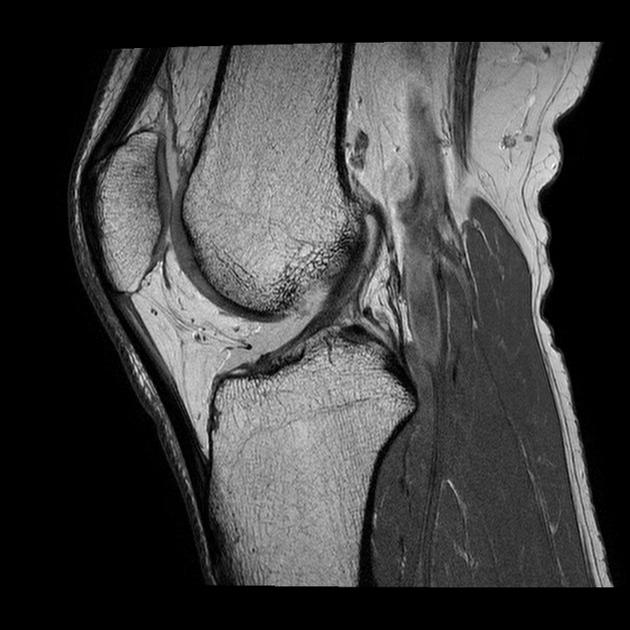
The true power of knee MRI lies in its ability to non-invasively unveil a multitude of pathologies that impact function and quality of life. The advanced technological capabilities discussed earlier directly enable this diagnostic precision.
Ligament and Tendon Injuries: ACL, PCL, MCL, LCL, and Patellar Tendon
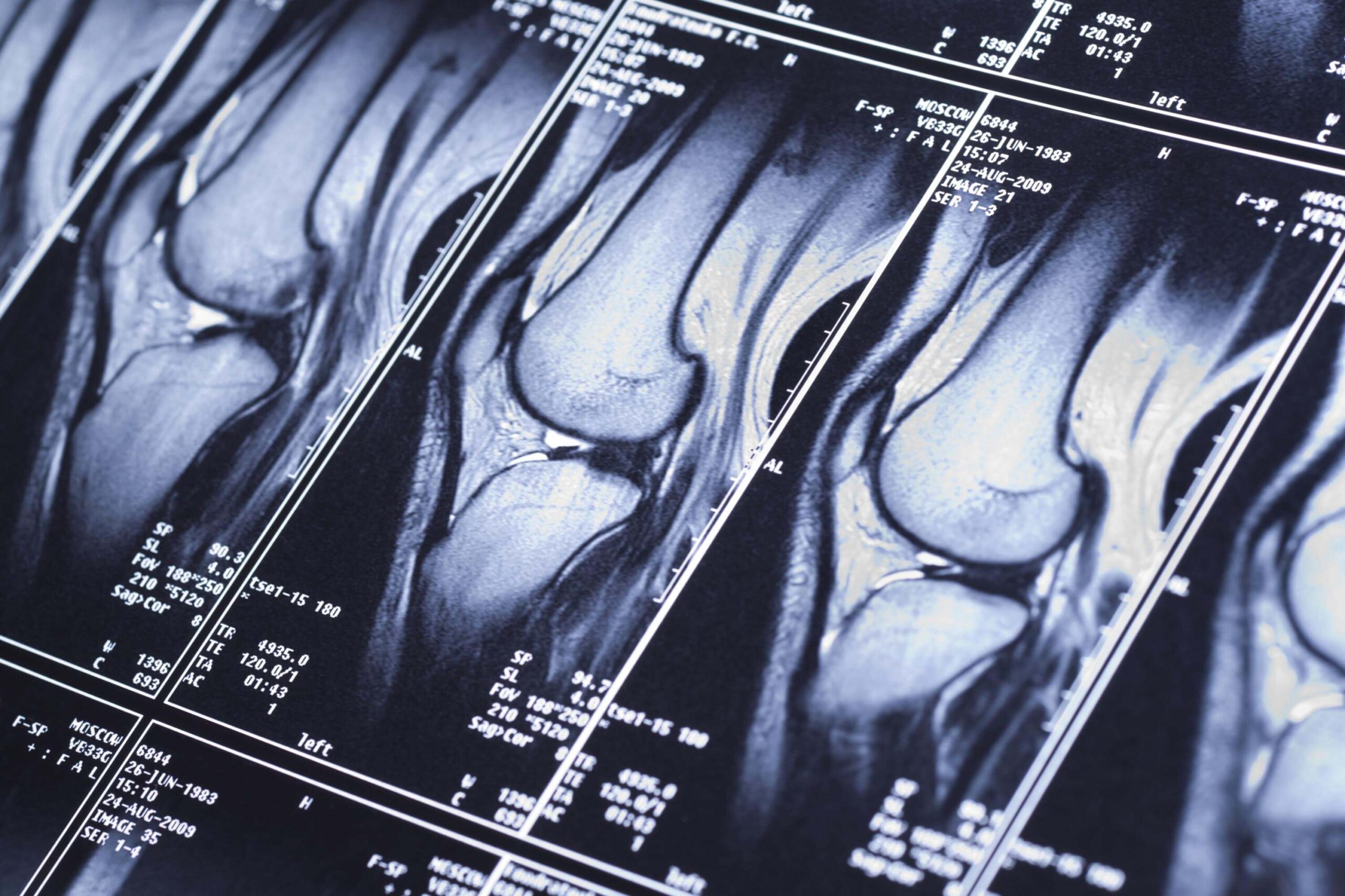
The knee is stabilized by a network of ligaments and tendons. MRI excels at visualizing these soft tissue structures.
- Cruciate Ligaments (ACL, PCL): MRI is the gold standard for diagnosing tears of the anterior cruciate ligament (ACL) and posterior cruciate ligament (PCL). It can show complete ruptures, partial tears, and associated bone bruising, which often accompanies ACL injuries. The detailed sagittal and coronal views provided by MRI software are crucial for surgical planning.
- Collateral Ligaments (MCL, LCL): Medial collateral ligament (MCL) and lateral collateral ligament (LCL) injuries are also clearly visualized, showing grades of sprain (stretching), partial tears, or complete ruptures. MRI distinguishes these from other medial or lateral knee pain causes.
- Tendons (Patellar, Quadriceps): Tears, tendinitis, and degeneration of the patellar tendon (below the kneecap) and quadriceps tendon (above the kneecap) are readily apparent. High-resolution images allow for precise localization and grading of these injuries.
Cartilage Damage: Meniscus Tears and Articular Cartilage Defects
Cartilage, which provides cushioning and smooth joint movement, is a prime target for MRI assessment in the knee.
- Meniscus Tears: The menisci are C-shaped cartilage pads that act as shock absorbers. MRI is highly accurate in detecting meniscal tears, identifying their location, type (e.g., radial, bucket-handle), and extent. This detail is vital for determining if surgical repair or removal is needed. Advanced MRI software can even help differentiate degenerative tears from acute traumatic tears.
- Articular Cartilage Defects: The smooth articular cartilage covering the ends of the femur and tibia can suffer from softening (chondromalacia), focal defects, or widespread osteoarthritis. MRI can evaluate the thickness and integrity of this cartilage, helping stage degenerative changes and guiding treatment for cartilage repair procedures. Newer quantitative MRI techniques, aided by specialized software, can even assess the biochemical composition of cartilage, providing earlier markers of degeneration.
Bone and Bone Marrow Abnormalities: Fractures, Edema, and Tumors
While X-rays are excellent for gross fractures, MRI offers superior sensitivity for subtle bone issues and changes within the bone marrow.
- Occult Fractures: Stress fractures or non-displaced fractures that may not be visible on X-ray are easily detected by MRI, which shows associated bone marrow edema.
- Bone Bruises: Traumatic bone bruises, common after twisting injuries, are well-visualized and indicate significant force transmission through the bone.
- Osteonecrosis: Avascular necrosis, or the death of bone tissue due to lack of blood supply, is accurately diagnosed at early stages by MRI, allowing for timely intervention.
- Bone Marrow Edema: Non-specific bone marrow edema can indicate inflammation, infection, or reactive changes, which MRI can pinpoint.
- Tumors and Lesions: Benign and malignant bone tumors, cysts, or other lesions within the knee can be characterized by MRI, providing crucial information for biopsy or surgical planning.
Inflammatory Conditions and Other Pathologies
MRI’s ability to visualize fluid and soft tissue inflammation makes it invaluable for various other knee conditions.
- Synovitis and Bursitis: Inflammation of the synovial lining (synovitis) or bursae (bursitis), causing swelling and pain, is clearly depicted.
- Joint Effusion (Fluid in the Joint): MRI can quantify the amount of fluid and sometimes suggest its nature (e.g., bloody, inflammatory).
- Plica Syndrome: Thickened folds of the joint lining (plica) can become inflamed and symptomatic.
- Cysts: Baker’s cysts (popliteal cysts) and other synovial cysts are readily identified.
- Foreign Bodies: Retained foreign bodies within the joint after trauma can also be seen.
The Future of Knee MRI: Trends and Innovations
The diagnostic landscape for knee injuries is continually evolving, with MRI at the forefront of innovation, driven by advancements that align perfectly with modern technology trends.
AI and Machine Learning in Image Interpretation
The integration of Artificial Intelligence (AI) and Machine Learning (ML) is perhaps the most exciting frontier for MRI. AI tools are being developed to:
- Automate Image Analysis: Algorithms can quickly identify and segment anatomical structures, measure lesion sizes, and even detect subtle abnormalities that might be missed by the human eye. This enhances “Productivity” for radiologists and can speed up diagnosis.
- Predictive Analytics: Beyond diagnosis, AI could help predict treatment response or disease progression based on imaging biomarkers. This contributes to personalized medicine, tailoring treatments to individual patient needs.
- Reduce Scan Time: AI-powered reconstruction algorithms can produce high-quality images from fewer data points, potentially reducing scan times and patient discomfort, making MRI more efficient. This is a direct application of “AI Tools” from the website’s themes.
Portable MRI and Enhanced Accessibility
While traditional MRI units are massive, stationary machines, there’s a growing trend towards developing more compact and even portable MRI scanners. These “gadgets” could potentially:
- Improve Accessibility: Bring MRI capabilities to remote areas, athletic fields, or even directly into emergency rooms, reducing delays in diagnosis and treatment.
- Reduce Costs: Smaller, lower-field systems might have lower acquisition and operational costs, making MRI more financially accessible for smaller clinics or specialized practices, potentially creating new “Business Finance” models and “Side Hustles” for mobile imaging units.
Integration with Digital Health Platforms and Telemedicine
The future of knee MRI also involves seamless integration into broader digital health ecosystems.
- Secure Data Sharing: Cloud-based platforms and secure network protocols will enable easier, yet secure, sharing of MRI images and reports between specialists, primary care physicians, and patients, adhering to “Digital Security” standards.
- Tele-radiology and Telemedicine: MRI images can be securely transmitted for remote interpretation by expert radiologists anywhere in the world, facilitating specialist consultations and second opinions through “Apps” and “Software” platforms designed for telemedicine. This expands access to specialized care and enhances global collaboration.
- Patient Engagement Apps: Mobile applications could allow patients to access their MRI reports and images, track their recovery progress, and engage more actively in their healthcare journey, fostering greater transparency and patient empowerment.
In conclusion, the MRI scan for the knee is far more than just a medical test; it’s a nexus where cutting-edge technology, strategic brand building, and crucial financial considerations converge. From the intricate software processing radio signals into detailed images to the way its diagnostic precision elevates a healthcare provider’s brand, and from the multi-million dollar investment required to its impact on individual financial planning and productivity, MRI exemplifies how interconnected modern fields truly are. As we look ahead, the continuous integration of AI, the development of more accessible hardware, and sophisticated digital health platforms will further enhance MRI’s role, ensuring it remains an indispensable tool for understanding and healing the complex human knee in an ever-evolving technological landscape.
aViewFromTheCave is a participant in the Amazon Services LLC Associates Program, an affiliate advertising program designed to provide a means for sites to earn advertising fees by advertising and linking to Amazon.com. Amazon, the Amazon logo, AmazonSupply, and the AmazonSupply logo are trademarks of Amazon.com, Inc. or its affiliates. As an Amazon Associate we earn affiliate commissions from qualifying purchases.