
In the landscape of modern medicine, few technological advancements have revolutionized internal diagnostics as profoundly as Computed Tomography (CT). When we ask what a CT scan shows in the abdomen, we are not merely discussing a static picture; we are exploring a sophisticated intersection of high-speed hardware, complex mathematics, and cutting-edge software. This technology provides a non-invasive window into the human torso, translating thousands of X-ray measurements into high-resolution digital data that clinicians use to map the complexities of human anatomy.
The abdominal CT scan serves as a primary diagnostic tool for assessing the internal organs, the gastrointestinal tract, and the intricate network of blood vessels. From a technological standpoint, the “vision” of a CT scanner is defined by its ability to differentiate between tissues of varying densities, a feat achieved through the precise manipulation of photons and the rapid processing of digital signals.

The Hardware Behind the Image: Multidetector Computed Tomography (MDCT)
The ability of a CT scan to show the fine details of the liver, kidneys, and spleen is fundamentally a product of advanced hardware engineering. Modern abdominal imaging relies on Multidetector Computed Tomography (MDCT), a technology that has evolved significantly from the single-slice scanners of previous decades.
High-Resolution Spatial Mapping
At the heart of the hardware is the gantry, which contains an X-ray tube and a curved array of digital detectors. As the gantry rotates at high speeds—often completing a full revolution in less than 0.3 seconds—the detectors capture thousands of “views” of the abdomen. The density of these detectors determines the spatial resolution of the final image. Modern 64-slice, 128-slice, and even 256-slice scanners allow for sub-millimeter isotropic resolution. This means the digital data points (voxels) are equal in all dimensions, allowing for a seamless reconstruction of the abdomen in any plane—axial, sagittal, or coronal—without any loss of detail.
Speed and Temporal Resolution
The “tech” of abdominal CT is also a race against motion. The organs within the abdomen, particularly the bowel and those affected by diaphragmatic movement during breathing, are prone to motion blur. High-speed rotation and rapid table movement (pitch) allow for the entire abdomen to be scanned during a single breath-hold. This temporal resolution ensures that the resulting digital data is crisp, allowing the software to differentiate between a small gallstone and a vessel wall with extreme precision.
Software-Driven Insights: 3D Reconstruction and AI Integration
While the hardware captures the raw data, the true magic of what an abdominal CT shows happens in the computer console. The raw data consists of “projections” that must be converted into images through a process called filtered back-projection or iterative reconstruction.
Volumetric Rendering and Virtual Endoscopy
One of the most impressive technological feats in abdominal imaging is 3D volumetric rendering. By assigning different color and opacity values to specific Hounsfield Units (the scale used to measure radiodensity), software can “peel away” layers of the abdomen. For example, a technician can digitally remove the skin and muscle layers to show only the skeletal structure or the vascular system.
Furthermore, “Virtual Colonoscopy” (CT Colonography) uses advanced software to simulate an endoscopic view of the interior of the large intestine. The software processes the data to create a fly-through animation, allowing radiologists to look for polyps or tumors without the need for an invasive physical scope. This is a prime example of how software algorithms can transform 2D slices into an immersive, navigable digital environment.
AI-Assisted Lesion Detection and Noise Reduction
Artificial Intelligence (AI) and Machine Learning (ML) are currently the most significant trends in diagnostic tech. In the context of an abdominal CT, AI tools are used for automated lesion detection. For instance, deep learning algorithms can scan through the thousands of images produced in an abdominal study to flag potential anomalies in the liver or kidneys that might be overlooked by the human eye.
Additionally, AI-driven iterative reconstruction has enabled “Low-Dose CT.” Historically, higher image quality required higher radiation doses. However, sophisticated AI models can now “denoise” images, allowing the scanner to use significantly less radiation while still producing high-fidelity digital output. This represents a major shift in the safety and efficiency of abdominal imaging technology.
Decoding the Digital Anatomy: Organs, Vasculature, and Lymphatics
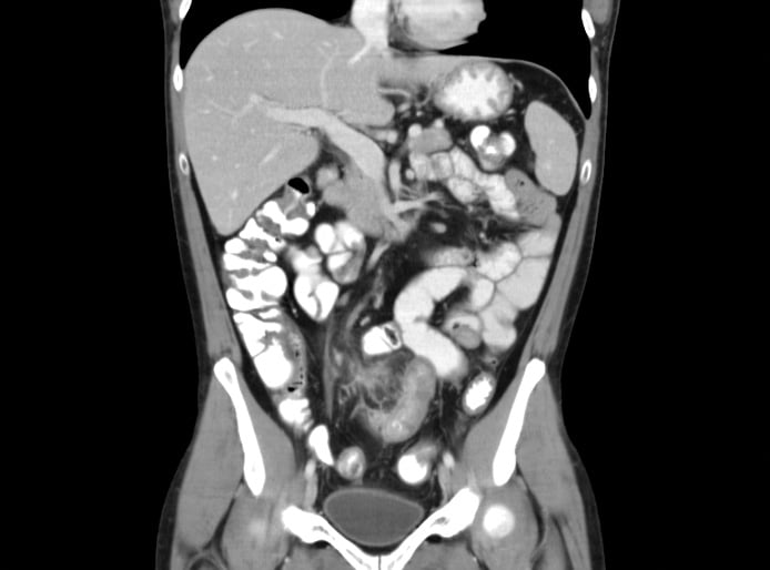
When we look at the digital output of an abdominal CT, we are viewing a density map of the body. The tech shows specific structures by highlighting the way they interact with X-ray photons, often enhanced by the use of contrast media.
Parenchymal Assessment
The CT scan provides a detailed view of the “parenchyma,” or the functional tissue of solid organs. In the liver, the technology can identify fatty infiltration, cysts, and hemangiomas by measuring their specific pixel density. For the kidneys, CT technology is the gold standard for identifying nephrolithiasis (kidney stones). Because stones are highly dense, they appear as bright white pixels against the darker, water-dense tissue of the renal pelvis. The software can even calculate the exact volume and density of the stone to help predict if it will pass naturally.
Vascular Imaging and Digital Angiography
Computed Tomography Angiography (CTA) is a specialized application of abdominal CT tech. By timing the scan to the exact moment an intravenous contrast agent enters the abdominal aorta, the scanner can map the entire vascular tree. This tech is used to identify abdominal aortic aneurysms (AAA), renal artery stenosis, and mesenteric ischemia. The software can then create a “Maximum Intensity Projection” (MIP), which highlights only the brightest pixels (the blood vessels), providing a clear digital map of the patient’s circulatory health.
Data Security and Interoperability in Abdominal Imaging
The output of an abdominal CT scan is not just an image; it is a massive data file. A single abdominal study can generate several gigabytes of data. Managing, storing, and sharing this data requires a robust technological infrastructure.
DICOM Standards and PACS Integration
All modern CT scanners output data in the DICOM (Digital Imaging and Communications in Medicine) format. This is the universal language of medical imaging. These files contain not only the image data but also extensive metadata, including scanner settings, patient demographics, and calibration data. These files are stored in a PACS (Picture Archiving and Communication System), a specialized server-client network designed to handle high-bandwidth image transfers.
The technology behind PACS allows a radiologist in one city to view the abdominal CT of a patient in another city in real-time. This interoperability is a cornerstone of modern digital health, enabling remote consultations and teleradiology.
Cloud-Based Diagnostics and Remote Analysis
The shift toward cloud computing is also hitting the diagnostic imaging space. Modern clinics are increasingly moving away from on-site servers to cloud-based PACS solutions. This allows for the integration of “Zero-Footprint Viewers,” which are web-based tools that allow clinicians to view high-resolution abdominal scans on any device—including tablets and smartphones—without needing to install heavy software. This mobility ensures that critical data regarding a patient’s internal health is available to the surgical team exactly when and where they need it.
The Future of Abdominal Imaging: From Spectral CT to Machine Learning
As we look toward the future, the technology of what an abdominal CT shows is set to become even more granular.
Spectral (Dual-Energy) CT
Spectral CT is an emerging technology that uses two different X-ray energy levels simultaneously. This allows the software to differentiate between materials that have the same density but different chemical compositions. For example, in a standard CT, it might be difficult to tell the difference between a small amount of hemorrhage and a concentrated contrast agent. Spectral CT tech can “subtract” the iodine from the image, allowing the software to show exactly what is blood and what is contrast. This provides a level of chemical insight that was previously impossible with traditional X-ray tech.

Generative AI and Predictive Analytics
The next frontier is the use of Generative AI to predict health outcomes based on abdominal CT data. Researchers are developing models that can analyze the texture of organ tissue—features invisible to the human eye—to predict the likelihood of future organ failure or the development of chronic diseases like diabetes. By treating the abdominal scan as a “Big Data” set rather than just a picture, the technology moves from being a reactive diagnostic tool to a proactive health management tool.
In summary, what a CT scan shows in the abdomen is a complex digital reconstruction of human biology. It is the result of high-speed photon detection, advanced algorithmic processing, and secure data management. As hardware becomes faster and AI becomes more integrated, the “digital window” into the abdomen will only become clearer, providing deeper insights into the silent workings of the human body.
aViewFromTheCave is a participant in the Amazon Services LLC Associates Program, an affiliate advertising program designed to provide a means for sites to earn advertising fees by advertising and linking to Amazon.com. Amazon, the Amazon logo, AmazonSupply, and the AmazonSupply logo are trademarks of Amazon.com, Inc. or its affiliates. As an Amazon Associate we earn affiliate commissions from qualifying purchases.