
Understanding the intricate nature of a brain tumor, particularly “what it feels like,” has been profoundly transformed by advancements in technology. While the subjective experience remains deeply personal and complex, modern tech tools offer unprecedented insights into the physiological and neurological underpinnings that manifest as symptoms. These technologies don’t just diagnose; they provide a window into the dynamic interplay between the tumor and the brain, helping medical professionals, researchers, and increasingly, patients themselves, comprehend the invisible shifts occurring within. This perspective focuses strictly on the technological innovations that help us interpret, predict, and manage the multifaceted “feeling” of a brain tumor, moving beyond mere anecdotal descriptions to data-driven understanding.
Unveiling the Unseen: Advanced Imaging Technologies
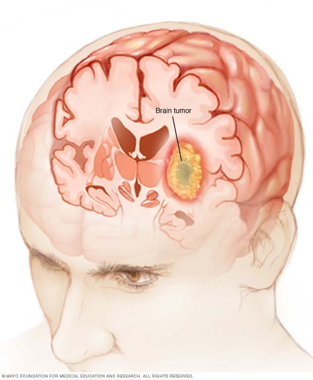
The most fundamental way technology has allowed us to grasp “what a brain tumor feels like” from an objective standpoint is through its ability to visualize the unseen. Before advanced imaging, understanding was limited to external symptoms and post-mortem examination. Today, cutting-edge diagnostic imaging provides a real-time, high-resolution map of the brain, revealing the tumor’s presence, size, location, and its impact on surrounding neural structures.
MRI and CT: Visualizing the Brain’s Internal Landscape
Magnetic Resonance Imaging (MRI) and Computed Tomography (CT) scans are the cornerstones of brain tumor diagnosis and monitoring. CT scans utilize X-rays to create cross-sectional images of the brain, quickly identifying significant structural changes, hemorrhages, or calcifications often associated with tumors. Its speed makes it invaluable in emergency situations where a rapid assessment is crucial. However, it’s the MRI that truly excels in detailing the soft tissues of the brain, offering superior contrast and resolution to delineate tumor margins, identify edema (swelling), and detect subtle changes that a CT might miss. Different MRI sequences can highlight various tissue properties, such as fluid content (FLAIR sequences) or blood flow (perfusion imaging), providing a multi-dimensional view.
The advanced capabilities of MRI allow neuro-radiologists to pinpoint exactly where a tumor is located and how it might be impinging on critical brain regions responsible for functions like movement, speech, or vision. For instance, a tumor pressing on the motor cortex might explain weakness or paralysis “felt” by a patient. One encroaching on the temporal lobe could explain memory disturbances. The visualization offered by these technologies bridges the gap between reported symptoms and their neurological origins, allowing clinicians to interpret “what it feels like” in terms of precise anatomical and pathological changes.
PET Scans and Functional Imaging: Mapping Metabolic and Activity Changes
Beyond structural imaging, Positron Emission Tomography (PET) scans offer a glimpse into the metabolic activity of brain tumors. By introducing a radioactive tracer (often a glucose analog), PET scans can highlight areas of increased metabolic activity, characteristic of rapidly growing tumor cells. This is crucial for differentiating between active tumor tissue and post-treatment changes like necrosis or scar tissue, which might appear similar on an MRI but have different metabolic profiles. PET can help determine the aggressiveness of a tumor and monitor its response to treatment at a cellular level, informing clinicians if treatments are truly altering the tumor’s “feel” at its most fundamental level of activity.
Furthermore, functional MRI (fMRI) takes imaging a step further by mapping brain activity. By detecting changes in blood flow associated with neural activity, fMRI can identify eloquent brain regions (those critical for language, motor function, etc.) in proximity to a tumor. This is invaluable for surgical planning, allowing surgeons to avoid damaging these areas and thus minimize post-operative deficits. For a patient, preserving these functions directly impacts “what it feels like” to recover from surgery, preventing devastating losses of capability. Diffusion Tensor Imaging (DTI), another advanced MRI technique, visualizes the white matter tracts of the brain, showing how the tumor might displace or infiltrate neural pathways, providing crucial information about potential neurological impairments.
Decoding Neurological Signals: The Role of Neuromonitoring and Data Analytics
While imaging provides static snapshots, neuromonitoring technologies offer dynamic, real-time data on brain function, translating the immediate “feel” of a tumor’s impact into quantifiable signals. When combined with sophisticated data analytics, these tools help discern patterns and predict events that might otherwise seem sudden or inexplicable.
EEG and Intracranial Pressure Monitoring: Real-time Data Insights
Electroencephalography (EEG) records the brain’s electrical activity, providing vital insights into neurological events such as seizures, which are a common symptom of brain tumors. Abnormal electrical patterns detected by EEG can indicate areas of irritation or dysfunction caused by the tumor. For patients experiencing focal seizures, the EEG can pinpoint the origin of these electrical storms, which directly correlates with the “feeling” of the seizure itself – whether it’s a sensory disturbance, a motor twitch, or a brief lapse in consciousness. Continuous EEG monitoring, particularly in an epilepsy monitoring unit, helps characterize seizure types and frequency, informing treatment strategies to mitigate this distressing symptom.
Intracranial Pressure (ICP) monitoring is another critical technology. Brain tumors can increase pressure within the rigid confines of the skull, leading to symptoms like severe headaches, nausea, vomiting, and altered consciousness – classic manifestations of “what a brain tumor feels like” when pressure builds. Invasive ICP monitors, surgically placed within the brain, provide continuous, real-time measurements of this pressure. This data allows clinicians to manage fluid balance, administer medications, or perform interventions to reduce pressure, directly alleviating symptoms that profoundly impact a patient’s comfort and neurological status. The ability to quantify and respond to ICP changes is vital for patient safety and comfort.
Big Data and Predictive Analytics in Symptom Understanding
Beyond individual patient monitoring, the aggregation of neuromonitoring data, imaging results, clinical notes, and genomic information into “big data” repositories is revolutionizing our understanding. Machine learning algorithms can sift through vast datasets to identify subtle correlations and predictive patterns that might be imperceptible to human analysis. For instance, analyzing thousands of patient records might reveal specific tumor locations or genetic markers that are strongly associated with particular types of headaches, seizure patterns, or cognitive deficits.
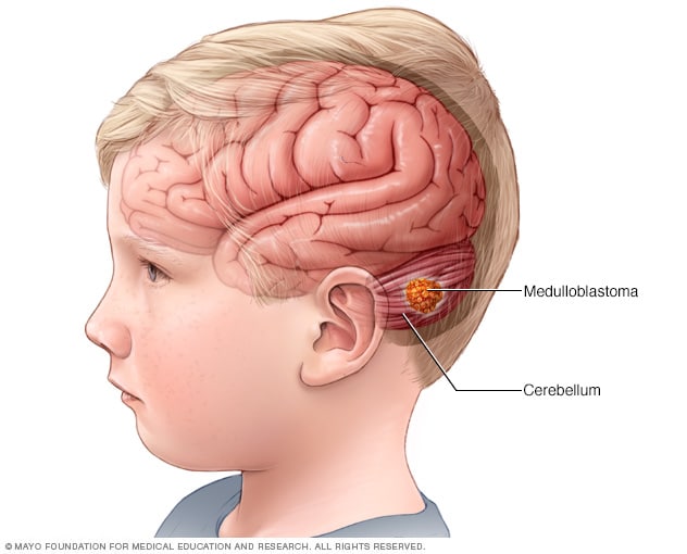
This data-driven approach allows for the development of predictive analytics models. These models can forecast the likelihood of certain symptoms developing, how rapidly a tumor might grow, or a patient’s response to specific therapies. By leveraging the collective “experience” of countless patients through their data, clinicians can gain a deeper, more granular understanding of “what a brain tumor feels like” for different individuals and tailor preventative or proactive interventions, thereby shifting from reactive treatment to predictive care.
AI and Machine Learning: Anticipating the Patient Experience
Artificial Intelligence (AI) and Machine Learning (ML) are rapidly advancing our capacity to diagnose, understand, and even anticipate the complex “feel” of a brain tumor. These computational powerhouses can process and interpret medical data at speeds and scales far beyond human capability, leading to more precise interventions and personalized care.
AI in Diagnostic Accuracy and Early Detection
AI algorithms are increasingly being trained on vast libraries of medical images (MRIs, CTs, PET scans) to assist in brain tumor diagnosis. These systems can identify tumors, classify their types (e.g., glioblastoma, meningioma), and even grade their aggressiveness with remarkable accuracy, sometimes surpassing human experts. Early detection, facilitated by AI’s ability to spot subtle anomalies, is paramount. Catching a tumor when it’s small and less symptomatic means treatment can begin before it causes significant neurological impairment, thus altering “what it feels like” to live with the condition from a state of severe disability to potential remission or effective management. AI-powered diagnostic tools can reduce diagnostic delays, prevent misdiagnoses, and streamline the path to appropriate care.
Furthermore, AI can integrate data from various sources – imaging, genomic sequencing, clinical symptoms – to provide a holistic diagnostic picture. This multi-modal data integration helps in understanding not just the tumor itself, but its specific biological fingerprint and its likely impact on brain function, thereby better predicting the spectrum of symptoms a patient might “feel.”
Predictive Models for Symptom Progression and Personalized Treatment
One of the most exciting applications of AI is its ability to build predictive models for symptom progression. Based on a patient’s unique tumor characteristics, genetic profile, and initial symptoms, AI can forecast how the tumor is likely to evolve and what new symptoms might arise. For example, an AI model might predict that a tumor in a specific location is highly likely to cause language difficulties within six months, allowing for proactive speech therapy or counseling. This foresight is invaluable in preparing both patients and caregivers for potential changes, making the experience less disorienting.
Moreover, AI is pivotal in personalizing treatment plans. By analyzing a patient’s tumor genomics and comparing it to vast databases of treatment outcomes for similar tumors, AI can recommend the most effective therapies with the fewest side effects. This precision medicine approach ensures that treatments are not a one-size-fits-all, but rather tailored to the individual, aiming to minimize adverse “feelings” (side effects) while maximizing therapeutic benefit. For example, AI can identify specific genetic mutations that make a tumor susceptible to a particular targeted drug, offering a more effective and less debilitating treatment course than traditional chemotherapy.
Technological Interventions: Managing the “Feel” of a Brain Tumor
Beyond diagnosis and prediction, technology plays a crucial role in the direct management and treatment of brain tumors, aiming to alleviate the symptoms and restore quality of life – fundamentally changing “what it feels like” to live with the condition.
Neuro-navigation and Robotic Surgery: Precision in Treatment
Surgical removal remains a primary treatment for many brain tumors. However, operating within the delicate confines of the brain requires extreme precision. Neuro-navigation systems, akin to a GPS for the brain, integrate pre-operative MRI and CT scans with real-time surgical instruments. These systems provide surgeons with a 3D map, allowing them to precisely localize the tumor, plan optimal surgical approaches, and avoid critical brain structures. This precision minimizes damage to healthy brain tissue, reducing post-operative neurological deficits (such as weakness, speech problems, or vision loss) that can profoundly impact a patient’s “feeling” of self and capability.
The advent of robotic surgery further enhances this precision. Robotic arms, controlled by surgeons, can execute micro-movements with unparalleled stability and accuracy, accessing deep-seated tumors with minimal invasiveness. Intraoperative imaging techniques, such as iMRI (intraoperative MRI), allow surgeons to update their navigational map during surgery, ensuring maximal safe resection of the tumor in real-time. These technologies are transformative, not only in improving outcomes but also in minimizing the trauma and neurological compromise associated with brain surgery, directly impacting the patient’s recovery and the “feel” of their post-surgical life.

Wearable Tech and Digital Health Apps for Symptom Management
The continuous “feel” of living with a brain tumor extends beyond the hospital. Wearable technologies and digital health applications empower patients to actively monitor and manage their symptoms in their daily lives. Smartwatches and fitness trackers can monitor vital signs, sleep patterns, and activity levels, which can provide early indicators of worsening symptoms or treatment side effects. For example, changes in heart rate variability or sleep disturbances might signal an impending seizure or an increase in intracranial pressure.
Digital health apps provide platforms for patients to log their symptoms, medication intake, and mood changes. This data, often shared with their healthcare team, creates a comprehensive picture of their day-to-day experience. Telehealth platforms and virtual consultations, enabled by digital technology, allow patients to receive expert medical advice and support from the comfort of their homes, reducing the burden of travel and making ongoing care more accessible. Some apps even incorporate cognitive exercises designed to help patients manage memory or attention deficits, directly addressing some of the challenging “feelings” associated with brain tumors. By harnessing these technologies, patients gain a greater sense of control and understanding over their condition, transforming the passive experience of illness into an active partnership in their care.
aViewFromTheCave is a participant in the Amazon Services LLC Associates Program, an affiliate advertising program designed to provide a means for sites to earn advertising fees by advertising and linking to Amazon.com. Amazon, the Amazon logo, AmazonSupply, and the AmazonSupply logo are trademarks of Amazon.com, Inc. or its affiliates. As an Amazon Associate we earn affiliate commissions from qualifying purchases.