
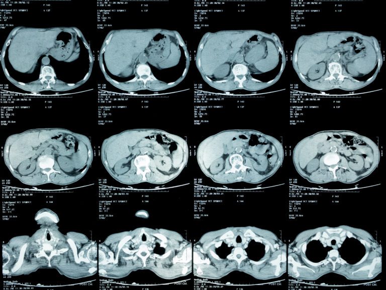
In the landscape of modern medicine, the Computed Tomography (CT) scan stands as a pinnacle of engineering and digital innovation. While a patient may see it as a simple medical procedure, from a technological standpoint, an abdominal CT scan is a masterclass in data acquisition, high-speed processing, and sophisticated software reconstruction. When we ask, “What does a CT of the abdomen show?” we are essentially asking how advanced hardware and software interpret millions of data points to create a high-definition digital map of the human interior.

To understand the output of a CT scan, one must look beyond the physical organs and focus on the digital layers: the voxels, the algorithms, and the artificial intelligence (AI) that transform raw X-ray attenuation into a diagnostic tool. This article explores the technological architecture of abdominal CT imaging, the role of AI in interpreting data, and the digital infrastructure required to manage these massive datasets.
The Evolution of Computed Tomography: From 2D Slices to 3D Digital Reconstructions
At its core, a CT scanner is a sophisticated hardware peripheral designed to capture the density of matter. Unlike a traditional X-ray, which produces a flat, two-dimensional image, a CT scan uses a rotating gantry to capture 360-degree data. In the context of the abdomen—a complex cavity containing the liver, kidneys, spleen, and gastrointestinal tract—this multi-dimensional approach is critical.
Multi-Detector Row CT (MDCT) Technology
The hardware advancement that revolutionized abdominal imaging is Multi-Detector Row CT (MDCT). Modern scanners are equipped with multiple rows of solid-state detectors that can capture dozens or even hundreds of “slices” in a single rotation. This high-speed acquisition is essential for the abdomen because it allows the scanner to capture images in between breaths, minimizing motion artifacts that could blur the digital output.
Dual-Energy CT and Spectral Imaging
One of the most exciting trends in imaging technology is Dual-Energy CT (DECT). By using two different X-ray energy spectra, the software can differentiate between materials based on their atomic number. In an abdominal scan, this allows the technology to “show” more than just shapes; it can distinguish between a kidney stone made of uric acid versus one made of calcium. It can also digitally “subtract” bone or contrast agents from the image, providing a clear view of the vascular system within the abdominal cavity.
The Role of Hounsfield Units in Data Visualization
The digital output of a CT scan is measured in Hounsfield Units (HU), a quantitative scale for describing radiodensity. The software assigns a numerical value to every pixel (or more accurately, every 3D “voxel”). For instance, water is calibrated at 0 HU, while air is -1000 HU and dense bone is +1000 HU. When the scan “shows” the abdomen, it is actually displaying a visual representation of these numerical values, allowing software to highlight specific densities that might indicate a lesion or a fluid collection.
AI and Machine Learning: Revolutionizing Automated Image Analysis
The sheer volume of data produced by a single abdominal CT scan—often consisting of thousands of images—can be overwhelming for human analysis. This is where AI tools and machine learning algorithms have become indispensable. Today, what a CT shows is increasingly filtered through a layer of intelligent software designed to augment human perception.
Computer-Aided Detection (CAD) Systems
AI-driven CAD systems are integrated into the diagnostic workflow to flag anomalies. In the abdomen, these tools are particularly effective at identifying small nodules or early-stage tumors in the liver or pancreas that might be invisible to the naked eye. By training on millions of previous scans, these AI tools can recognize patterns of density that correlate with specific pathologies, effectively “highlighting” areas of interest for the radiologist.
Deep Learning for Noise Reduction
One of the primary challenges in CT technology is balancing image quality with radiation dose. Higher doses produce clearer images, but they also increase patient risk. New AI software utilizes “Deep Learning Iterative Reconstruction” (DLIR). This technology can take a “noisy,” low-dose image and use neural networks to predict what the high-resolution version should look like. This allows the CT to show crystalline detail of the abdominal organs while utilizing significantly less raw data (and radiation).
Automated Organ Segmentation
Modern software can now perform “automated segmentation.” This means the computer can automatically identify and trace the boundaries of the liver, kidneys, and spleen. This tech is used for volumetric analysis—measuring the exact volume of an organ or a tumor over time. If a patient is undergoing treatment, the software can compare two scans and calculate the precise percentage of change in a digital model, providing a level of accuracy that manual measurement could never achieve.
The Digital Pipeline: How Software Processes Raw X-Ray Data into Diagnostic Insights
The journey from a physical scan to a viewable image is a complex software process known as reconstruction. When a CT “shows” the abdomen, it is showing a digital reconstruction of “sinograms”—the raw, overlapping data captured by the detectors.
Iterative Reconstruction Algorithms
The transition from Filtered Back Projection (FBP) to Iterative Reconstruction (IR) has been a major milestone in imaging software. IR algorithms are mathematically intensive processes that compare a predicted image with the actual measured data, refining the image through multiple “iterations.” This process requires massive computational power, often handled by high-end GPUs, to produce the final, sharp image of the abdominal structures.
Cinematic Rendering and 3D Visualization
Advancements in graphics processing have led to “Cinematic Rendering.” This software technique uses complex lighting models—similar to those used in high-end movie animation—to create photorealistic 3D models of the abdominal interior. This tech allows surgeons to “fly through” the abdominal cavity virtually before an operation, visualizing the spatial relationship between blood vessels and organs in a way that traditional 2D slices cannot convey.
Post-Processing and Multiplanar Reformation (MPR)
Once the raw data is reconstructed, post-processing software allows the user to view the abdomen from any angle. Multiplanar Reformation (MPR) allows a vertical or diagonal “slice” to be generated from the original horizontal data. This is crucial for visualizing the long path of the ureters or the complex branching of the mesenteric arteries. The tech isn’t just showing a picture; it’s providing a manipulatable 3D environment.
Data Security and Interoperability: Protecting the Digital Human
As CT scans become more detailed, the files they produce become larger. A high-resolution abdominal CT can produce several gigabytes of data. Managing, storing, and securing this information is a significant challenge within the tech infrastructure of healthcare.
DICOM Standards and Interoperability
The universal language of medical imaging is DICOM (Digital Imaging and Communications in Medicine). This protocol ensures that an abdominal CT taken on a GE scanner can be read by software developed by Siemens, Agfa, or an open-source viewer. This interoperability is what allows for the seamless transfer of data between specialists, ensuring that the “view” of the abdomen remains consistent across different platforms.
PACS and Cloud-Based Storage
The storage of these massive datasets is handled by PACS (Picture Archiving and Communication Systems). Modern tech trends are moving toward “Cloud PACS,” where the heavy lifting of image rendering is done on remote servers rather than local workstations. This allows a specialist to view a high-resolution abdominal CT on a mobile tablet or a remote laptop without sacrificing performance, using streaming technologies similar to those used by high-end gaming services.
Cyber Security and Patient Data Privacy
Because a CT scan is a digital representation of a person’s internal anatomy, it is considered highly sensitive Biometric Data. Protecting this data from breaches is a major focus of medical tech. Encryption at rest and in transit, multi-factor authentication for access to PACS, and the anonymization of datasets for research purposes are all critical components of the technology surrounding the scan. What the CT “shows” must only be visible to those with the digital keys to see it.

Conclusion: The Future of Abdominal Imaging
When we analyze “what a CT of the abdomen shows,” we are witnessing the convergence of physical hardware, complex mathematics, and cutting-edge AI. We are no longer looking at simple shadows; we are looking at a high-fidelity digital twin of the patient’s internal anatomy.
As we move forward, the integration of 5G technology will allow for real-time remote “telementoring,” where a specialist on the other side of the world can manipulate a 3D abdominal reconstruction in real-time. Furthermore, the rise of “Radiomics”—the extraction of large amounts of quantitative data from medical images—means that in the future, a CT might show us molecular-level changes before they are even visible as structural anomalies. The abdomen, once a “black box” of the human body, has been fully illuminated by the relentless march of digital technology.
aViewFromTheCave is a participant in the Amazon Services LLC Associates Program, an affiliate advertising program designed to provide a means for sites to earn advertising fees by advertising and linking to Amazon.com. Amazon, the Amazon logo, AmazonSupply, and the AmazonSupply logo are trademarks of Amazon.com, Inc. or its affiliates. As an Amazon Associate we earn affiliate commissions from qualifying purchases.