
The human brain remains one of the most complex structures in the known universe, but the rapid evolution of medical technology is beginning to decode its intricate pathways. At the center of this revolution is Magnetic Resonance Imaging (MRI). While once viewed simply as a high-tech camera for the body, the modern brain MRI has evolved into a sophisticated data-gathering platform. Through a combination of advanced hardware engineering, high-performance computing, and artificial intelligence, brain MRIs now show us more than just anatomy; they show us the functional and structural “software” of human consciousness.
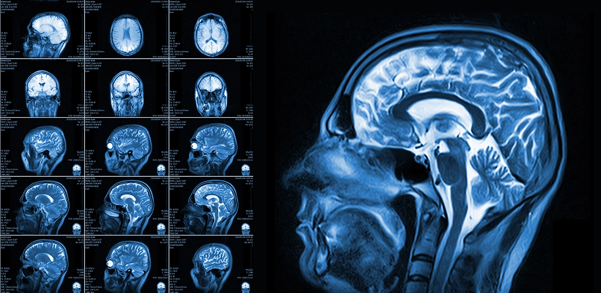
The Engineering Behind the Image: How MRI Hardware Maps the Mind
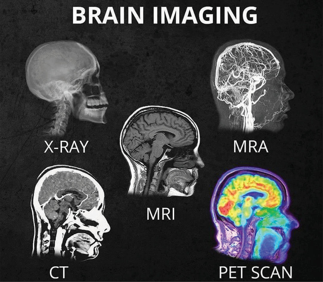
To understand what a brain MRI can show, one must first understand the technological infrastructure required to generate these images. Unlike X-rays or CT scans, which use ionizing radiation, an MRI relies on powerful magnets and radiofrequency (RF) pulses.
High-Field Magnetism: The Tesla Advantage
The “strength” of an MRI machine is measured in Teslas (T). Most standard clinical scanners operate at 1.5T or 3.0T. However, the tech frontier has moved toward 7.0T and even 11.7T systems. These high-field magnets align the hydrogen protons in the brain with extreme precision. The higher the Tesla rating, the higher the signal-to-noise ratio, allowing clinicians to see structures as small as a fraction of a millimeter. This tech reveals the delicate layers of the cerebral cortex and the tiny vascular networks that support neural health.
Gradient Coils and Signal Acquisition
If the main magnet is the engine, the gradient coils are the steering wheel. These coils allow the system to “slice” the brain into 3D voxels (volumetric pixels). Modern gradient technology has become faster and more efficient, reducing the time a patient must remain still while increasing the temporal resolution of the data. This allows for the visualization of rapid changes in the brain, such as the pulsating of blood vessels or the subtle shifts in tissue density.
RF Pulse Sequences: The Software of the Scan
The “pulse sequence” is essentially the code that tells the MRI machine how to behave. By varying the timing and intensity of radio waves, technicians can highlight different types of tissue. For example, T1-weighted sequences are excellent for looking at brain anatomy, while T2-weighted sequences are better for identifying inflammation or fluid buildup. The ability to switch between these “digital filters” is what makes MRI the most versatile diagnostic tool in the tech stack of modern neurology.
Beyond Anatomy: Functional and Diffusion Imaging Software
In the early days of neuroimaging, we could only see the “hardware”—the physical structures of the brain. Today, advanced software protocols allow us to see the “network traffic.”
Functional MRI (fMRI) and Brain Activity
fMRI technology measures the Blood Oxygen Level Dependent (BOLD) signal. When a specific part of the brain is active, it consumes more oxygen. The MRI software detects these hemodynamic changes in real-time. This reveals which parts of the brain are “lighting up” during specific tasks, such as speaking, moving, or even dreaming. In a clinical tech context, this is used for “brain mapping” before surgery, ensuring that surgeons do not disrupt critical functional zones.
Diffusion Tensor Imaging (DTI): Mapping the Wiring
While standard MRI shows the gray matter (the processors), DTI shows the white matter (the wiring). DTI tracks the movement of water molecules along myelinated axons. Because water moves more easily parallel to nerve fibers than across them, software can reconstruct 3D maps of the brain’s communication pathways. This “tractography” reveals how different regions of the brain are interconnected, providing insights into traumatic brain injuries (TBI) and neurodegenerative diseases that were previously invisible on standard scans.
Magnetic Resonance Spectroscopy (MRS)
MRS is essentially a non-invasive “virtual biopsy.” Instead of producing an image, this tech produces a frequency spectrum that identifies the chemical composition of brain tissue. It can show the levels of metabolites like choline, creatine, and lactate. For a technician, this is like running a diagnostic script on a computer to see if the internal chemistry is balanced or if there are signs of tumor growth or metabolic failure.
The AI Revolution in Brain MRI Analysis
The sheer volume of data produced by a single high-resolution brain MRI is staggering. A typical scan can produce thousands of images. This is where Artificial Intelligence (AI) and Machine Learning (ML) have become indispensable tools in the radiology workflow.
Automated Lesion Detection and Segmentation
AI algorithms, trained on millions of previous scans, can now identify abnormalities with superhuman speed. In cases of Multiple Sclerosis (MS) or oncology, AI software can automatically segment tumors or lesions, measuring their volume down to the microliter. This allows for precise longitudinal tracking—comparing a scan from six months ago to today to see if a treatment is working with mathematical certainty.
Predictive Modeling and Early Diagnostics
The tech industry is currently focusing on “biomarkers” identified through AI. By analyzing subtle patterns in brain atrophy or white matter changes that are invisible to the human eye, ML models can predict the onset of Alzheimer’s disease or Parkinson’s years before clinical symptoms appear. This shift from “diagnostic” to “predictive” tech is one of the most significant trends in digital health.
Deep Learning for Image Reconstruction
One of the biggest limitations of MRI tech has always been scan time. Patients must stay still for 30 to 45 minutes, and any movement blurs the data. New deep-learning tools, such as NVIDIA’s specialized healthcare AI or GE’s AIR Recon DL, can “clean up” noisy images and reconstruct high-resolution outputs from shorter scans. This not only improves the user experience for the patient but also increases the throughput of the imaging center.
Data Security and the Digital Infrastructure of Neuroimaging
As brain MRIs become higher in resolution, they also become larger in file size. Managing this data requires a robust digital infrastructure and a heavy focus on cybersecurity.
The DICOM Standard and Cloud Integration
Medical images are stored in a specialized format called DICOM (Digital Imaging and Communications in Medicine). Modern neuroimaging tech is moving away from local “on-premise” servers toward secure, encrypted cloud storage. This allows for “teleradiology,” where a specialist in London can analyze a brain scan taken in a rural clinic in real-time. The integration of cloud computing ensures that high-compute tasks, like 3D rendering of the brain, can be offloaded to powerful remote servers.
Privacy and the “Neural Fingerprint”
A burgeoning topic in digital security is the concept of the “neural fingerprint.” Recent studies have shown that the structural connectivity of a person’s brain (as revealed by DTI) is unique enough to identify them. As brain MRI data becomes more detailed, the tech industry must grapple with how to “de-identify” this data for research while protecting the biometric privacy of the individual. This involves advanced encryption and the use of “synthetic data” to train AI models without compromising real patient identities.
The Future: Portable MRI and Quantum Sensors
The current trend in imaging technology is moving toward democratization and miniaturization. The goal is to take the capabilities of a room-sized MRI machine and make them accessible in any environment.
Point-of-Care (POC) MRI Tech
Companies like Hyperfine have introduced the first portable, “Swoop” MRI scanners. These devices operate on a much lower magnetic field and can be wheeled to a patient’s bedside in an Intensive Care Unit (ICU). While they don’t yet offer the ultra-high resolution of a 7T research magnet, the software-driven image enhancement allows them to show critical issues like strokes or large hemorrhages instantly, bypassing the need for a dedicated imaging suite.
Hyperpolarized Imaging and Quantum Sensing
The next “gadget” in the neuroimaging world involves hyperpolarization tech. By “over-polarizing” certain molecules (like Carbon-13) before injecting them, researchers can increase the MRI signal by more than 10,000 times. This allows us to see the actual metabolism of the brain—the “burning” of glucose—in real-time. Furthermore, quantum sensors are being developed to detect the tiny magnetic fields produced by the brain’s electrical activity, potentially merging the spatial resolution of an MRI with the temporal resolution of an EEG.
In summary, a brain MRI shows far more than just “the brain.” It shows the intersection of biology and extreme-scale technology. From the superconducting magnets that align our atoms to the AI algorithms that interpret our thoughts, the MRI is the ultimate tool in the digital transformation of human health. As these technologies continue to converge, the “black box” of the human mind will become increasingly transparent, paved by innovations in hardware, software, and data science.
aViewFromTheCave is a participant in the Amazon Services LLC Associates Program, an affiliate advertising program designed to provide a means for sites to earn advertising fees by advertising and linking to Amazon.com. Amazon, the Amazon logo, AmazonSupply, and the AmazonSupply logo are trademarks of Amazon.com, Inc. or its affiliates. As an Amazon Associate we earn affiliate commissions from qualifying purchases.