
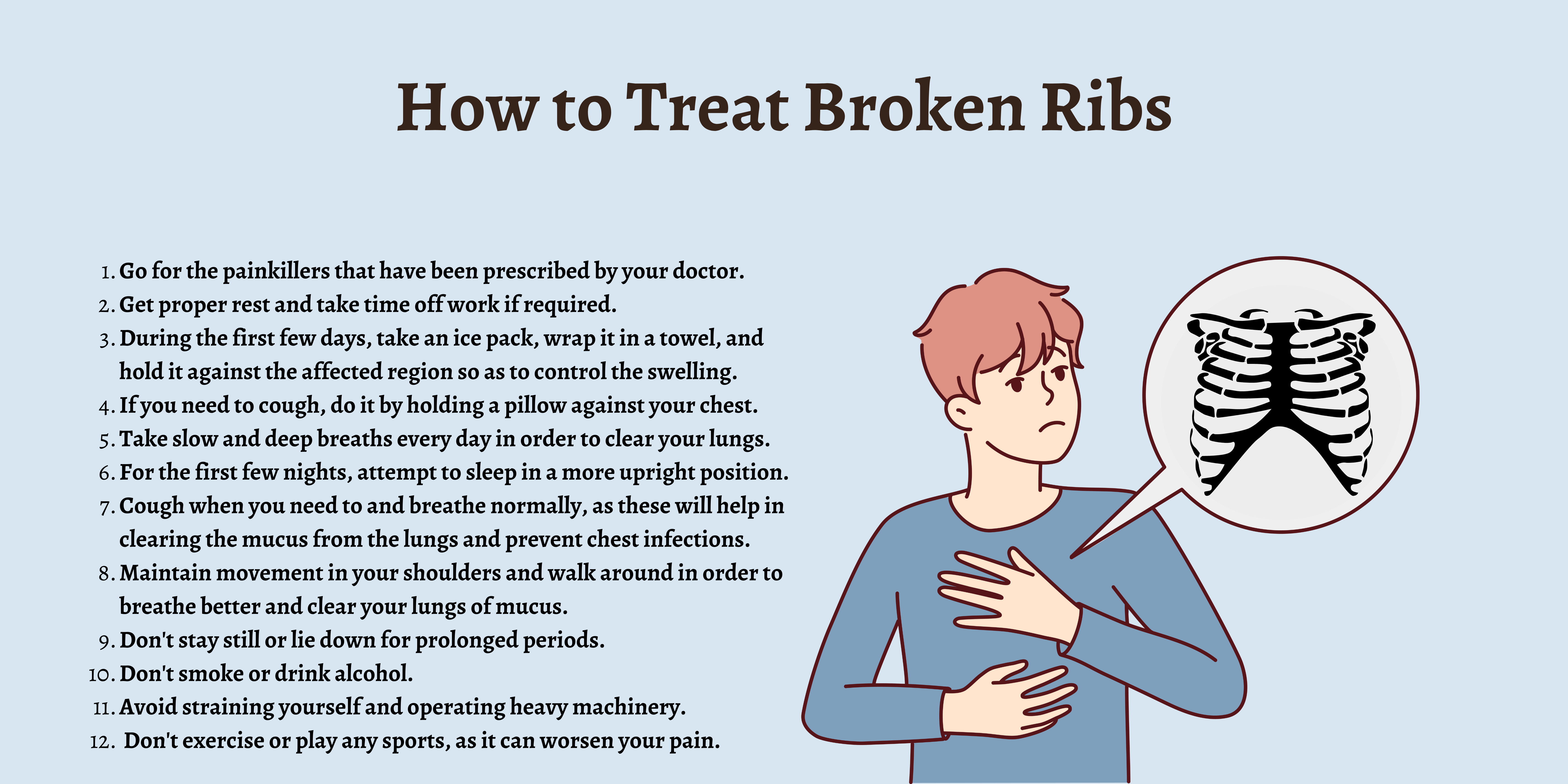
For decades, the standard medical response to a fractured rib was underwhelmingly low-tech: “Rest, take some pain medication, and wait.” Unlike a broken arm that can be immobilized in a plaster cast, the ribs are in constant motion to facilitate breathing, making traditional stabilization nearly impossible. However, the landscape of thoracic injury management is undergoing a seismic shift. Driven by breakthroughs in artificial intelligence, additive manufacturing, and wearable sensors, the question of “what can be done for a fractured rib” is no longer answered with passive patience, but with high-tech intervention.
The convergence of digital health and mechanical engineering is providing clinicians with tools that were once the province of science fiction. From AI-driven diagnostics that catch microscopic hairline fractures to 3D-printed titanium implants tailored to a patient’s unique anatomy, technology is reducing recovery times, minimizing chronic pain, and preventing the pulmonary complications that often follow thoracic trauma.
Digital Diagnostics: The AI Revolution in Rib Fracture Detection
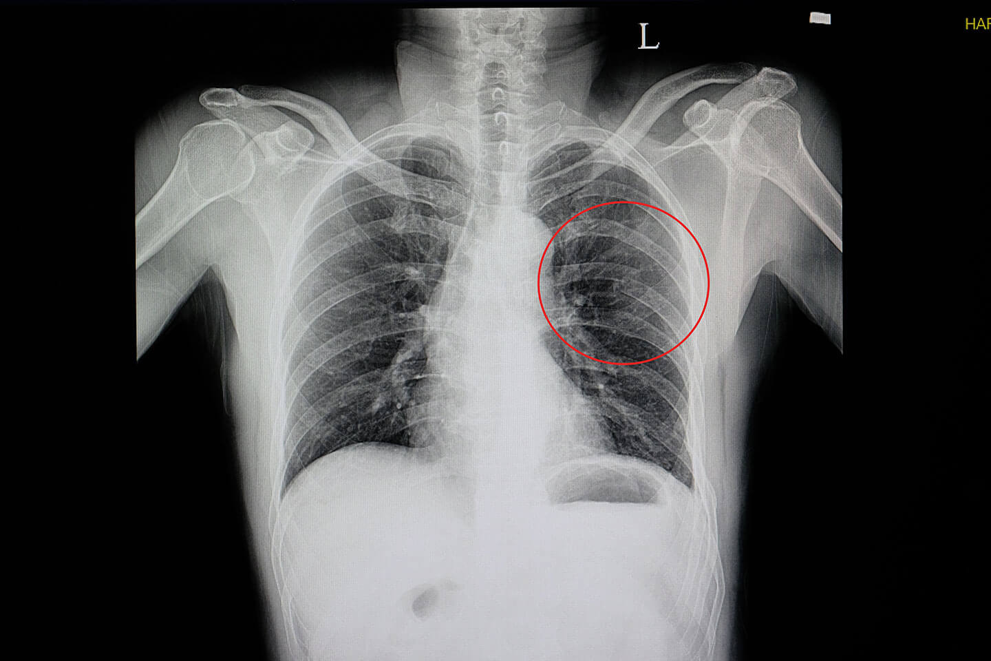
The first step in addressing a fractured rib is accurate identification, a task that has historically been surprisingly difficult. Standard chest X-rays are notorious for missing up to 50% of rib fractures, particularly those in the cartilage or behind the heart and diaphragm. This diagnostic gap can lead to improper management and prolonged suffering. Today, technology is closing this gap through advanced imaging and machine learning.
Neural Networks and Automated Lesion Identification
Artificial Intelligence (AI) is now being integrated directly into radiology workflows to assist in the detection of rib fractures. Deep learning algorithms, trained on hundreds of thousands of CT scans, can scan a patient’s thoracic cage in seconds, highlighting potential fractures that the human eye might overlook. These AI tools act as a “second pair of eyes,” providing a probability score for each identified lesion.
In trauma centers, where speed is critical, these algorithms prioritize scans with positive findings, ensuring that patients with severe internal injuries receive immediate attention. This software doesn’t just find the break; it categorizes the fracture type—whether it is displaced, non-displaced, or a “flail chest” segment—enabling a more precise treatment plan from the moment the patient enters the emergency department.
Advanced 3D Reconstruction Software
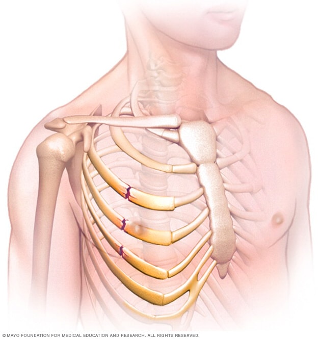
Beyond simple detection, the “Tech” niche has introduced sophisticated 3D reconstruction software. By taking the two-dimensional slices of a CT scan, these programs can render a fully rotatable, high-definition 3D model of a patient’s rib cage. This allows surgeons to visualize the exact geometry of the fracture from every angle. This spatial data is crucial for determining whether a patient is a candidate for surgical stabilization or if the fracture is likely to heal correctly through conservative management. This digital “digital twin” of the patient’s skeletal structure serves as the blueprint for all subsequent technological interventions.
Engineering the Cure: 3D Printing and Bio-Mechanical Innovations
When a rib fracture is severe enough to require surgery—a process known as Surgical Stabilization of Rib Fractures (SSRF)—technology moves from the screen to the operating table. The development of advanced materials and manufacturing processes has turned rib repair into an exercise in high-precision engineering.
Patient-Specific Implants (PSI) and CAD/CAM Workflows
The most significant leap in treating complex rib fractures is the move away from “one-size-fits-all” hardware. Historically, surgeons used generic metal plates that they had to manually bend and shape during surgery to fit the patient’s rib. This was time-consuming and often resulted in a sub-optimal fit.
Today, Computer-Aided Design (CAD) and Computer-Aided Manufacturing (CAM) allow for the creation of Patient-Specific Implants (PSI). Using the 3D models generated during the diagnostic phase, engineers can 3D-print titanium plates that perfectly match the contour of the specific rib being repaired. These custom implants are lighter, stronger, and can be fixed with greater precision, significantly reducing the time a patient spends under anesthesia and improving the long-term structural integrity of the chest wall.
Smart Materials and Shape Memory Alloys
Research is also progressing into the use of “smart materials” for rib repair. One of the most promising areas involves Nitinol—a shape-memory alloy composed of nickel and titanium. Nitinol can be programmed to “remember” a specific shape. When used in rib staples or clips, the material can be applied in a flexible state and then, when it reaches body temperature, contract to provide a constant, dynamic compression across the fracture site. This tech-driven approach ensures that the bones remain in contact even as the rib cage expands and contracts with every breath, accelerating the natural ossification process.
Connected Recovery: IoT, Wearables, and Remote Monitoring
The danger of a fractured rib doesn’t end once the patient leaves the hospital. The primary risk during recovery is shallow breathing due to pain, which can lead to atelectasis (collapsed lung) and pneumonia. Technology is now stepping in to monitor patients in their own homes, ensuring that the recovery phase is as data-driven as the surgical one.
Smart Garments and Respiratory Telemetry
The “Internet of Medical Things” (IoMT) has birthed a new generation of wearable sensors designed specifically for thoracic health. Smart vests, embedded with textile-based sensors and accelerometers, can monitor a patient’s respiratory rate, tidal volume, and cough frequency in real-time.
These wearables transmit data via Bluetooth to a smartphone app, which then uploads the information to a cloud-based platform for clinician review. If the technology detects that a patient’s breathing is becoming dangerously shallow or that their oxygen saturation levels are dipping, it can trigger an automated alert. This allows for early intervention, such as adjusting pain medication or initiating physical therapy, before a minor respiratory issue turns into a life-threatening infection.
The Role of Mobile Health (mHealth) in Post-Fracture Protocols
Software applications—often referred to as Digital Therapeutics (DTx)—are also playing a vital role. Specialized apps guide patients through incentive spirometry exercises, which are essential for keeping the lungs clear. These apps use gamification to encourage compliance, turning the tedious task of deep breathing into a digital challenge with progress tracking and rewards. By transforming the smartphone into a recovery coach, tech is empowering patients to take an active role in their healing, reducing readmission rates and improving overall outcomes.
Virtual Frontiers: Augmented Reality and Non-Invasive Pain Management
As we look toward the future of what can be done for a fractured rib, the boundaries between the physical and digital worlds continue to blur. Augmented Reality (AR) and Virtual Reality (VR) are moving beyond the gaming industry and into the center of thoracic trauma care.
VR-Based Analgesia: Disrupting Traditional Pain Management
One of the greatest challenges in treating fractured ribs is pain management, particularly given the risks associated with long-term opioid use. High-tech solutions are offering a non-pharmacological alternative. Virtual Reality (VR) “analgesia” uses immersive digital environments to distract the brain’s pain-processing centers.
When a patient is engaged in a highly immersive VR experience, the brain’s bandwidth for processing pain signals is reduced. Clinical studies have shown that VR can significantly lower perceived pain scores in trauma patients, allowing them to perform necessary breathing exercises and movements that would otherwise be too painful. This “digital sedative” represents a major shift in how the industry views pain—not just as a chemical problem, but as a neurological one that can be managed via tech.
AR-Guided Surgical Precision
For surgeons, Augmented Reality (AR) is becoming a standard tool in the operating suite. Using AR headsets, a surgeon can “see through” the patient’s skin by overlaying the 3D CT reconstruction directly onto the patient’s body in real-time. This allows for “GPS-guided” surgery, where the surgeon knows exactly where the fracture is located before the first incision is made.
This level of precision minimizes tissue damage, reduces the size of incisions, and ensures that hardware is placed in the biomechanically optimal position. The integration of AR into the surgical workflow is a prime example of how software is enhancing human capability to provide better, faster, and safer care.
The Future of Thoracic Tech
The question of “what can be done for a fractured rib” has evolved from a matter of basic first aid to a sophisticated technological ecosystem. We are entering an era where rib fractures are diagnosed by AI, repaired with custom 3D-printed bio-materials, monitored by IoT sensors, and managed through virtual reality.
As these technologies continue to mature and become more accessible, the “fractured rib” will no longer be synonymous with weeks of debilitating pain and the threat of pneumonia. Instead, it will be a manageable condition where digital tools and medical expertise converge to restore the patient’s quality of life with unprecedented speed and precision. The future of recovery is digital, and for those suffering from thoracic injuries, that future can’t arrive soon enough.
aViewFromTheCave is a participant in the Amazon Services LLC Associates Program, an affiliate advertising program designed to provide a means for sites to earn advertising fees by advertising and linking to Amazon.com. Amazon, the Amazon logo, AmazonSupply, and the AmazonSupply logo are trademarks of Amazon.com, Inc. or its affiliates. As an Amazon Associate we earn affiliate commissions from qualifying purchases.