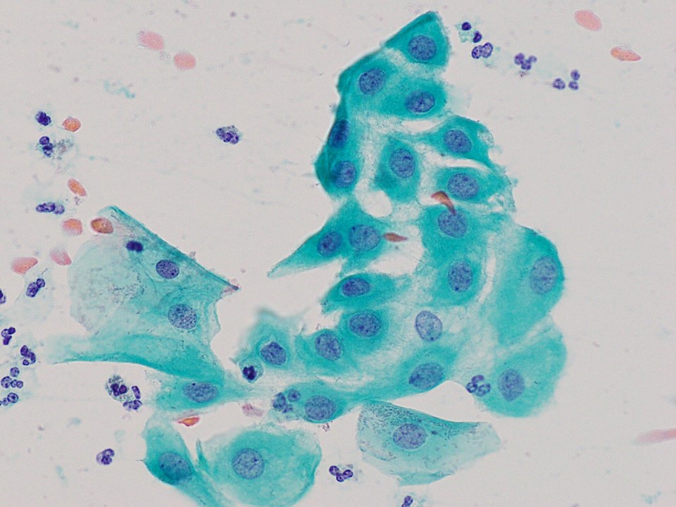
In the rapidly evolving landscape of health technology, the intersection of biological science and digital innovation has birthed a new era of diagnostic precision. At the heart of this transformation is the study of cytology—the examination of individual cells—and one specific cellular phenomenon that has become a primary focus for advanced AI algorithms: squamous metaplastic cells. While traditionally a subject for pathologists peering through glass slides, these cells are now at the center of a technological revolution involving machine learning, high-resolution imaging, and cloud-based diagnostic platforms.
Understanding squamous metaplastic cells through the lens of modern technology requires an appreciation for how software and hardware have converged to interpret complex biological transitions. Metaplasia, in a biological sense, is a process where one mature cell type is replaced by another in response to environmental stressors. In the digital age, identifying these changes is no longer just a human skill; it is a data-driven challenge that tech companies are solving with unprecedented accuracy.

The Digital Transformation of Cytopathology
The shift from manual microscopy to digital pathology represents one of the most significant tech trends in the medical sector. This transition involves the digitization of physical tissue samples into high-resolution, terabyte-scale images, allowing for deep analysis through specialized software.
High-Resolution Whole Slide Imaging (WSI)
The first step in the tech-heavy analysis of squamous metaplastic cells is the use of Whole Slide Imaging (WSI) scanners. These gadgets are not merely cameras; they are sophisticated robotic systems that capture thousands of individual image “tiles” at 40x or 100x magnification. The software then stitches these tiles together using complex alignment algorithms to create a seamless digital representation of a biological sample. For a tech specialist, this is akin to creating a Google Maps-style interface for the human body, where one can zoom from a macro view of tissue down to the micro-details of a single squamous cell’s nucleus.
Digital Annotation and Ground Truth Data
In the development of diagnostic apps and software, “ground truth” is essential. Tech platforms now allow pathologists to use digital pens and touch-sensitive interfaces to annotate squamous metaplastic cells directly on the screen. These annotations serve as the training data for neural networks. This collaborative environment—often hosted on SaaS (Software as a Service) platforms—enables specialists from around the globe to contribute to a centralized database, refining the software’s ability to distinguish between benign metaplasia and potentially precancerous transitions.
AI and Machine Learning: Automating Metaplastic Recognition
The most exciting tech application in this niche is the deployment of Artificial Intelligence, specifically Convolutional Neural Networks (CNNs), to identify and categorize squamous metaplastic cells. These AI tools are designed to mimic the human visual cortex, identifying patterns that might be invisible to the naked eye.
Feature Extraction and Pattern Recognition
AI models for cytology are trained to perform “feature extraction.” When looking at squamous metaplastic cells, the software analyzes specific parameters: the nuclear-to-cytoplasmic ratio, the texture of the chromatin, and the integrity of the cell borders. Advanced AI tools use deep learning to assign a probability score to each cell. If a cell displays the characteristics of squamous metaplasia—such as a “cobblestone” appearance or specific staining patterns—the algorithm flags it for review. This automation significantly reduces the “search time” for human technicians, allowing them to focus on the most complex cases.
Minimizing False Positives with Deep Learning
One of the hurdles in medical software development is the high cost of errors. Squamous metaplastic cells are often reactive and can mimic more serious conditions like high-grade dysplasia. Modern tech solutions utilize “ensemble learning,” where multiple AI models cross-reference each other’s findings to minimize false positives. By layering different algorithmic approaches, developers can ensure that the software understands the nuance of metaplasia as a normal physiological response rather than a pathological threat, thereby preventing unnecessary medical interventions.
The Infrastructure of Modern Diagnostics: Cloud and Edge Computing

The sheer volume of data generated when analyzing cellular structures requires a robust technological infrastructure. A single digital slide can exceed 2 GB in size, making local storage and processing inefficient for large-scale clinical use.
Cloud-Based Diagnostic Workflows
SaaS platforms in the pathology space are moving toward cloud-native architectures. By leveraging AWS, Azure, or Google Cloud, diagnostic labs can process thousands of slides simultaneously. These platforms offer “Pathology-as-a-Service,” where images of squamous metaplastic cells are uploaded to the cloud, analyzed by AI in a matter of seconds, and the results are delivered to a web-based dashboard accessible by clinicians anywhere in the world. This democratization of tech allows rural clinics to access the same high-level diagnostic tools as major metropolitan hospitals.
Edge Computing in the Lab
While the cloud provides scale, edge computing provides speed. Modern scanners are now equipped with “on-device” AI processing. By performing initial analysis at the “edge” (on the scanner itself), the system can instantly identify areas of interest on a slide. If the scanner detects a cluster of squamous metaplastic cells, it can automatically trigger a higher-resolution scan of that specific area, optimizing data usage and reducing the time required for a full diagnostic report.
Data Security and Interoperability in Cellular Analysis
As with any digital tool involving sensitive information, the technology used to track and analyze squamous metaplastic cells must adhere to strict security protocols. Digital security is a cornerstone of the modern health tech industry.
Encryption and HIPAA Compliance
The software used to store and transmit cellular images must employ end-to-end encryption. In the United States, adherence to HIPAA (Health Insurance Portability and Accountability Act) is mandatory. Tech developers focus on “data anonymization,” where the images of squamous metaplastic cells are stripped of personally identifiable information (PII) before being used for AI training or shared across research networks. This ensures that while the technology learns, the patient’s privacy remains intact.
Interoperability and HL7 Standards
A major trend in software development is ensuring that different systems can “talk” to each other. Diagnostic platforms for cellular analysis are increasingly built on HL7 (Health Level Seven) and FHIR (Fast Healthcare Interoperability Resources) standards. This allows the findings regarding squamous metaplastic cells to be seamlessly integrated into a patient’s Electronic Health Record (EHR). When a tech tool identifies a specific cellular change, it doesn’t just sit in a silo; it updates the patient’s digital history, alerts their primary care provider, and can even trigger automated follow-up scheduling.
The Future Trend: Augmented Reality and Predictive Analytics
Looking forward, the tech surrounding cellular analysis is moving beyond static screens and into more immersive and predictive territories.
AR-Enabled Microscopes
The “smart microscope” is a trending gadget in pathology labs. By using Augmented Reality (AR), a digital overlay is projected onto the physical view of the microscope. As the pathologist looks at the slide, the AR software highlights squamous metaplastic cells in real-time, providing digital “hints” or diagnostic data points directly in the field of vision. This hybrid approach combines the reliability of traditional optics with the processing power of modern AI.
Predictive Modeling and Longitudinal Data
The next frontier for tech in this space is predictive analytics. By analyzing the digital history of squamous metaplastic cells in a specific patient over several years, machine learning models can begin to predict the likelihood of future cellular changes. This shifts the paradigm from “reactive” diagnostics to “proactive” health management. Using big data, tech companies are identifying environmental and genetic markers that correlate with metaplastic transitions, offering a level of insight that was impossible in the pre-digital era.
In conclusion, the question of “what are squamous metaplastic cells” is no longer confined to biology textbooks. In the modern tech ecosystem, these cells are complex data sets that drive innovation in AI, cloud computing, and digital imaging. As software continues to refine its ability to interpret the nuances of cellular life, the synergy between technology and pathology will only grow stronger, leading to faster, more accurate, and more accessible healthcare for everyone.
aViewFromTheCave is a participant in the Amazon Services LLC Associates Program, an affiliate advertising program designed to provide a means for sites to earn advertising fees by advertising and linking to Amazon.com. Amazon, the Amazon logo, AmazonSupply, and the AmazonSupply logo are trademarks of Amazon.com, Inc. or its affiliates. As an Amazon Associate we earn affiliate commissions from qualifying purchases.