
In the realm of modern medicine, the term “brain lesion” often sounds like a finality—a definitive physical scar on the most complex organ in the human body. However, from a technological standpoint, a brain lesion is essentially a data point. It is an area of tissue that has been damaged through injury, infection, or disease, and its identification is a triumph of engineering and software development. For decades, the “black box” of the human skull was impenetrable, but through the lens of advanced neuroimaging, artificial intelligence, and high-performance computing, we are now able to map, categorize, and treat these abnormalities with unprecedented precision.
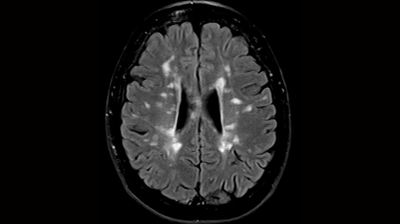
Understanding lesions on the brain today is less about general biology and more about the sophisticated tech stack that allows us to see what was once invisible.
The Hardware of Visualization: Evolution of Neuroimaging Tech
The primary way we define and understand brain lesions is through the hardware used to visualize them. Without high-resolution imaging technology, a lesion is merely a theoretical cause for a clinical symptom.
Magnetic Resonance Imaging (MRI) and High-Field Magnets
The gold standard for identifying brain lesions is the MRI. Unlike X-rays, which use ionizing radiation, MRIs leverage powerful magnetic fields and radio waves to align hydrogen protons in the body. When we talk about “seeing” a lesion, we are actually discussing the software’s ability to interpret the relaxation times of these protons.
In recent years, the industry has moved from 1.5 Tesla (T) to 3T and even 7T scanners. These higher “Tesla” ratings represent stronger magnetic fields, which translate to a higher signal-to-noise ratio. For technologists, this means higher resolution. At 7T, neuroradiologists can see lesions so small they were previously categorized as “idiopathic” (of unknown cause). This leap in hardware allows for the visualization of micro-hemorrhages and tiny demyelinating plaques associated with Multiple Sclerosis (MS) that older tech simply could not render.
Computed Tomography (CT) and Rapid Processing
While MRI is the high-definition choice, CT technology remains the “fast-response” hardware of neurology. Modern CT scanners utilize helical scanning and multi-detector arrays to capture hundreds of “slices” of the brain in seconds. The tech behind CT has evolved to include “Perfusion CT,” which uses sophisticated algorithms to measure blood flow in real-time. This is critical for identifying ischemic lesions—areas where the brain is starved of oxygen during a stroke—allowing software to calculate the “penumbra,” or the salvageable brain tissue surrounding a lesion.
Diffusion Tensor Imaging (DTI)
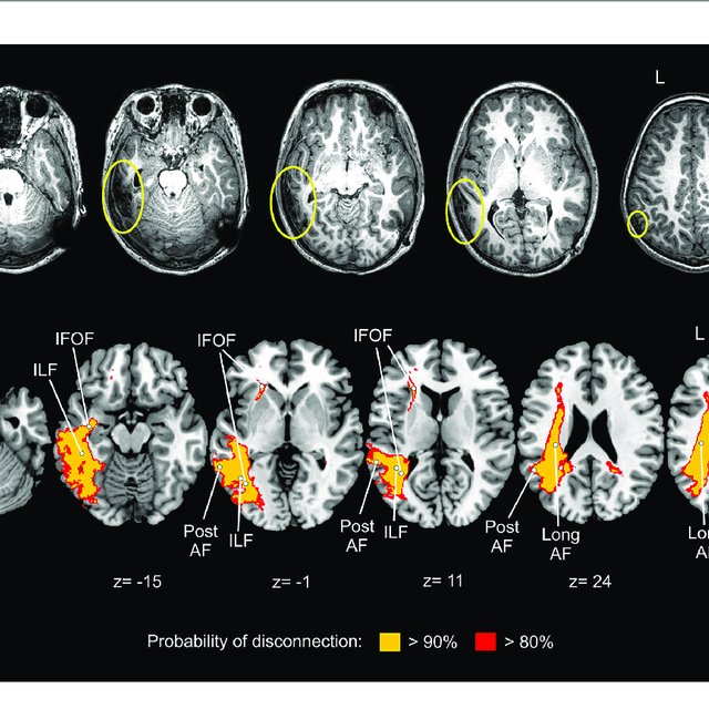
DTI is a specialized MRI technique that represents a massive leap in how we map the brain’s “wiring” (white matter). It measures the diffusion of water molecules. In healthy white matter, water moves along the path of the axons. A lesion disrupts this flow. DTI software creates color-coded maps of these pathways, allowing tech-driven surgical planning to avoid critical tracts during lesion removal.
The AI Revolution in Lesion Detection and Classification
The bottleneck in neurology has historically been the human element: the radiologist. A single MRI scan can produce thousands of images. This is where Artificial Intelligence (AI) and Machine Learning (ML) have become the most transformative tools in identifying brain lesions.
Automated Segmentation and Deep Learning
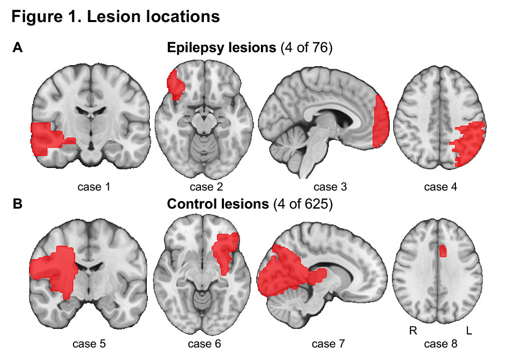
Computer Vision, a subset of AI, is now used to perform “automated segmentation.” In the past, a doctor would have to manually trace the outline of a tumor or a lesion to measure its volume. Today, deep learning algorithms—trained on millions of annotated brain scans—can identify and segment lesions in seconds.
These AI tools use Convolutional Neural Networks (CNNs) to recognize patterns that might be too subtle for the human eye. For instance, in patients with Multiple Sclerosis, AI can compare a new scan with one from six months ago, pixel by pixel, to detect “silent” lesions—new areas of damage that haven’t yet caused physical symptoms but indicate a need for a change in medication.
Predictive Analytics and Radiomics
We are moving beyond simple identification into the era of “Radiomics.” This technology treats medical images as data. By extracting a large number of features from a brain lesion image (such as texture, shape, and intensity), AI can predict the molecular subtype of a brain tumor without a physical biopsy.
This tech-heavy approach allows for “virtual biopsies.” If an algorithm can identify a lesion as a specific type of glioblastoma based on its digital signature, the treatment protocol can begin days or weeks earlier than if the patient had to wait for surgical pathology.
Reducing “Noisy” Data
One of the greatest challenges in neuro-tech is “motion artifact.” If a patient moves even slightly during a 45-minute MRI, the data becomes blurred. New AI-driven reconstruction algorithms, such as Deep Learning Reconstruction (DLR), can now “clean” this data. These tools can distinguish between actual brain tissue (a lesion) and digital noise, allowing for clear images even when hardware conditions are less than ideal.
Software Ecosystems and the Data Infrastructure of Neurology
A brain lesion is not just a picture; it is a massive file. Managing the data generated by neuroimaging requires a robust technological infrastructure, from cloud storage to interoperable software platforms.
PACS and Cloud-Based Imaging
Picture Archiving and Communication Systems (PACS) are the backbone of digital radiology. Modern PACS have migrated to the cloud, allowing for the instantaneous sharing of high-resolution brain scans across the globe. For a patient with a rare brain lesion, this means their “digital twin” (their scan data) can be analyzed by a specialist in Switzerland and a software tool in Silicon Valley simultaneously.
The integration of “DICOM” (Digital Imaging and Communications in Medicine) standards ensures that whether a scan is taken on a Siemens machine or a GE machine, the data remains readable and actionable across different software environments.
The Rise of the “Digital Twin” in Neurosurgery
One of the most exciting applications of lesion technology is the creation of a digital twin. Surgeons now use VR (Virtual Reality) and AR (Augmented Reality) software to “walk through” a patient’s brain before the first incision is made.
By layering MRI and DTI data into a 3D rendering engine (often the same engines used in high-end video games, like Unity or Unreal Engine), software creates a 1:1 map of the patient’s brain. The lesion is highlighted in high contrast, and the “tech-surgeon” can practice the optimal approach, identifying which digital “vessels” are at risk. This reduces surgical time and significantly improves post-operative outcomes.
Longitudinal Tracking and Big Data
How does a lesion change over ten years? This question is now answered through big data. Large-scale neurological databases, such as the UK Biobank, store hundreds of thousands of brain scans. Researchers use high-performance computing clusters to run longitudinal studies, identifying how certain lifestyle factors or genetic markers correlate with the development of brain lesions over time. This is “Preventative Tech”—using historical data to predict future neurological risks.
The Future of Lesion Management: Wearables and Neural Interfaces
As we look toward the horizon, the technology for managing brain lesions is moving outside the hospital and into the realm of “Everyday Tech.”
Remote Monitoring and Digital Biomarkers
For patients with chronic lesions (like those from a stroke or MS), the “check-up” is moving to the smartphone. Apps now use “digital biomarkers”—such as typing speed, gait analysis via accelerometers, and voice pattern recognition—to monitor neurological stability. If a patient’s typing rhythm changes, it may indicate that a lesion is progressing, triggering an automated alert to their neurologist to schedule a new scan.
Brain-Computer Interfaces (BCI)
In cases where a lesion has caused permanent damage—such as a lesion in the motor cortex resulting in paralysis—technology is providing a workaround. Companies like Neuralink, Synchron, and Blackrock Neurotech are developing BCIs. These are arrays of micro-electrodes that “bypass” the lesion.
If the brain’s natural “hardware” (the neurons) is damaged by a lesion, these chips pick up the electrical signals and translate them into digital commands. This allows a patient to control a computer cursor or a robotic limb using only their thoughts. In this context, the lesion is no longer a dead end; it is simply a broken circuit that has been rerouted through external hardware.
Nanotechnology and Targeted Delivery
The future of treating lesions lies in nanotechnology. Engineers are developing “nanobots” and lipid nanoparticles that can cross the blood-brain barrier—a feat that most traditional drugs cannot achieve. These tech-driven delivery systems can be programmed to release their payload only when they detect the specific chemical signature of a lesion, such as the acidic environment of a tumor. This is “Programmable Medicine,” where the treatment is as much a piece of software as it is a chemical compound.
Conclusion: From Scars to Signals
A brain lesion, once a mystery shrouded in medical jargon, has become a focal point for the world’s most advanced technology. We no longer view these abnormalities as mere physical defects; we view them as complex challenges in imaging, data analysis, and signal processing.
Through the power of high-field MRI magnets, the cognitive leap of artificial intelligence, and the connectivity of cloud computing, our ability to detect and define brain lesions is sharper than ever. As we integrate wearables and neural interfaces into the mix, the goal is shifting from merely identifying damage to reversing its effects. In the digital age, the “lesion” is just another problem for which we are rapidly developing a high-tech solution.
aViewFromTheCave is a participant in the Amazon Services LLC Associates Program, an affiliate advertising program designed to provide a means for sites to earn advertising fees by advertising and linking to Amazon.com. Amazon, the Amazon logo, AmazonSupply, and the AmazonSupply logo are trademarks of Amazon.com, Inc. or its affiliates. As an Amazon Associate we earn affiliate commissions from qualifying purchases.