
In the constantly evolving landscape of medical diagnostics, few technologies have had as profound and widespread an impact as Computed Tomography (CT) scans. Far more sophisticated than conventional X-rays, CT technology leverages advanced computational power and precise hardware engineering to create detailed cross-sectional images of the body. Initially developed in the 1970s, CT scanning has undergone relentless innovation, transforming from a revolutionary but rudimentary tool into an indispensable, high-tech cornerstone of modern healthcare.
At its core, a CT scan is a non-invasive medical imaging procedure that uses specialized X-ray equipment combined with powerful computers to generate multiple images of the inside of the body. These images are then layered and digitally manipulated to create comprehensive 2D slices or even intricate 3D representations of organs, bones, soft tissues, and blood vessels. This capability offers clinicians an unprecedented view into the human anatomy, enabling rapid and accurate diagnosis of a vast array of conditions that might otherwise remain hidden. From urgent trauma assessment to meticulous cancer staging and guided interventional procedures, the applications of CT technology are as diverse as they are critical. This article delves into the technological marvels that underpin CT scans and explores their multifaceted uses, firmly positioning them within the realm of cutting-edge diagnostic technology.
The Technological Marvel Behind CT Scans
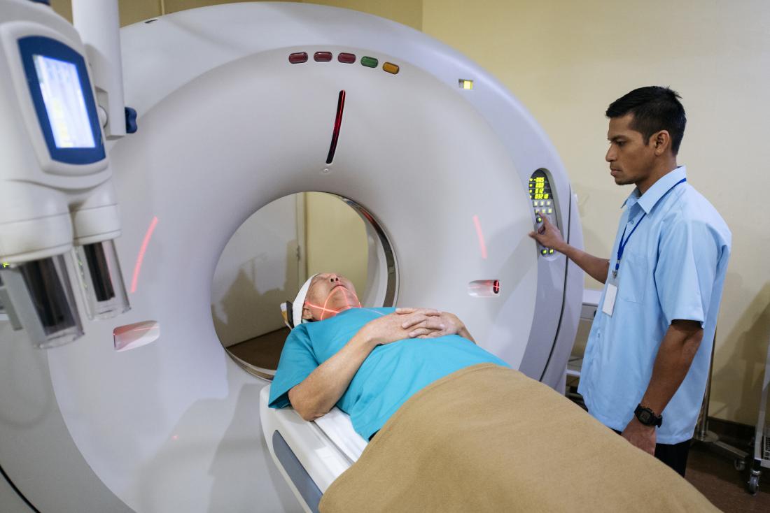
The utility and diagnostic power of CT scans stem directly from the sophisticated engineering and computational algorithms that drive them. Unlike a standard X-ray which produces a single, flat image, a CT scanner uses a rotating X-ray tube and an array of detectors that move around the patient, acquiring hundreds or thousands of individual X-ray projections from different angles. This deluge of raw data is then fed into powerful computers, which employ complex mathematical algorithms to reconstruct detailed cross-sectional images.
How CT Technology Works: From X-rays to Digital Slices
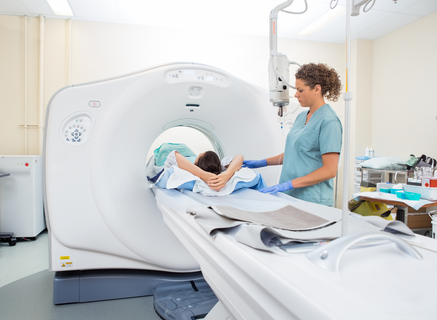
The fundamental principle involves a gantry—a large, doughnut-shaped machine—through which the patient passes. Inside the gantry, an X-ray tube rapidly rotates, emitting a narrow fan beam of X-rays that pass through the body. On the opposite side, a ring of detectors measures the X-rays that successfully penetrate the tissues. Different tissues absorb X-rays at varying rates; bones, being dense, absorb more, while air and soft tissues absorb less. The detectors convert the received X-ray signals into electrical impulses, which are then digitized.
Early CT scanners acquired data one slice at a time, requiring the patient to move slightly for each new image. Modern systems employ helical (or spiral) CT, where the X-ray tube and detectors rotate continuously as the patient table moves smoothly through the gantry. This “spiral” acquisition significantly reduces scan time, minimizes motion artifacts, and allows for much larger anatomical coverage in a single breath-hold, making it ideal for restless patients or emergency situations. Furthermore, the advent of multi-slice (or multi-detector) CT (MDCT) scanners, featuring dozens or even hundreds of detector rows, has revolutionized image acquisition, allowing for even faster scans and the ability to capture very thin slices (sub-millimeter resolution) simultaneously. This leap in hardware capability dramatically improves diagnostic accuracy and opens doors for advanced applications.
Beyond 2D: The Power of 3D Reconstruction and Visualization
One of the most significant technological advantages of CT is its ability to move beyond simple 2D slices. The vast amount of data collected during a helical or multi-slice CT scan is inherently volumetric. Advanced image processing software can take these individual axial (cross-sectional) images and reconstruct them into sagittal (side-to-side) and coronal (front-to-back) planes without needing to re-scan the patient. More importantly, these digital slices can be stacked and rendered to create highly detailed 3D volumetric reconstructions.
This 3D capability is a game-changer for medical visualization. Surgeons can use 3D models to meticulously plan complex operations, such as orthopedic surgeries or tumor resections, visualizing anatomical relationships in an intuitive manner. Radiologists can rotate and manipulate these 3D models to better understand the spatial extent of diseases, detect subtle anomalies, or evaluate vascular structures with greater precision. Volume Rendering (VR) and Maximum Intensity Projection (MIP) are common software techniques used to create these compelling 3D images, highlighting specific structures like blood vessels (CT angiography) or bones. This advanced visualization is a testament to the sophisticated software and computational power inherent in modern CT systems, elevating diagnostic imaging from mere picture-taking to comprehensive anatomical mapping.
Software and AI: Enhancing Image Quality and Diagnostic Efficiency
The raw data collected by a CT scanner is just the beginning. The quality and interpretability of the final images rely heavily on sophisticated software algorithms. Iterative reconstruction (IR) algorithms, for instance, have replaced older filtered back projection methods as the standard for image reconstruction. IR algorithms use complex mathematical models that simulate the X-ray acquisition process, comparing estimated data with actual measured data and iteratively refining the image until a high-quality, low-noise result is achieved. This technological advancement allows for significant reductions in radiation dose without compromising image clarity, a critical patient safety feature.
Beyond reconstruction, Artificial Intelligence (AI) and Machine Learning (ML) are rapidly integrating into CT workflows, pushing the boundaries of what’s possible. AI-powered software can assist in automated lesion detection, flagging potential abnormalities like lung nodules or bone fractures, thereby increasing diagnostic efficiency and reducing radiologist workload. AI also plays a role in image enhancement, noise reduction, and artifact suppression, further improving image quality. Furthermore, AI algorithms can segment organs and tissues automatically, providing quantitative measurements (e.g., organ volume, fat quantification) that aid in disease monitoring and treatment response assessment. This integration of AI represents the next frontier in CT technology, promising faster, more precise, and even more personalized diagnostic capabilities.
Radiation Dose Management: Smart Tech for Patient Safety
While incredibly valuable, CT scans involve exposure to ionizing radiation. A significant focus of technological innovation in CT has been on minimizing this dose while maintaining diagnostic image quality. Modern CT scanners incorporate numerous advanced features designed to optimize radiation exposure:
-
Automatic Exposure Control (AEC): This intelligent system dynamically adjusts the X-ray tube current during the scan based on the patient’s size and anatomy, ensuring just enough radiation is used for diagnostic clarity.
-
Dose Modulation: Techniques that vary the X-ray tube current based on the angular position of the tube around the patient (e.g., lower dose when X-rays pass through less dense tissues) or along the patient’s long axis.
-
Iterative Reconstruction (IR): As mentioned, IR algorithms allow for significant dose reductions (up to 50-80% in some cases) compared to traditional methods, while maintaining or even improving image quality by effectively suppressing noise.
-
Organ-Based Dose Modulation: Shielding specific radiosensitive organs (e.g., breasts, eyes) where possible without obscuring diagnostic information.
-
Pediatric Protocols: Specific, lower-dose protocols tailored for children, who are more sensitive to radiation.
These technological safeguards underscore the commitment to patient safety within the realm of high-tech medical imaging, continually balancing diagnostic necessity with the imperative of minimizing risk.
Diverse Applications Across Medical Disciplines
The versatility and diagnostic precision of CT scans make them invaluable across virtually every medical specialty. Their ability to rapidly acquire detailed anatomical information has revolutionized patient care in numerous scenarios.
Emergency Medicine and Trauma Assessment
In emergency rooms, CT scans are often the first line of advanced imaging due to their speed and comprehensiveness. For patients with severe trauma, CT can quickly identify internal injuries such as organ damage, internal bleeding, complex bone fractures, and head injuries (e.g., intracranial hemorrhage, skull fractures). A “trauma CT” protocol might involve scanning the head, chest, abdomen, and pelvis in a single, rapid acquisition, providing critical information to guide immediate life-saving interventions. This rapid assessment capability is a prime example of high-tech diagnostics directly impacting critical care outcomes.
Oncology: Detection, Staging, and Monitoring Cancer
CT plays a pivotal role in the entire cancer journey. It is widely used for:
-
Detection: Identifying suspicious masses or lesions in various organs.
-
Staging: Determining the size of a tumor and whether it has spread (metastasized) to nearby lymph nodes or distant organs, which is crucial for treatment planning.
-
Biopsy Guidance: Providing real-time imaging guidance for percutaneous (through the skin) biopsies, allowing radiologists to accurately sample tissue from a tumor for pathological analysis.
-
Treatment Planning: Assisting in radiation therapy planning by precisely mapping the tumor and surrounding healthy tissues.
-
Monitoring Treatment Response: Comparing scans over time to assess whether a tumor is shrinking, growing, or remaining stable in response to chemotherapy or radiation.
-
Surveillance: Following up with patients post-treatment to detect recurrence.
The detailed spatial resolution of CT allows for precise evaluation of tumor characteristics and extent, profoundly influencing treatment strategies and prognoses.
Neurological Conditions: Peering into the Brain and Spine
CT scans are indispensable for diagnosing a wide range of neurological disorders. They can rapidly detect:
-
Strokes: Differentiating between ischemic stroke (blood clot) and hemorrhagic stroke (bleeding) is critical for determining immediate treatment.
-
Brain Hemorrhages: Identifying bleeding within the brain from trauma or aneurysms.
-
Tumors: Locating brain and spinal cord tumors, assessing their size, and relationship to critical structures.
-
Spinal Injuries: Providing detailed views of vertebral fractures, dislocations, and spinal cord compression.
-
Infections and Inflammation: Detecting abscesses or inflammation within the central nervous system.
-
Hydrocephalus: Diagnosing the buildup of fluid in the brain.
While MRI often provides superior soft tissue contrast for subtle neurological lesions, CT’s speed and ability to detect acute blood and bone abnormalities make it a vital tool, especially in emergency neuroimaging.
Cardiovascular Imaging: Assessing Heart and Vessels

CT Angiography (CTA) is a specialized application of CT that uses an intravenous contrast agent to visualize blood vessels throughout the body. It has become a crucial non-invasive tool for:
-
Coronary Artery Disease: Detecting blockages or narrowing in the arteries supplying the heart, often used as an alternative to invasive catheter angiography in select patients.
-
Aneurysms: Identifying and characterizing bulges in arterial walls (e.g., aortic aneurysms in the chest or abdomen, cerebral aneurysms).
-
Pulmonary Embolism (PE): Rapidly diagnosing blood clots in the arteries of the lungs, a life-threatening condition.
-
Peripheral Artery Disease: Visualizing blockages in arteries supplying the limbs.
-
Congenital Heart Defects: Assessing structural abnormalities in the heart and great vessels, particularly in pediatric patients.
The ability to create high-resolution 3D reconstructions of complex vascular networks is a testament to the advanced imaging and processing capabilities of modern CT systems.
Musculoskeletal Imaging: Detailed Bone and Joint Analysis
While conventional X-rays are excellent for basic bone imaging, CT provides far superior detail for complex orthopedic problems. It is used for:
-
Complex Fractures: Precisely delineating comminuted (shattered) fractures, intra-articular fractures (involving joint surfaces), and fractures of anatomically complex bones (e.g., pelvis, spine, wrist, ankle).
-
Joint Abnormalities: Assessing degenerative joint disease, bone tumors, and infections (osteomyelitis) with greater clarity than X-rays.
-
Surgical Planning: Guiding surgeons in pre-operative planning for intricate reconstructive surgeries.
-
Bone Density Measurement: Specialized CT techniques (e.g., QCT) can provide quantitative assessment of bone mineral density, aiding in osteoporosis diagnosis.
The ability to reconstruct images in multiple planes and in 3D is particularly valuable in orthopedics, offering surgeons a detailed roadmap for intervention.
Interventional Procedures: CT-Guided Precision
CT technology extends beyond pure diagnostics into interventional medicine. Radiologists use real-time CT guidance to perform minimally invasive procedures with pinpoint accuracy. This includes:
-
Biopsies: Guiding a needle to precisely target and sample suspicious lesions in organs like the lung, liver, or bone, minimizing risks to surrounding structures.
-
Abscess Drainage: Placing drainage catheters into collections of pus to treat infections.
-
Pain Management: Delivering steroid injections or nerve blocks to specific areas of the spine or joints for chronic pain relief.
-
Tumor Ablation: Guiding probes to precisely ablate (destroy) small tumors using heat (radiofrequency or microwave ablation) or cold (cryoablation).
These procedures leverage CT’s high spatial resolution to ensure accuracy and reduce patient recovery times compared to traditional open surgical approaches, highlighting the technological convergence of imaging and therapy.
The Evolution and Future of CT Technology
The journey of CT technology is one of continuous innovation, driven by advances in computing power, detector technology, and AI. The future promises even more sophisticated capabilities, further enhancing diagnostic accuracy and patient safety.
Advancements in Speed and Resolution
Modern CT scanners are astonishingly fast. A complete body scan can be performed in seconds, minimizing patient discomfort and motion artifacts. This speed is crucial in trauma or cardiac imaging where rapid acquisition is essential. Simultaneously, resolution continues to improve, with sub-millimeter isotropic voxels (3D pixels) becoming standard, allowing for exquisite detail in small anatomical structures. Large-bore scanners accommodate a wider range of patients, and photon-counting CT detectors, currently under development and early clinical adoption, promise to revolutionize image quality by directly counting individual X-ray photons, leading to even lower noise, higher resolution, and more spectral information than current energy-integrating detectors. This next-generation hardware is set to push the boundaries of diagnostic imaging even further.
Dual-Energy CT and Spectral Imaging
Dual-Energy CT (DECT) is a rapidly evolving technology that uses two different X-ray energy spectra simultaneously or sequentially. By analyzing how tissues absorb X-rays at different energies, DECT can differentiate materials based on their atomic number, providing more information than conventional CT. For instance, DECT can:
-
Characterize kidney stones (e.g., uric acid vs. calcium oxalate).
-
Improve differentiation between iodine (contrast agent) and hemorrhage.
-
Remove bone artifacts from images, improving visualization of structures near bone (e.g., spinal cord).
-
Quantify tissue composition (e.g., fat, protein, calcium).
This “spectral” information adds a new dimension to diagnostic imaging, moving beyond mere anatomical visualization to tissue characterization, embodying a significant leap in data acquisition and analytical capability.
AI and Machine Learning Integration
The future of CT will be profoundly shaped by AI and ML. Beyond automated detection and image enhancement, AI is poised to revolutionize several aspects:
-
Personalized Protocols: AI algorithms will optimize scan parameters for individual patients based on their body habitus, clinical indication, and medical history, minimizing dose while maximizing diagnostic yield.
-
Predictive Analytics: ML models could analyze CT images to predict disease progression, identify patients at risk for certain conditions (e.g., cardiovascular events), or forecast treatment response.
-
Automated Reporting: AI-powered tools may generate preliminary reports, highlight critical findings, and ensure consistency in radiological interpretations, streamlining workflow and reducing reporting times.
-
Research and Drug Development: AI can sift through vast datasets of CT images to identify biomarkers and accelerate the development of new therapies.
The synergy between advanced CT hardware and intelligent software represents a paradigm shift in how medical images are acquired, processed, and interpreted.
Portability and Accessibility
While full-scale CT scanners are large, expensive installations, there is a growing trend towards developing more compact and portable CT systems. These smaller units, sometimes designed for specific body parts (e.g., head CT for stroke detection, extremity CT for orthopedic injuries), could bring advanced imaging capabilities directly to emergency departments, operating rooms, intensive care units, or even remote clinics. This enhanced accessibility, driven by miniaturization and efficiency in design, could dramatically improve access to rapid, high-quality diagnostics, particularly in underserved areas or critical care settings where patient transport is risky.
Challenges and Considerations in CT Scan Deployment
Despite its immense benefits and continuous technological advancement, the deployment and utilization of CT technology come with inherent challenges that healthcare systems and technology providers must address.
Managing Radiation Exposure
While technological advancements have significantly reduced radiation doses, the cumulative effect of multiple CT scans over a patient’s lifetime remains a concern. The principle of “As Low As Reasonably Achievable” (ALARA) guides all CT imaging. Radiologists and referring physicians must carefully weigh the diagnostic benefit against the radiation risk for each patient, utilizing advanced dose reduction techniques and alternative imaging modalities (e.g., ultrasound, MRI) when appropriate. This ongoing ethical and technological challenge requires continuous research into ultra-low-dose protocols and improved shielding.
Cost and Accessibility
CT scanners are complex, high-tech devices, representing a substantial capital investment for healthcare facilities. This cost, along with the expenses of maintenance, specialized training for technologists and radiologists, and power consumption, contributes to the overall cost of healthcare. In many parts of the world, limited financial resources restrict access to these advanced diagnostic tools, creating disparities in healthcare delivery. Future innovations aim not only to improve performance but also to reduce the cost of acquisition and operation, making CT technology more globally accessible.
Data Management and Cybersecurity
Modern CT scanners generate enormous volumes of data—tens to hundreds of gigabytes per study. Managing this data efficiently, from storage and retrieval to transmission and archiving, requires robust IT infrastructure. The secure transmission and storage of sensitive patient data are paramount, necessitating advanced cybersecurity measures to protect against breaches and ensure patient privacy. As AI integration grows, the challenge of managing and securing large datasets for training and deployment will become even more critical, requiring sophisticated cloud solutions, advanced encryption, and stringent compliance with data protection regulations.
Conclusion
The CT scan stands as a monumental achievement in medical technology, bridging the gap between fundamental physics and sophisticated computational power to offer unparalleled insights into the human body. From its foundational principles of X-ray absorption and digital reconstruction to its cutting-edge applications in 3D visualization, AI integration, and spectral imaging, CT technology continually evolves. Its indispensable role in emergency medicine, oncology, neurology, cardiology, and orthopedics has profoundly impacted patient diagnosis, treatment planning, and overall outcomes. As we look to the future, ongoing advancements promise even faster, safer, and more diagnostically powerful systems, further cementing the CT scan’s status as a cornerstone of modern, high-tech healthcare and a testament to the relentless pursuit of precision in medicine.
aViewFromTheCave is a participant in the Amazon Services LLC Associates Program, an affiliate advertising program designed to provide a means for sites to earn advertising fees by advertising and linking to Amazon.com. Amazon, the Amazon logo, AmazonSupply, and the AmazonSupply logo are trademarks of Amazon.com, Inc. or its affiliates. As an Amazon Associate we earn affiliate commissions from qualifying purchases.