Ultrasound technology has revolutionized medical diagnostics, offering a non-invasive and real-time window into the human body. When a physician suspects a growth or abnormality, an ultrasound is often one of the first imaging modalities employed. While the term “cyst” is commonly used, understanding its appearance on an ultrasound requires appreciating the underlying technological principles and how they translate visual information. This article will delve into the characteristic visual signatures of cysts as depicted by ultrasound, exploring the nuances that allow medical professionals to differentiate them from other formations.
The Principles of Ultrasound Imaging: A Visual Foundation
To understand what a cyst “looks like” on an ultrasound, we must first grasp how the technology functions. Ultrasound machines emit high-frequency sound waves into the body. These waves travel through different tissues and organs, encountering varying densities and interfaces. When these sound waves strike a boundary between two different types of tissue (e.g., fluid and solid mass, or fluid and air), they are reflected back to the ultrasound transducer, which acts as both a transmitter and receiver.
The transducer then converts these returning sound waves, or echoes, into electrical signals. A sophisticated computer processes these signals, taking into account the time it took for the echoes to return and their intensity. This information is then translated into a visual image displayed on a monitor. The brightness of a pixel on the ultrasound screen corresponds to the strength of the returning echo.
Echogenicity: The Key to Ultrasound Visualization
The concept of “echogenicity” is central to interpreting ultrasound images. Echogenicity refers to how well a tissue reflects sound waves. We can categorize tissues based on their echogenicity:
- Anechoic: This means “without echoes.” Tissues that are completely anechoic appear uniformly black on an ultrasound image. This is because sound waves pass through them with minimal reflection.
- Hypoechoic: These tissues reflect sound waves weakly and appear darker than surrounding tissues.
- Isoechoic: Tissues that have similar echogenicity to surrounding tissues appear with a similar brightness.
- Hyperechoic: These tissues reflect sound waves strongly and appear brighter than surrounding tissues.
- Echogenic: This is a general term for tissues that reflect sound waves.
Understanding these terms is crucial because they directly dictate how different structures, including cysts, will manifest on an ultrasound screen. The composition of a cyst, particularly its fluid content, plays a significant role in its echogenicity.
Artifacts and Their Impact on Interpretation
It’s also important to acknowledge that ultrasound images are not always perfect representations of reality. Various artifacts can occur, which are visual distortions or misleading appearances caused by the interaction of sound waves with the body and the ultrasound equipment. Common artifacts include:
- Shadowing: This occurs when a dense object (like a calcification or bone) absorbs or reflects almost all the sound waves, creating a dark shadow behind it.
- Enhancement: Conversely, fluid-filled structures can sometimes cause sound waves to travel through them with less attenuation, leading to a brighter appearance behind the structure.
- Edge Shadowing: This artifact can occur at the curved edges of fluid-filled structures, creating a dark shadow that can sometimes be mistaken for a solid component.
- Reverberation: This occurs when sound waves bounce back and forth between two reflective surfaces, creating multiple, equally spaced, and progressively fainter echoes that can appear as linear streaks.
Recognizing and differentiating these artifacts from actual anatomical structures is a critical skill for sonographers and radiologists.
The Ultrasound Signature of a Typical Cyst
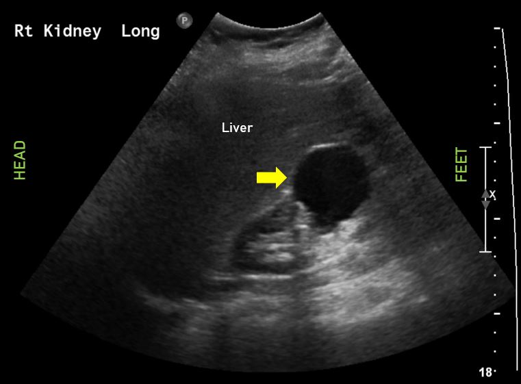
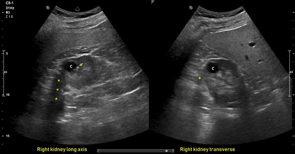
A simple cyst is a fluid-filled sac with a thin, smooth wall. On an ultrasound, these characteristics translate into a very specific visual appearance.
Anechoic Structure with Posterior Acoustic Enhancement
The most defining feature of a simple cyst on ultrasound is its anechoic nature. Since the cyst is filled with fluid (like water, which has very little echogenicity), the sound waves pass through it with minimal reflection. This results in a well-defined, uniformly black area within the ultrasound image.
Furthermore, simple cysts typically exhibit posterior acoustic enhancement. Because the fluid within the cyst allows sound waves to travel through it with less attenuation (weakening) than the surrounding tissues, the echoes returning from structures behind the cyst are stronger. This makes the tissue posterior to the cyst appear brighter or more echogenic than it would otherwise. This enhancement is a strong indicator of a fluid-filled structure.
Well-Defined, Smooth Margins
Another hallmark of a simple cyst is its well-defined and smooth margins. The fluid-filled sac is typically encapsulated by a thin, uniform wall. This smooth, even border is clearly delineated from the surrounding tissues on the ultrasound image. This sharp demarcation is a key differentiator from more complex or irregular masses.
Spherical or Oval Shape
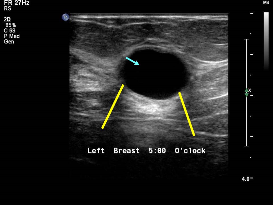
Simple cysts generally assume a spherical or oval shape. This is due to the pressure exerted by the fluid within the sac, which tends to form the most energy-efficient shape. While variations can occur due to external pressure or anatomical constraints, a generally rounded form is characteristic.
Differentiating Cysts from Other Lesions: Complexities and Nuances
While simple cysts have a predictable appearance, not all cysts are simple. The term “complex cyst” encompasses a wider range of formations that deviate from the simple anechoic, well-defined model. Understanding these complexities is vital for accurate diagnosis.
Complex Cysts: Introducing Internal Features
Complex cysts are characterized by the presence of internal structures or characteristics that alter their anechoic appearance. These can include:
- Internal Septations: These are thin, linear echoes within the cyst that divide it into smaller compartments. They represent thin walls or membranes within the cyst. The presence of septations can range from a single thin line to multiple, intricate divisions.
- Internal Echogenic Material: This refers to the presence of debris, blood clots, pus, or other solid components within the cyst. These elements will appear as brighter, more echogenic areas within the typically anechoic or hypoechoic fluid. The texture and distribution of this material can vary significantly.
- Solid Components: In some cases, a complex cyst may have areas that appear to be solid rather than purely fluid-filled. These solid components will be more echogenic than the surrounding fluid and may have irregular shapes and margins.
- Cysts with Irregular or Thickened Walls: While simple cysts have thin, smooth walls, complex cysts can have walls that are thicker, irregular, or nodular. This can be due to inflammation, infection, or neoplastic processes.
Differentiating from Solid Masses
The presence of these internal features is what distinguishes a complex cyst from a solid mass. Solid masses are generally composed of tissue rather than fluid and will therefore appear more uniformly echogenic, often with irregular borders and internal heterogeneity that is different from the patterns seen in cystic structures. The absence of posterior acoustic enhancement is also a clue that a structure might be solid. However, some complex cystic lesions can mimic solid masses, requiring careful evaluation of all features.
The Role of Doppler Ultrasound
Doppler ultrasound is a crucial adjunct in evaluating cystic and solid lesions. It measures the flow of blood within tissues.
- Absence of Blood Flow: A simple cyst, being fluid-filled, will typically show no blood flow within it when examined with Doppler.
- Presence of Blood Flow: The presence of blood flow within septations, solid components, or the wall of a lesion can indicate increased vascularity. This is often a sign of inflammation, infection, or, in some cases, malignancy. For instance, increased vascularity within a solid component of a complex cyst can raise concern.
By combining gray-scale imaging with Doppler assessment, physicians gain a more comprehensive understanding of the nature of the lesion.
Beyond Simple and Complex: Other Considerations in Cyst Ultrasound
While the distinction between simple and complex cysts is foundational, other factors influence how a cyst is visualized and interpreted on ultrasound.
Location and Associated Findings
The location of a cyst within the body can provide important contextual information. For example, a cyst identified in the liver might be evaluated differently than a cyst in the breast or ovary. Associated findings, such as inflammation or enlargement of surrounding structures, can also be important clues. The presence of ascites (fluid in the abdominal cavity) alongside a complex ovarian cyst, for instance, would be a significant finding.
Size and Morphology
While size alone is not always indicative of benignity or malignancy, it is a critical parameter to measure and monitor. Changes in size over time are often more significant than a single measurement. The overall morphology, including the presence of any protruding papillary structures or internal calcifications, also contributes to the assessment.
The Human Element: Sonographer Skill and Radiologist Expertise
It is paramount to remember that ultrasound interpretation is not solely an objective process dictated by algorithms. The skill and experience of the sonographer performing the examination are crucial. Their ability to optimize image acquisition, identify subtle features, and manipulate the transducer to gain different views directly impacts the quality of the diagnostic information.
Similarly, the expertise of the radiologist in interpreting these images is indispensable. They integrate the visual information with the patient’s clinical history, other imaging modalities (if available), and their vast knowledge base to arrive at an accurate diagnosis. The “look” of a cyst on ultrasound is a language understood through a combination of technological capabilities and human interpretation.
In conclusion, understanding what a cyst looks like in an ultrasound involves appreciating the physics of sound wave reflection and the subsequent translation into visual data. While simple cysts present a clear anechoic, well-defined, and enhanced appearance, complex cysts introduce variations with internal septations, solid components, or thickened walls. The judicious use of Doppler ultrasound and the integration of clinical context, along with the expertise of skilled medical professionals, are essential for accurate diagnosis and appropriate patient management.
aViewFromTheCave is a participant in the Amazon Services LLC Associates Program, an affiliate advertising program designed to provide a means for sites to earn advertising fees by advertising and linking to Amazon.com. Amazon, the Amazon logo, AmazonSupply, and the AmazonSupply logo are trademarks of Amazon.com, Inc. or its affiliates. As an Amazon Associate we earn affiliate commissions from qualifying purchases.