
In the vast landscape of scientific instrumentation, few tools possess the transformative power to reveal the atomic blueprint of materials quite like the X-ray diffractometer (XRD). This sophisticated analytical instrument is not merely a device for observation; it is a gateway to understanding the fundamental structure of matter, enabling researchers, engineers, and scientists across a myriad of disciplines to probe the crystalline order within solid substances. At its core, an XRD leverages the unique interaction of X-rays with the electron clouds of atoms arranged in a periodic lattice. By analyzing the diffraction patterns generated from this interaction, an XRD system can precisely determine the atomic and molecular structure of materials, offering unparalleled insights into their properties, behavior, and potential applications. From pioneering advancements in materials science to ensuring the quality of pharmaceuticals and uncovering the secrets of our planet’s geology, the X-ray diffractometer stands as an indispensable cornerstone of modern scientific inquiry, constantly evolving to meet the complex demands of technological progress.
Unveiling the Microscopic World: The Science Behind XRD
The ability of an X-ray diffractometer to reveal the atomic arrangement of materials is rooted in a fascinating interplay of physics and crystallography. The principle relies on the wave-like nature of X-rays and their specific interaction with the regularly spaced atoms within crystalline solids. Unlike visible light, which has much longer wavelengths and interacts with larger features, X-rays possess wavelengths comparable to the interatomic distances in crystals, typically in the angstrom range (0.1-10 Å). This precise alignment of scales is what makes X-rays the ideal probe for atomic-level structural analysis.
The Fundamental Principle: Bragg’s Law
The cornerstone of X-ray diffraction is Bragg’s Law, formulated by Sir William Henry Bragg and Sir William Lawrence Bragg in 1913. This law describes the conditions under which X-rays, when striking a crystal lattice, will constructively interfere to produce a diffracted beam. Essentially, when a beam of monochromatic X-rays interacts with the planes of atoms in a crystal, scattering occurs. If the path difference between X-rays scattered from successive planes is an integer multiple of the X-ray wavelength, constructive interference results, leading to a strong diffracted signal. Mathematically, Bragg’s Law is expressed as:
nλ = 2d sinθ
Where:
nis an integer (the order of diffraction, typically 1)λis the wavelength of the incident X-ray beamdis the interplanar spacing (distance between parallel atomic planes in the crystal)θis the Bragg angle (half the angle between the incident and diffracted beams)
By varying the angle θ and measuring the intensity of the diffracted X-rays, an XRD instrument can determine the d spacing, which is characteristic of the crystal structure. Each diffracted peak corresponds to a specific set of atomic planes within the material.
The Interaction of X-Rays with Crystalline Materials
When an X-ray beam impinges upon a crystalline sample, the X-rays penetrate the material and interact primarily with the electron clouds surrounding the atoms. Each electron scatters a small portion of the incident X-ray radiation. Because atoms in a crystal are arranged in a highly ordered, periodic fashion, the scattered X-ray waves from multiple atoms can interfere with each other. In most directions, this interference is destructive, and the waves cancel out. However, at specific angles—those satisfying Bragg’s Law—the scattered waves reinforce each other constructively, leading to detectable “diffraction peaks.” The position, intensity, and shape of these peaks in a diffraction pattern provide a unique fingerprint of the material’s atomic structure. Amorphous materials, lacking this long-range atomic order, produce broad, diffuse patterns instead of sharp peaks.
Key Components of an XRD System
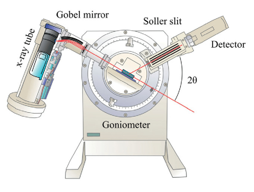
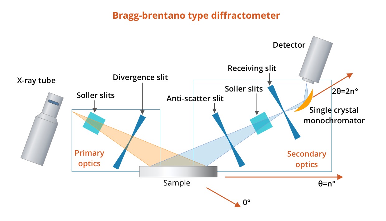
A typical X-ray diffractometer comprises several critical components working in concert:
- X-Ray Source: Generates a stable and intense beam of monochromatic X-rays. Common sources include sealed X-ray tubes (e.g., copper anode) or more powerful synchrotron radiation sources for advanced applications.
- Goniometer: A precision mechanical assembly that holds the sample and detector, allowing them to be rotated with high accuracy relative to the X-ray source. This movement enables the collection of diffraction data across a range of angles (
2θ). - Sample Holder: Secures the sample in a precise position within the X-ray beam path.
- Detector: Measures the intensity of the diffracted X-rays at various angles. Modern detectors are typically highly sensitive and fast, ranging from point detectors to linear or area detectors that can capture diffraction patterns simultaneously.
- Optics: Components such as slits, monochromators, and mirrors are used to shape, filter, and focus the X-ray beam, ensuring high data quality and reducing background noise.
- Computer and Software: Controls the instrument, collects raw data, and provides sophisticated algorithms for data processing, analysis, and interpretation of the diffraction patterns.
Applications Across Industries: Where XRD Makes a Difference
The insights gleaned from X-ray diffraction are invaluable across an astonishing array of scientific and industrial sectors. Its ability to characterize crystalline structures provides critical information that influences research, development, quality control, and problem-solving.
Materials Science and Engineering: Unlocking Properties
Perhaps the most fundamental application of XRD is in materials science. Researchers use XRD to identify unknown crystalline phases, determine crystallite size, quantify the degree of crystallinity in polymers, and analyze texture (preferred orientation of crystallites). This information is crucial for developing new materials with desired properties, such as stronger alloys, more efficient semiconductors, or novel catalytic surfaces. For instance, understanding how processing affects crystal structure can lead to improved manufacturing techniques for everything from aerospace components to consumer electronics.
Pharmaceuticals: Ensuring Purity and Performance
In the pharmaceutical industry, XRD is a cornerstone of quality control and drug development. Polymorphism—where a chemical compound can exist in more than one crystalline form—is a critical concern as different polymorphs can have varying solubility, stability, and bioavailability, directly impacting drug efficacy and patentability. XRD is used to identify and quantify specific polymorphic forms, monitor phase transformations, and ensure the purity and consistency of active pharmaceutical ingredients (APIs) and excipients throughout manufacturing.
Geology and Mineralogy: Decoding Earth’s Composition
Geologists and mineralogists rely on XRD to identify mineral phases in rocks, soils, and sediments. This is vital for understanding geological processes, exploring for natural resources (e.g., oil, gas, minerals), and assessing environmental impacts. For example, characterizing clay minerals can reveal information about soil fertility or the stability of geological formations, while analyzing ore samples can guide mining operations.
Forensics and Art Conservation: Aiding Investigation and Preservation
The unique “fingerprint” provided by XRD is also utilized in more niche, yet critical, fields. Forensic scientists use it to analyze trace evidence, such as paint chips, fibers, or soil samples, to link suspects to crime scenes. In art conservation, XRD helps identify pigments, binders, and corrosion products in artworks and artifacts, aiding in authentication, understanding degradation mechanisms, and informing restoration strategies. This non-destructive nature is particularly valuable for irreplaceable objects.
Beyond Traditional Fields: Emerging Applications
The versatility of XRD continues to drive its adoption into new areas. It plays a role in environmental science for analyzing particulate matter and pollutants, in battery technology for characterizing electrode materials and understanding degradation, and in cement chemistry for optimizing formulations and studying hydration processes. The technique is also fundamental in nanotechnology for characterizing nanoparticles and thin films, where crystal structure directly dictates function.
Types of X-Ray Diffractometers: Tailoring Analysis to Needs
While the fundamental principles remain consistent, X-ray diffractometers come in various configurations, each optimized for specific sample types and analytical objectives. The choice of instrument depends on the material’s form, the desired level of detail, and the specific property to be investigated.
Powder X-Ray Diffractometry (PXRD)
Powder X-ray diffractometry is the most common and widely accessible form of XRD. It is used to analyze polycrystalline materials, which consist of a vast number of tiny, randomly oriented single crystals (crystallites). In PXRD, a finely ground sample is prepared, ensuring that there are always crystallites oriented correctly to satisfy Bragg’s Law for every possible diffraction plane as the sample is rotated or oscillated. The resulting diffraction pattern is a plot of X-ray intensity versus the diffraction angle (2θ), characterized by a series of sharp peaks that represent the unique structural fingerprint of the material. PXRD is excellent for phase identification, quantifying mixtures, determining crystallite size, and assessing residual stress.
Single Crystal X-Ray Diffractometry (SCXRD)
In contrast to PXRD, single crystal X-ray diffractometry is used to determine the precise three-dimensional atomic and molecular structure of individual crystals. This technique provides the most detailed structural information, including bond lengths, bond angles, and atom connectivity. A small, high-quality single crystal (typically less than 1 mm) is mounted on a goniometer and rotated through a range of angles while a detector records the diffracted spots. Sophisticated software then reconstructs the full 3D electron density map, allowing for the unambiguous determination of the crystal structure. SCXRD is indispensable in chemistry for characterizing new compounds, determining absolute configurations, and understanding intermolecular interactions.
Advanced and Specialized XRD Techniques
Beyond the fundamental powder and single-crystal methods, numerous specialized XRD techniques have emerged to address complex research questions:
- High-Resolution X-Ray Diffraction (HRXRD): Used for characterizing epitaxial thin films and semiconductor superlattices, providing insights into strain, thickness, and interface quality.
- Grazing Incidence X-Ray Diffraction (GIXRD): Designed for analyzing the crystal structure of surfaces and thin films by keeping the incident X-ray beam at a very shallow angle to the sample surface.
- Small-Angle X-Ray Scattering (SAXS): While related to diffraction, SAXS measures scattering at very small angles to probe larger-scale structures, typically in the nanometer range, such as pores, particles, and polymers.
- In-Situ/Operando XRD: Allows for the study of structural changes in materials under real-world conditions (e.g., varying temperature, pressure, humidity, gas atmospheres, or applied electric fields) as a reaction or process occurs. This provides dynamic insights into material behavior.
- Texture Analysis (Pole Figure Measurement): Used to quantify the preferred orientation of crystallites in polycrystalline materials, which can significantly influence mechanical properties.
The XRD Process: From Sample to Data Interpretation
Performing an X-ray diffraction experiment is a multi-step process that requires careful preparation, precise execution, and sophisticated data analysis to yield meaningful results.
Sample Preparation: The Crucial First Step
The quality of the diffraction data is heavily dependent on the sample preparation. For PXRD, a representative sample must be finely ground (to minimize preferred orientation and ensure a good distribution of crystallite orientations) and packed uniformly into a holder, often as a thin layer on a zero-background substrate or a well-packed pellet. For SCXRD, obtaining a high-quality, defect-free single crystal of appropriate size is paramount and can often be the most challenging part of the experiment. Improper sample preparation can lead to broadened peaks, shifts in peak positions, or an inaccurate representation of the bulk material.
Data Acquisition: Scanning and Signal Detection
Once the sample is prepared and loaded onto the goniometer, the X-ray source is activated, and the detector begins collecting data. In a typical PXRD scan, the goniometer sweeps through a range of 2θ angles, usually from a few degrees up to 90-150 degrees, with the detector moving in conjunction with the sample. At each angular step, the detector measures the intensity of the diffracted X-rays. For SCXRD, the crystal is rotated, and multiple images of diffracted spots are collected, often over the course of several hours. The raw data consists of an intensity measurement for each angle (PXRD) or each recorded image frame (SCXRD).
Data Analysis and Interpretation: Unveiling Structural Secrets
The raw diffraction data is then processed and interpreted using specialized software. For PXRD, the primary steps involve:
- Peak Identification: Locating the positions (2θ values) and intensities of the diffraction peaks.
- Phase Identification: Comparing the experimental diffraction pattern to vast databases of known crystal structures (e.g., ICDD PDF database). A match confirms the identity of the crystalline phases present.
- Quantitative Analysis: Determining the relative proportions of different crystalline phases in a mixture.
- Structural Refinement (Rietveld Analysis): A powerful method for refining crystal structure parameters (lattice parameters, atomic positions, site occupancies) directly from powder diffraction data.
- Crystallite Size and Strain Analysis: Analyzing peak broadening to estimate average crystallite size and microstrain within the material.
For SCXRD, the data analysis is more complex, involving indexing the diffraction spots to determine the unit cell parameters, integrating spot intensities, correcting for various experimental factors, and finally solving and refining the atomic structure. The ultimate output is a detailed 3D atomic model of the molecule or material.
The Future of X-Ray Diffractometry: Innovations and Challenges
X-ray diffractometry is a mature technique, yet it continues to evolve at a rapid pace, driven by advancements in X-ray source technology, detector design, computational power, and the ever-increasing complexity of materials science challenges.
Miniaturization and Portability
One significant trend is the development of more compact and portable XRD instruments. While traditional laboratory diffractometers are large and stationary, smaller, benchtop, and even handheld devices are emerging. These allow for in-field analysis, on-site quality control, and educational applications where space and cost are limiting factors. Such developments democratize access to structural characterization, extending its reach beyond specialized research labs.
Enhanced Data Processing and AI Integration
The sheer volume and complexity of data generated by modern XRD experiments, especially those involving advanced detectors or in-situ studies, necessitate sophisticated computational tools. Artificial intelligence and machine learning algorithms are increasingly being integrated into XRD software for tasks such as automated peak identification, pattern matching, structural prediction, and even autonomous experiment design. This promises faster, more accurate data interpretation and the ability to extract hidden correlations that might be missed by traditional methods.
Addressing Sample Limitations and New Materials
Future innovations in XRD will also focus on overcoming current limitations. This includes developing techniques for analyzing extremely small samples, highly disordered or nanocrystalline materials, and challenging in-situ environments. Advancements in focusing optics and brighter X-ray sources (such as fourth-generation synchrotrons and X-ray free-electron lasers) will enable faster data acquisition and the study of transient phenomena, pushing the boundaries of what can be observed at the atomic scale. Furthermore, the integration of XRD with complementary techniques (e.g., spectroscopy, microscopy) into multimodal analytical platforms will provide a more comprehensive understanding of complex materials.
In conclusion, the X-ray diffractometer remains an unparalleled tool for probing the atomic and molecular architecture of crystalline materials. Its foundational principles, diverse applications across science and industry, and continuous technological evolution ensure its enduring relevance as a cornerstone of materials characterization and a key enabler of technological progress. As our understanding of matter deepens and new materials are engineered, the XRD will undoubtedly continue to be at the forefront of discovery, providing the structural insights essential for innovation.
aViewFromTheCave is a participant in the Amazon Services LLC Associates Program, an affiliate advertising program designed to provide a means for sites to earn advertising fees by advertising and linking to Amazon.com. Amazon, the Amazon logo, AmazonSupply, and the AmazonSupply logo are trademarks of Amazon.com, Inc. or its affiliates. As an Amazon Associate we earn affiliate commissions from qualifying purchases.