Down Syndrome, a genetic disorder characterized by a unique set of physical and developmental traits, has long been a subject of intense scientific inquiry. While its observable manifestations are well-documented, the underlying cause – a specific type of genetic mutation – has become a focal point for groundbreaking research, driven by increasingly sophisticated technological advancements. This article delves into the precise genetic alterations responsible for Down Syndrome, exploring the technological tools and methodologies that have illuminated our understanding of this complex condition.
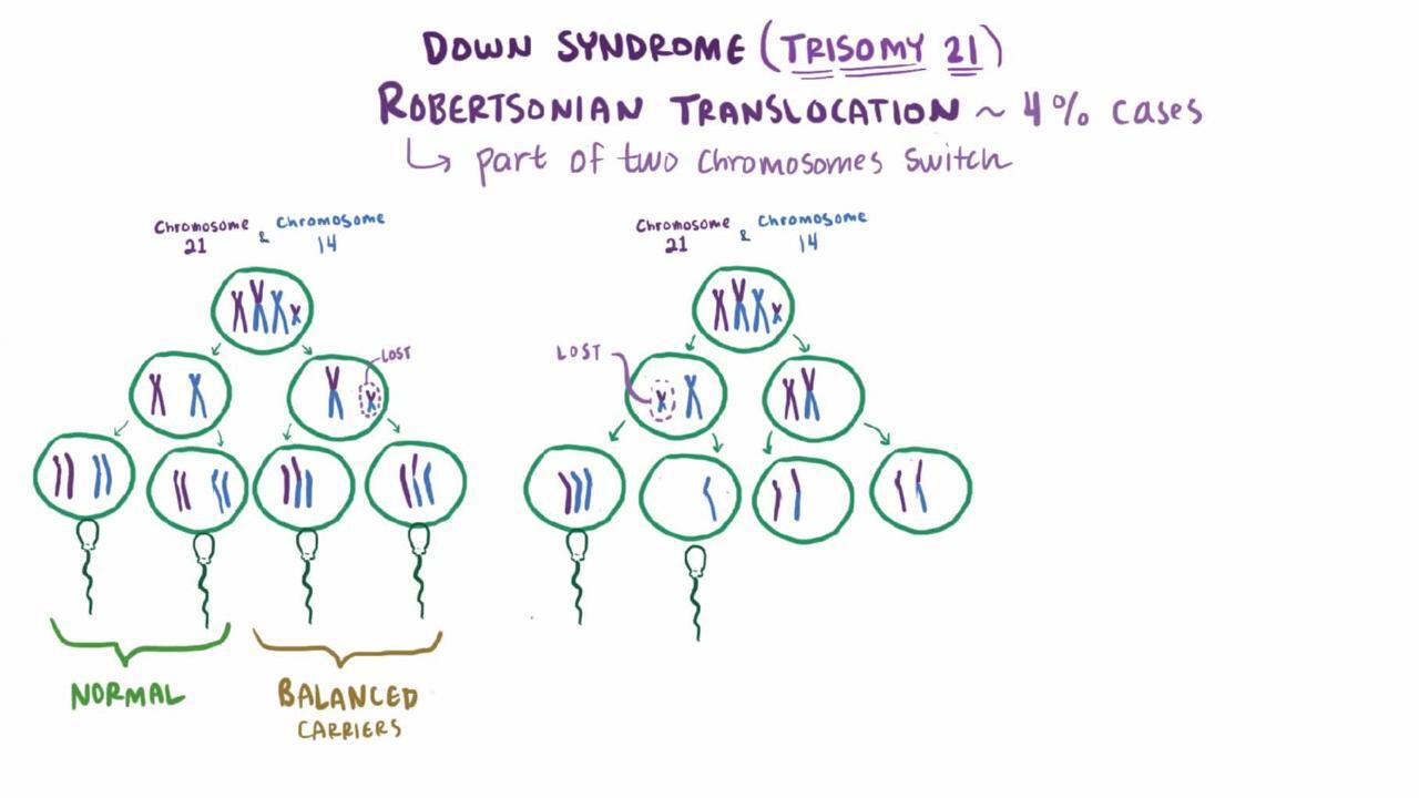
The answer to “what type of mutation causes Down Syndrome” lies not in a single gene mutation, as is often the case with other genetic disorders, but rather in the presence of an entire extra chromosome. This phenomenon, known as trisomy, is where technology plays a crucial role in its identification, analysis, and the ongoing development of diagnostic and therapeutic strategies.
Understanding the Genetic Architecture of Down Syndrome
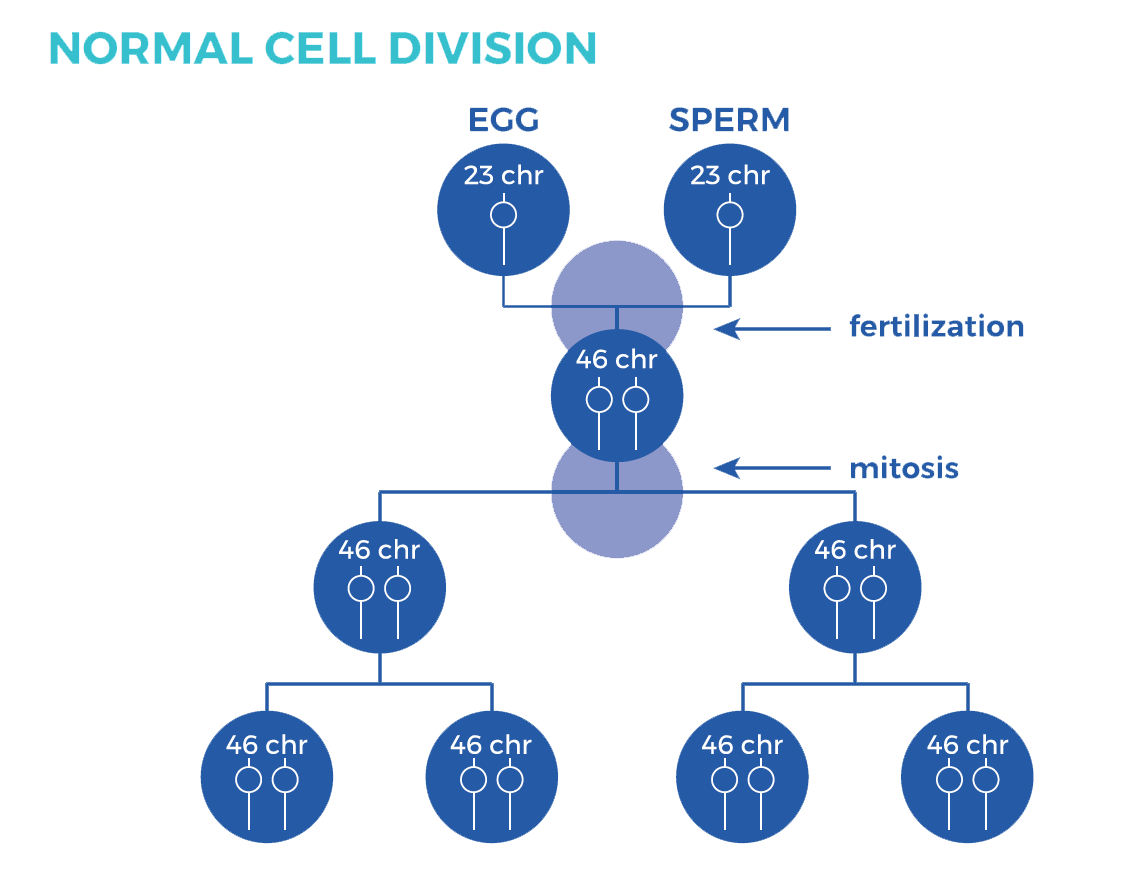
At its core, Down Syndrome is a chromosomal abnormality. Humans typically have 23 pairs of chromosomes, totaling 46. These chromosomes carry our genetic blueprint, dictating everything from physical characteristics to predispositions to certain diseases. In individuals with Down Syndrome, there is an extra copy of chromosome 21. This means they have three copies of this chromosome instead of the usual two. This extra genetic material fundamentally alters the course of development, leading to the characteristic features associated with the condition.
The Role of Karyotyping in Chromosomal Analysis
The foundational technology for diagnosing chromosomal abnormalities like Down Syndrome has historically been karyotyping. This technique, a cornerstone of cytogenetics, involves analyzing a person’s chromosomes under a microscope.
The Karyotyping Process: From Cell Culture to Chromosome Banding
The process begins with obtaining a sample of cells, typically from blood. These cells are then cultured in a laboratory to encourage them to divide. During the metaphase stage of cell division, when chromosomes are most condensed and visible, the cells are treated with a chemical agent to halt the process. The chromosomes are then stained and spread out onto a slide.
The staining process, often using a technique called Giemsa banding, reveals distinct light and dark bands across each chromosome. These banding patterns are unique to each chromosome and allow geneticists to identify and organize them into homologous pairs. By examining the karyotype, which is essentially a photograph of an individual’s complete set of chromosomes arranged in order, clinicians can readily identify the presence of an extra chromosome 21. This visual confirmation remains a critical diagnostic tool, allowing for early identification and enabling families to access appropriate support and resources from the outset.
Beyond Karyotyping: Advanced Genetic Technologies for Precision
While karyotyping provides a broad overview, it has limitations in detecting smaller chromosomal rearrangements or mosaicism (where an individual has a mix of cells with and without the extra chromosome). This is where more advanced technological solutions have become indispensable.
Fluorescence In Situ Hybridization (FISH) and its Applications
Fluorescence In Situ Hybridization (FISH) represents a significant leap forward in chromosomal analysis. This technique utilizes fluorescent probes that bind specifically to certain DNA sequences on chromosomes. In the context of Down Syndrome, a probe designed to target a region of chromosome 21 can be used to “light up” the chromosome under a fluorescence microscope.
FISH offers several advantages over traditional karyotyping. It is faster and can detect smaller chromosomal abnormalities, including deletions, duplications, and translocations that might be missed by visual inspection alone. For instance, in cases where a translocation involves chromosome 21, FISH can precisely pinpoint the location of the extra genetic material, providing a more detailed understanding of the chromosomal makeup. This level of precision is invaluable for genetic counseling and risk assessment.
Microarray-Based Comparative Genomic Hybridization (array CGH) for Uncovering Subtle Deletions and Duplications
Perhaps the most powerful technological tool for detecting copy number variations – the gain or loss of DNA segments – is microarray-based comparative genomic hybridization (array CGH). This technology allows for a genome-wide analysis of chromosomal imbalances with much higher resolution than karyotyping or even standard FISH.

Array CGH works by comparing the DNA of a patient to that of a reference control, labeled with different fluorescent dyes. The DNA is then hybridized to a microarray chip containing thousands of DNA probes that cover the entire genome. Regions where the patient has an extra copy of DNA (like the extra chromosome 21 in Down Syndrome) will show a stronger fluorescent signal compared to the control, while regions with a deletion would show a weaker signal.
This technology is particularly adept at identifying submicroscopic deletions and duplications that are too small to be seen with a microscope. While Down Syndrome is primarily characterized by a whole extra chromosome, array CGH can be crucial in identifying other, less common chromosomal anomalies that might be associated with similar phenotypes or in cases of mosaicism where the extra chromosome is not present in all cells. The data generated by array CGH provides an unprecedented level of detail about an individual’s genetic makeup, aiding in the diagnosis of a wider spectrum of genetic conditions.
The Technological Evolution of Prenatal Diagnosis
The ability to diagnose Down Syndrome before birth has been revolutionized by technological advancements, offering expectant parents crucial information and the opportunity for informed decision-making.
Ultrasound and Nuchal Translucency Screening
Early prenatal screening for Down Syndrome often begins with ultrasound. Specialized ultrasound techniques can detect certain physical markers in the fetus that are associated with an increased risk of Down Syndrome. One such marker is increased nuchal translucency (NT), which is the accumulation of fluid in the tissue at the back of the baby’s neck during the first trimester of pregnancy. While not a definitive diagnosis, a thickened NT is a strong indicator that further genetic testing might be warranted. Technological improvements in ultrasound resolution and the development of standardized measurement protocols have significantly enhanced the accuracy of NT screening.
Biochemical Screening and the Rise of Non-Invasive Prenatal Testing (NIPT)
Complementing ultrasound, biochemical screening analyzes specific substances in the mother’s blood, such as pregnancy-associated plasma protein-A (PAPP-A) and human chorionic gonadotropin (hCG). Abnormal levels of these markers can also indicate an increased risk of Down Syndrome.
The most significant technological breakthrough in prenatal diagnosis, however, is Non-Invasive Prenatal Testing (NIPT). This remarkable advancement analyzes small fragments of fetal DNA that circulate in the mother’s bloodstream. By using advanced sequencing technologies and sophisticated bioinformatics algorithms, NIPT can accurately detect the presence of an extra copy of chromosome 21 with very high sensitivity and specificity. This method eliminates the need for invasive procedures like amniocentesis or chorionic villus sampling, which carry a small risk of miscarriage. NIPT has fundamentally transformed prenatal genetic screening, offering a safer and more accessible option for millions of expectant parents worldwide.
Implications for Research and Future Technologies
The ability to precisely identify the genetic basis of Down Syndrome has profound implications for ongoing research and the development of future technologies aimed at understanding and potentially mitigating its effects.
Gene Expression Analysis and the Search for Therapeutic Targets
The extra genetic material on chromosome 21 leads to an overexpression of numerous genes. Technologies that allow for gene expression analysis, such as RNA sequencing, are crucial for understanding how this genetic imbalance impacts cellular function and development. By quantifying the levels of messenger RNA (mRNA) produced from different genes, researchers can identify which genes are abnormally regulated in individuals with Down Syndrome. This understanding is a critical step in identifying potential therapeutic targets. If specific gene pathways are consistently dysregulated, it opens avenues for developing interventions that could normalize gene expression or ameliorate the resulting cellular defects.
Advances in Gene Editing and Personalized Medicine
Emerging technologies like CRISPR-Cas9 gene editing hold immense promise for the future of genetic medicine. While still in its early stages for complex conditions like Down Syndrome, the ability to precisely edit DNA offers the potential to correct genetic errors. Researchers are exploring whether gene editing could be used to downregulate the expression of specific genes on chromosome 21 that contribute to the condition’s symptoms, or even to correct the chromosomal anomaly itself.
Furthermore, the detailed genetic information gathered through advanced sequencing and microarray technologies is paving the way for personalized medicine. By understanding the unique genetic profile of an individual with Down Syndrome, healthcare providers can tailor interventions, therapies, and support strategies to their specific needs, optimizing outcomes and enhancing quality of life.
In conclusion, the question of “what type of mutation causes Down Syndrome” leads us to the intricate world of chromosomal abnormalities, specifically trisomy 21. The journey from basic visual identification through karyotyping to the sophisticated genomic analyses offered by FISH, array CGH, and NIPT highlights the transformative power of technology. These advancements not only provide crucial diagnostic capabilities but also fuel ongoing research, promising a future where our understanding of genetic disorders like Down Syndrome translates into more effective interventions and improved lives for those affected. The ongoing evolution of genetic technologies continues to unlock new frontiers in unraveling the complexities of the human genome and its impact on health.
aViewFromTheCave is a participant in the Amazon Services LLC Associates Program, an affiliate advertising program designed to provide a means for sites to earn advertising fees by advertising and linking to Amazon.com. Amazon, the Amazon logo, AmazonSupply, and the AmazonSupply logo are trademarks of Amazon.com, Inc. or its affiliates. As an Amazon Associate we earn affiliate commissions from qualifying purchases.