
The human heart, a marvel of biological engineering, operates as a relentless pump, circulating blood throughout the body to deliver oxygen and nutrients while removing waste products. At the core of its efficiency are four intricate structures: the heart valves. While their fundamental biological purpose—to ensure unidirectional blood flow—remains constant, our understanding, diagnosis, and treatment of valve-related conditions have been profoundly revolutionized by advancements in technology. Viewing the heart’s valves through a technological lens allows us to appreciate not just their intrinsic biological design but also the sophisticated tools and innovations that have emerged to safeguard this vital aspect of human physiology.
The Biomechanical Genius of Heart Valves: A Blueprint for Innovation
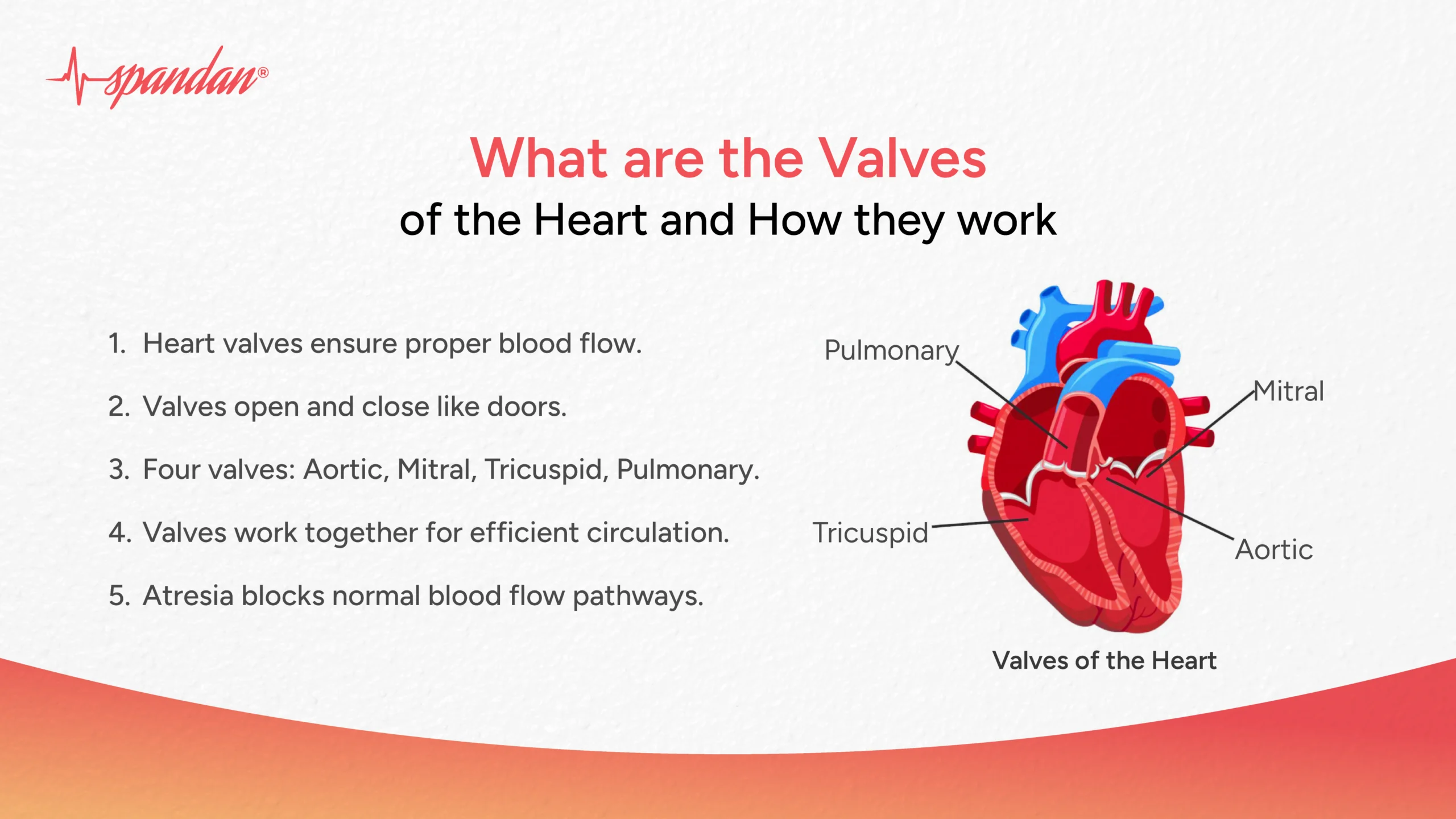
At a macroscopic level, the purpose of the valves inside the heart is elegantly simple yet critically complex: to prevent the backflow of blood, ensuring it moves in the correct direction through the heart’s chambers and out to the body. This seemingly straightforward task is executed through an exquisitely synchronized dance of opening and closing, powered by pressure gradients and the structural integrity of the valve leaflets.
Ensuring Unidirectional Flow: The Core Function
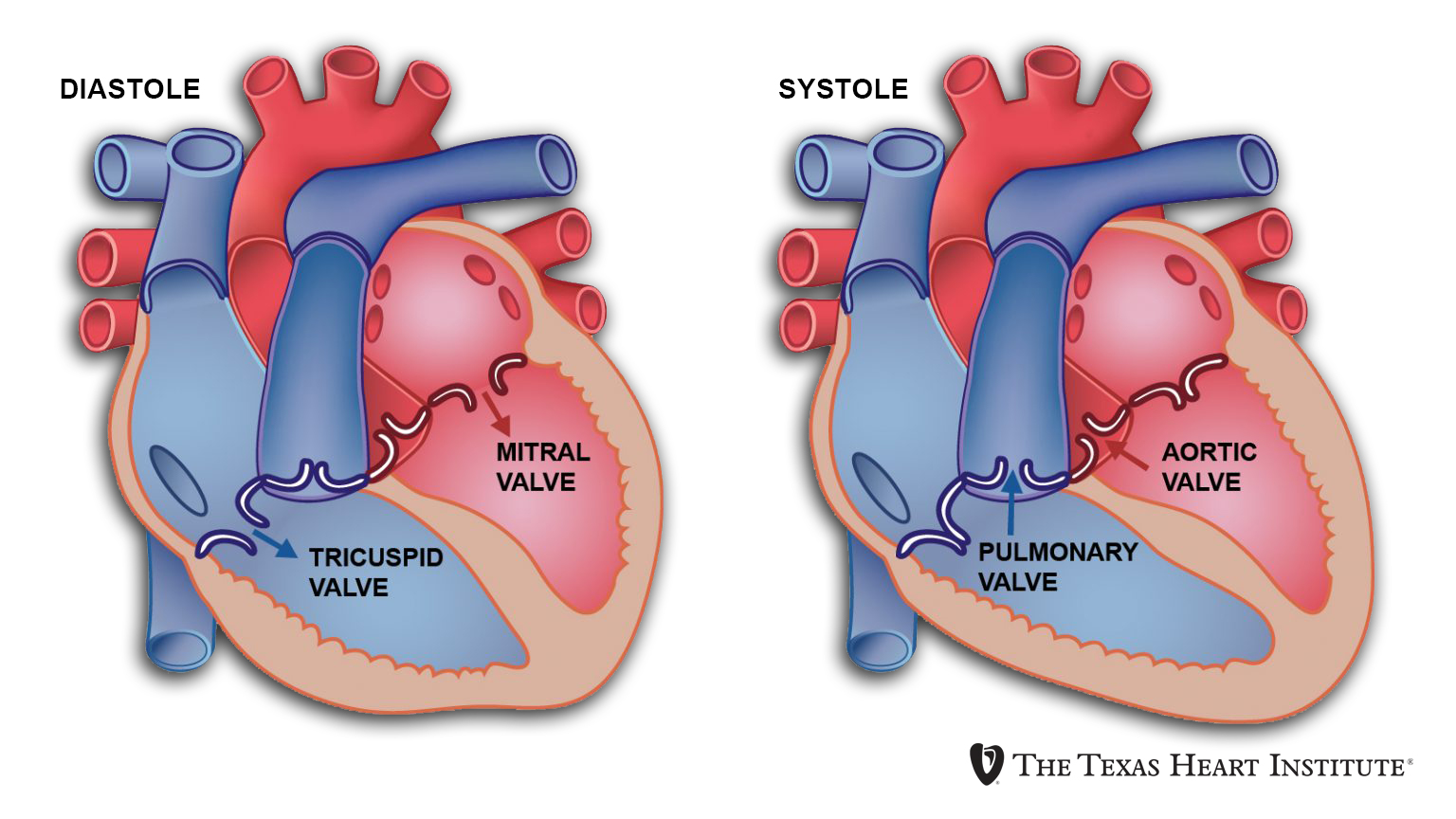
Imagine a series of one-way gates within a complex plumbing system. This is essentially what heart valves are. As the heart muscle contracts and relaxes, pressure changes occur within its chambers. These pressure differences dictate the opening and closing of the valves. When a chamber contracts, the pressure within it rises, pushing blood forward and forcing the downstream valve open, while simultaneously snapping the upstream valve shut to prevent regurgitation. This precise timing and mechanical action are fundamental to maintaining efficient circulation, preventing blood from pooling or flowing backward, which would severely compromise the heart’s pumping efficiency and overall body function. Any disruption to this unidirectional flow, such as a valve failing to open fully (stenosis) or failing to close completely (regurgitation or insufficiency), places enormous strain on the heart and can lead to serious cardiovascular disease.
The Four Guardians: A Brief Overview of Aortic, Mitral, Pulmonary, and Tricuspid Valves
The heart houses two sets of valves: the atrioventricular (AV) valves, located between the atria and ventricles, and the semilunar valves, situated between the ventricles and the major arteries.
-
Mitral Valve: Positioned between the left atrium and left ventricle, this bicuspid (two-leaflet) valve ensures oxygenated blood flows from the lungs into the left ventricle, preventing backflow into the atrium during ventricular contraction.
-
Tricuspid Valve: Located between the right atrium and right ventricle, this tricuspid (three-leaflet) valve prevents deoxygenated blood from flowing back into the right atrium during the contraction of the right ventricle.
-
Aortic Valve: A semilunar valve with three leaflets, it controls the outflow of oxygenated blood from the left ventricle into the aorta, the body’s main artery. Its proper function is paramount for systemic blood circulation.
-
Pulmonary Valve: Also a semilunar valve with three leaflets, it regulates the flow of deoxygenated blood from the right ventricle into the pulmonary artery, which carries blood to the lungs.
Understanding the unique architecture and function of each valve is not merely an academic exercise; it’s the foundational knowledge upon which all modern diagnostic tools, surgical interventions, and innovative medical devices are built. The biological “engineering” of these valves serves as a constant source of inspiration for bio-engineers and medical innovators.
Diagnostic Technologies: Unveiling Valve Function and Dysfunction
While the biological purpose of heart valves remains unchanged, our ability to assess their condition, identify malfunctions, and predict future problems has been revolutionized by cutting-edge technology. Diagnostic advancements have moved beyond rudimentary methods to offer unprecedented insights into valve anatomy and hemodynamics.
Advanced Imaging: From Echocardiography to CT Scans
Echocardiography, or ultrasound of the heart, stands as the cornerstone of cardiac valve diagnosis. Utilizing high-frequency sound waves, this non-invasive technology provides real-time images of the heart’s structure, including valve leaflets, and allows for precise measurement of blood flow velocity and direction. Doppler echocardiography, in particular, can quantify the severity of valve stenosis or regurgitation, helping clinicians make informed decisions.
Beyond ultrasound, more sophisticated imaging modalities provide complementary information. Cardiac Magnetic Resonance Imaging (MRI) offers exquisite detail of valve morphology, tissue characterization, and quantification of regurgitant volumes, especially beneficial in complex cases. Cardiac Computed Tomography (CT) scans, particularly with contrast, are invaluable for assessing valve calcification, annular dimensions, and planning for transcatheter valve interventions, providing a high-resolution 3D roadmap for surgeons and interventional cardiologists. These technologies transform abstract physiological concepts into tangible, visual data, enabling precise diagnosis and intervention planning.
AI in Early Detection and Prognosis
The advent of Artificial Intelligence (AI) and machine learning (ML) is rapidly transforming cardiac diagnostics. AI algorithms can analyze vast datasets from echocardiograms, MRIs, and CT scans with greater speed and sometimes even greater accuracy than the human eye. They can identify subtle patterns indicative of early valve disease, predict disease progression, and stratify patient risk. For instance, AI can be trained to detect early signs of valve calcification or quantify regurgitation fractions with high precision, aiding in the timely initiation of therapy or surveillance. Furthermore, AI-powered predictive models are being developed to forecast the likelihood of adverse events in patients with specific valve pathologies, enabling personalized treatment strategies.
Wearable Tech and Remote Monitoring
The proliferation of wearable technology, from smartwatches to advanced biosensors, is extending diagnostic capabilities beyond the clinic. While not directly imaging the valves, these devices can monitor heart rate, rhythm, and activity levels, detecting irregularities that might signal underlying valve issues or worsening heart function. Continuous remote monitoring allows for early detection of decompensation in patients with known valve disease, enabling timely intervention and potentially preventing acute cardiac events. The integration of data from wearables with AI platforms could create powerful early warning systems, shifting the paradigm from reactive treatment to proactive, preventative care.
Interventional and Therapeutic Innovations
Understanding valve function and diagnosing dysfunction are only the first steps. The ultimate purpose of technological advancement in this field is to provide effective treatments. Here, technology has pushed the boundaries from traditional open-heart surgery to minimally invasive and even regenerative approaches.
Surgical Excellence: Open-Heart vs. Minimally Invasive Procedures (TAVI/TMVR)
For decades, severe valve disease necessitated open-heart surgery, a major procedure involving sternotomy (splitting the breastbone) to repair or replace the affected valve. This highly effective but invasive method carries significant recovery times and risks. However, technological breakthroughs have introduced minimally invasive options.
Transcatheter Aortic Valve Implantation (TAVI), also known as Transcatheter Aortic Valve Replacement (TAVR), is a prime example. Developed initially for high-risk patients unsuitable for open surgery, TAVI involves delivering a new artificial valve to the heart via a catheter inserted through a small incision, typically in the groin. This technology has dramatically reduced recovery times and improved outcomes for countless patients. Similar transcatheter techniques are now emerging for other valves, such as Transcatheter Mitral Valve Repair (TMVR) and Replacement (TMVR), expanding the scope of minimally invasive interventions and making life-saving procedures accessible to a broader patient population. These procedures are a testament to advanced catheter design, imaging guidance, and novel prosthetic valve structures.
The Rise of Artificial Heart Valves: Materials and Design
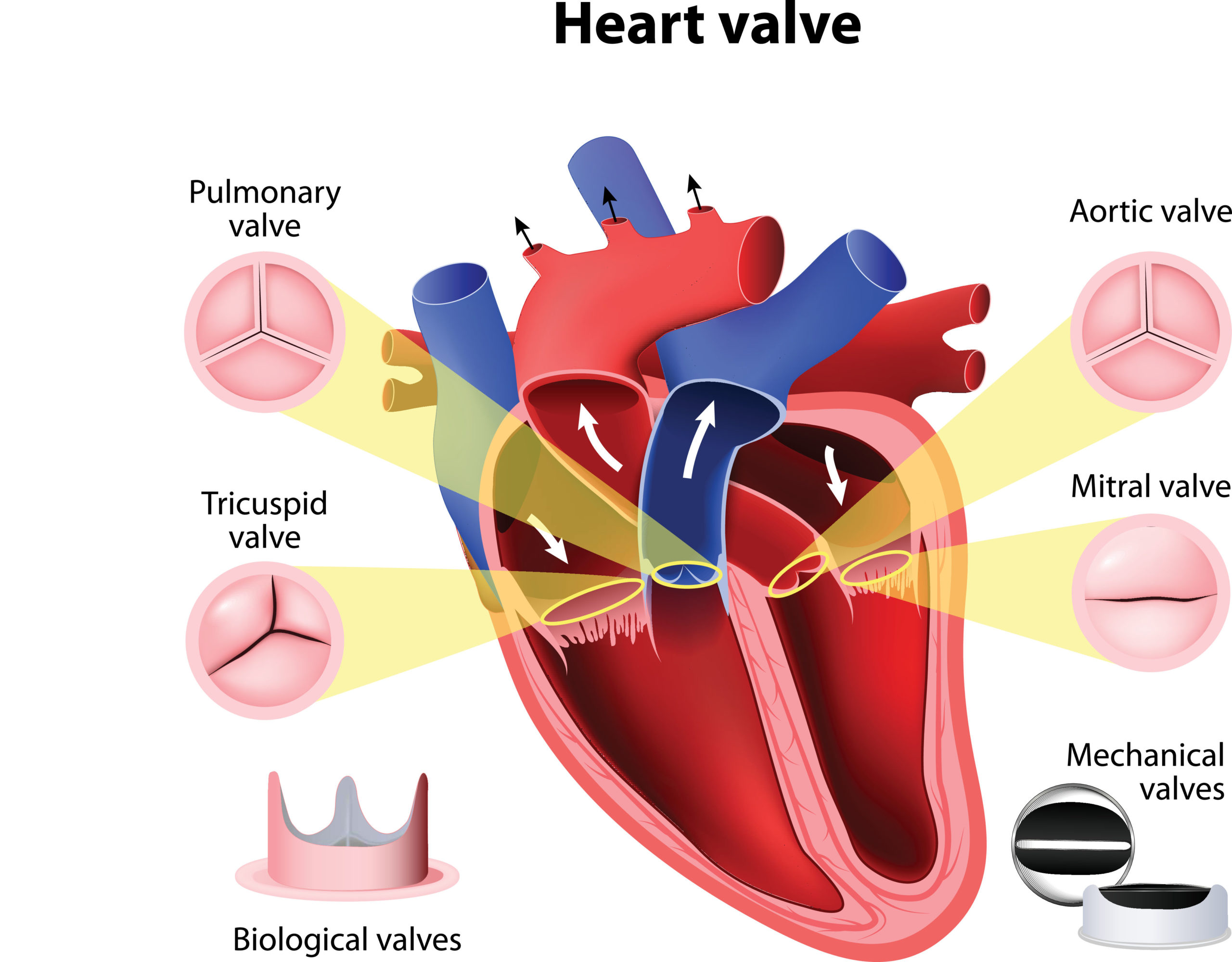
When a diseased valve cannot be repaired, replacement becomes necessary. The development of artificial heart valves is a triumph of materials science and biomechanical engineering. There are two primary types:
-
Mechanical Valves: Constructed from durable materials like pyrolytic carbon, these valves are designed for longevity. They offer excellent durability but require patients to take lifelong anticoagulant medication to prevent blood clots.
-
Bioprosthetic (Tissue) Valves: Derived from animal tissue (porcine or bovine pericardium), these valves are treated and mounted on a frame. They have a lower risk of clot formation, eliminating the need for lifelong anticoagulation, but typically have a shorter lifespan (10-20 years) due to structural deterioration.
Ongoing research in artificial valve design focuses on improving durability, reducing thrombosis risk, and developing valves that can grow or adapt within the patient, particularly for pediatric applications. Advanced computational modeling and simulation play a crucial role in testing and optimizing new valve designs before clinical trials.
Bio-Engineering and Regenerative Medicine
The frontier of valve therapy lies in bio-engineering and regenerative medicine. Researchers are exploring ways to grow new heart valves using a patient’s own cells, reducing the risk of rejection and the need for artificial materials. Techniques like tissue engineering involve seeding biodegradable scaffolds with patient-specific stem cells, which then mature into functional valve tissue. This approach aims to create “living” valves that can repair themselves and potentially last a lifetime, eliminating the limitations of current mechanical and bioprosthetic valves. While still largely experimental, the promise of such regenerative solutions is immense, representing the ultimate fusion of biological purpose and advanced technological intervention.
The Future of Cardiovascular Valve Technology
The journey of understanding and treating heart valve diseases is far from over. The rapid pace of technological innovation promises an even more sophisticated future, pushing the boundaries of what is possible in cardiovascular care.
AI-Driven Personalized Treatment Plans
Future cardiac care will increasingly leverage AI to create highly personalized treatment plans. By integrating patient-specific data—genetics, lifestyle, comorbidities, imaging results, and real-time physiological monitoring—AI algorithms can identify the optimal time for intervention, select the most appropriate valve type, and predict individual responses to different therapies. This move towards precision medicine will optimize outcomes, minimize risks, and enhance quality of life for patients with valve disease.
Advanced Robotics in Surgery
Robotics is already present in some surgical fields, and its role in cardiac surgery, particularly for valve procedures, is set to expand significantly. Robotic systems offer enhanced precision, dexterity, and visualization for surgeons, particularly in minimally invasive approaches. This can translate to smaller incisions, reduced blood loss, faster recovery, and fewer complications. The next generation of robotic surgical platforms may incorporate AI guidance, haptic feedback, and even autonomous or semi-autonomous capabilities for certain aspects of valve repair or replacement.
Towards Bioprinted and Self-Repairing Valves
The ultimate goal in heart valve technology is the creation of living, self-repairing valves that seamlessly integrate with the body. 3D bioprinting, a technique that uses “bio-inks” containing living cells to print complex biological structures layer by layer, holds immense potential. Imagine printing a new, custom-fit heart valve from a patient’s own cells, capable of growing and remodeling in response to physiological demands. This vision, once science fiction, is slowly moving towards reality, offering the prospect of lifelong solutions for valve disease without the drawbacks of current prosthetic options.
In conclusion, the fundamental purpose of the valves inside the heart is a testament to nature’s elegant design: to maintain the unidirectional flow of blood, sustaining life itself. However, it is through the relentless pursuit of technological advancement—from sophisticated imaging and AI diagnostics to groundbreaking surgical techniques and bio-engineered solutions—that we gain deeper insights into this purpose and overcome its failures. As technology continues to evolve, our ability to preserve, repair, and even regenerate these critical components of the heart will undoubtedly lead to healthier, longer lives for countless individuals around the globe, transforming the landscape of cardiovascular medicine.
aViewFromTheCave is a participant in the Amazon Services LLC Associates Program, an affiliate advertising program designed to provide a means for sites to earn advertising fees by advertising and linking to Amazon.com. Amazon, the Amazon logo, AmazonSupply, and the AmazonSupply logo are trademarks of Amazon.com, Inc. or its affiliates. As an Amazon Associate we earn affiliate commissions from qualifying purchases.