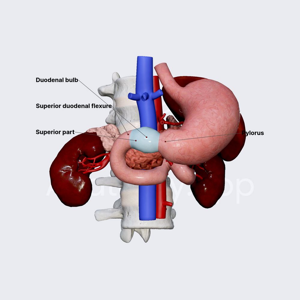
The duodenal bulb, often referred to as the first part of the duodenum, represents a critical juncture in the human digestive system. While its anatomical definition remains constant, our understanding, diagnosis, and management of this delicate structure have been profoundly revolutionized by advancements in technology. From high-resolution imaging to AI-driven diagnostics and sophisticated software solutions, technology provides an unprecedented window into the duodenal bulb’s function and pathology, transforming how medical professionals perceive and interact with this vital organ segment.
Unveiling Anatomy Through Digital Imaging
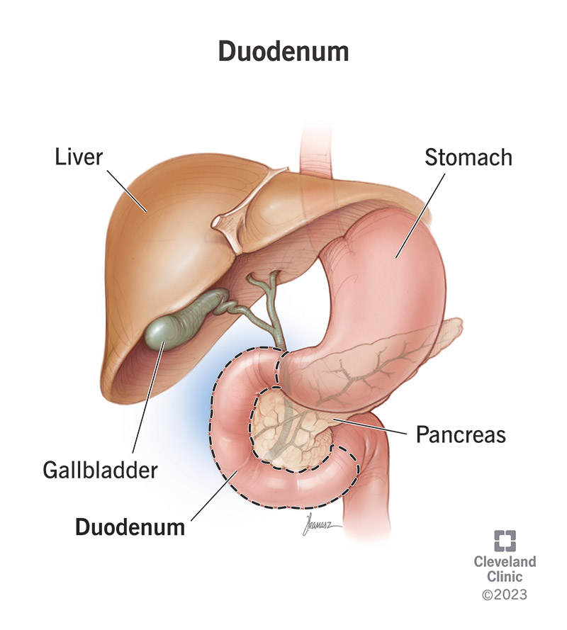
Understanding “what is the duodenal bulb” in the modern medical landscape is intrinsically linked to the technological marvels that allow us to visualize it with stunning clarity. Traditional anatomical diagrams provide a foundational understanding, but digital imaging techniques offer dynamic, real-time, and cross-sectional views that unveil its intricate details and functional dynamics. These technologies are not just tools for observation; they are extensions of our diagnostic capabilities, enabling precise identification of structure, potential anomalies, and physiological activity.
High-Resolution Endoscopy and Its Evolution
Endoscopy stands as a cornerstone in the direct visualization of the duodenal bulb. Modern endoscopes, equipped with high-definition cameras and advanced optical components, provide unparalleled clarity. These flexible tubes, guided through the esophagus and stomach, offer a direct, magnified view of the duodenal bulb’s mucosal lining. What was once a blurry, grainy image has evolved into crisp, vibrant visualizations, allowing gastroenterologists to detect subtle changes in color, texture, and vascular patterns that may indicate inflammation, ulcers, polyps, or early neoplastic lesions.
Beyond mere visual inspection, contemporary endoscopes integrate various enhanced imaging modalities. Narrow-Band Imaging (NBI) filters specific wavelengths of light to highlight mucosal and vascular patterns, proving invaluable for characterizing suspicious lesions and improving the detection rate of dysplasia. Confocal Laser Endomicroscopy (CLE) takes this a step further, providing real-time, “virtual biopsies” at a cellular level during the endoscopic procedure itself. This technology projects a laser onto the tissue and captures fluorescent light emitted by cells, presenting images comparable to conventional histology without the need for immediate tissue removal. This immediate cellular feedback can guide targeted biopsies, reduce unnecessary procedures, and accelerate diagnostic pathways. The evolution of endoscopic technology has transformed the definition of the duodenal bulb from a simple anatomical segment into a dynamic, observable, and diagnostically accessible entity.
CT and MRI: Non-Invasive Insights
While endoscopy offers direct luminal visualization, Computed Tomography (CT) and Magnetic Resonance Imaging (MRI) provide invaluable cross-sectional views of the duodenal bulb and its surrounding structures. These non-invasive imaging modalities offer complementary information, particularly concerning extramural pathology, mural thickening, or the relationship of the duodenal bulb to adjacent organs like the pancreas and gallbladder.
CT scans, leveraging X-rays and sophisticated computer processing, generate detailed cross-sectional images. For the duodenal bulb, CT is often used to assess for inflammation, perforations, or mass lesions, particularly in emergency settings or when evaluating the extent of inflammatory bowel disease (e.g., Crohn’s disease affecting the duodenum). Multi-detector CT (MDCT) scanners offer rapid acquisition and thinner slices, enabling multiplanar reconstructions and 3D renderings that provide a comprehensive spatial understanding of the duodenal bulb’s anatomy and any associated pathology.
MRI, utilizing strong magnetic fields and radio waves, offers superior soft tissue contrast without ionizing radiation. It excels in differentiating fluid collections, inflammation, and certain types of tumors. For the duodenal bulb, MRI can be particularly useful in cases of chronic inflammation, identifying subtle mucosal changes, or evaluating for fistulas. Functional MRI techniques are also emerging, capable of assessing duodenal motility and the movement of chyme, adding a physiological dimension to the anatomical insights. These advanced imaging tools augment our understanding of “what is the duodenal bulb” by integrating its internal structure with its external relationships and functional characteristics, creating a holistic digital model for medical practitioners.
AI-Powered Diagnostics and Predictive Analytics
The sheer volume of data generated by modern imaging and patient monitoring technologies necessitates advanced analytical capabilities. Artificial Intelligence (AI) and Machine Learning (ML) are rapidly emerging as transformative forces in gastroenterology, particularly in enhancing diagnostic accuracy and predicting outcomes related to the duodenal bulb. These intelligent systems process vast datasets, learn patterns imperceptible to the human eye, and provide invaluable support for clinical decision-making.
Early Detection of Duodenal Conditions
AI algorithms are being trained on extensive databases of endoscopic images and radiological scans of the duodenal bulb, learning to identify subtle signs of pathology. For instance, AI can assist endoscopists in real-time by highlighting suspicious polyps, areas of metaplasia, or early signs of inflammation or malignancy that might be missed during a rapid manual examination. These systems can analyze hundreds of image frames per second, providing an objective and consistent second opinion, thereby increasing diagnostic yield and reducing inter-observer variability.
Beyond visual pattern recognition, AI can integrate data from various sources—patient history, laboratory results, genetic markers, and imaging findings—to create a comprehensive risk assessment. For conditions like celiac disease, which often manifests with specific duodenal bulb changes, AI models can analyze biopsy images with greater precision, potentially aiding pathologists in challenging cases or screening large volumes of samples. In ulcerative or inflammatory conditions affecting the duodenal bulb, AI can quantify the severity of inflammation more objectively, leading to more standardized assessments and treatment responses. This technology essentially refines our definition of “what is the duodenal bulb” by providing a deeper, data-driven understanding of its health status.
Personalized Treatment Pathways
The application of AI extends beyond diagnosis to the realm of personalized medicine. By analyzing individual patient data—including genetic profiles, microbiome data (if available), and detailed responses to previous treatments—AI algorithms can predict which therapies are most likely to be effective for specific duodenal bulb conditions. For patients with refractory ulcers or inflammatory disorders of the duodenal bulb, AI could help identify optimal drug dosages, combinations, or even predict the likelihood of recurrence based on a multitude of factors.
Predictive analytics, powered by ML models, can forecast the progression of chronic duodenal conditions, identify patients at high risk for complications, or determine the optimal surveillance intervals. This personalized approach minimizes trial-and-error in treatment, reduces adverse drug reactions, and ultimately leads to more efficient and effective patient management. The duodenal bulb, once treated with a one-size-fits-all approach, can now benefit from tailored interventions guided by intelligent data analysis, fundamentally changing how we understand and approach its care.
Software Solutions for Digestive Health Management
The operational backbone of modern healthcare, especially concerning specialized areas like gastroenterology, relies heavily on sophisticated software platforms. These digital tools streamline workflows, enhance communication, and empower both clinicians and patients in managing conditions related to the duodenal bulb. From electronic record-keeping to remote patient engagement, software solutions are integral to a holistic, technologically driven approach to digestive health.
Electronic Health Records and Data Integration
Electronic Health Records (EHRs) are more than just digital versions of paper charts; they are comprehensive platforms for managing patient information. For conditions affecting the duodenal bulb, EHRs integrate all relevant data: endoscopic findings, biopsy reports, radiological images, medication histories, and treatment plans. This centralized data allows for a longitudinal view of a patient’s duodenal health, enabling clinicians to track disease progression, assess the long-term efficacy of treatments, and make informed decisions based on a complete clinical picture.
Beyond mere storage, advanced EHR systems facilitate data integration and interoperability. They can pull data from various diagnostic machines, lab systems, and even patient-reported outcomes, creating a unified data ecosystem. This interconnectedness ensures that specialists have access to the most current and relevant information, preventing redundant tests and improving coordination of care, especially when multiple disciplines are involved in managing complex duodenal conditions. The ability to quickly search, analyze, and share this information through secure digital channels elevates the understanding and management of the duodenal bulb to an unprecedented level of efficiency.
Telemedicine and Remote Monitoring
The rise of telemedicine platforms has profoundly impacted how patients with duodenal conditions interact with healthcare providers. Virtual consultations allow for follow-up appointments, medication reviews, and symptom assessments from the comfort of a patient’s home, reducing geographical barriers and improving access to specialized care. These platforms often incorporate secure video conferencing, chat functionalities, and digital document sharing, maintaining a high standard of communication and privacy.
Remote monitoring technologies further enhance this capability. For patients with chronic duodenal issues, digital health apps and connected devices can track symptoms (e.g., pain, nausea, stool frequency), dietary intake, and medication adherence. This real-time data can be securely transmitted to healthcare teams, allowing for proactive interventions and personalized adjustments to treatment plans. For instance, if a patient reports an increase in duodenal pain, the system can alert their physician, who can then initiate a virtual consultation or recommend further investigation. This continuous feedback loop ensures that the management of the duodenal bulb is not limited to sporadic clinic visits but becomes an ongoing, responsive process, driven by technological connectivity.
Future Tech Trends in Gastrointestinal Care
The trajectory of technological innovation suggests even more profound changes in our understanding and management of the duodenal bulb. Emerging technologies promise to make diagnosis less invasive, treatment more precise, and patient engagement more intuitive, redefining the future of gastrointestinal healthcare.
Smart Pills and Biosensors
The concept of a “smart pill” capable of navigating the digestive tract and transmitting data is already a reality, with capsule endoscopy widely used. However, future iterations promise even greater sophistication. Next-generation smart pills are envisioned to not only capture images but also measure pH levels, temperature, and specific biomarkers directly within the duodenal bulb. Some prototypes are even designed to deliver targeted medication precisely to affected areas or to perform micro-biopsies, offering a less invasive alternative to traditional endoscopy for certain applications.
Similarly, external and implantable biosensors are under development that could continuously monitor physiological parameters relevant to duodenal health. These could include non-invasive glucose monitoring in interstitial fluid, detection of inflammatory markers in sweat, or even subtle changes in abdominal sounds indicative of motility issues. The integration of such data with AI algorithms will provide unprecedented insights into the duodenal bulb’s physiological state, allowing for early detection of deviations from health and highly personalized preventive strategies.

Virtual and Augmented Reality for Training and Procedures
Virtual Reality (VR) and Augmented Reality (AR) are poised to transform medical education and procedural training in gastroenterology. VR simulations can offer immersive, risk-free environments for aspiring endoscopists to practice navigating the duodenal bulb, identifying anatomical landmarks, and performing complex maneuvers. This allows for repeated practice and mastery of skills before interacting with real patients, significantly improving proficiency and patient safety.
AR applications, which overlay digital information onto the real world, hold immense potential during actual procedures. An AR-enabled endoscope could project 3D reconstructions of the duodenal bulb, highlight areas of interest identified by AI, or even display real-time physiological data directly onto the endoscopist’s field of view. This “augmented vision” could enhance precision, reduce procedural time, and lead to more effective interventions within the duodenal bulb, pushing the boundaries of what is possible in gastrointestinal diagnostics and therapy.
aViewFromTheCave is a participant in the Amazon Services LLC Associates Program, an affiliate advertising program designed to provide a means for sites to earn advertising fees by advertising and linking to Amazon.com. Amazon, the Amazon logo, AmazonSupply, and the AmazonSupply logo are trademarks of Amazon.com, Inc. or its affiliates. As an Amazon Associate we earn affiliate commissions from qualifying purchases.