
The terms “ultrasound” and “sonogram” are often used interchangeably in everyday conversation and even in some medical contexts. While both refer to the same fundamental imaging technology, understanding the subtle nuances in their usage can enhance clarity, particularly within the technological domain where precision of language is paramount. This article will delve into the technical underpinnings and applications of this imaging modality, exploring how the terms are employed and the precise technological processes they represent. For clarity and accuracy, we will focus on the technical aspects of this technology.

The Core Technology: Acoustic Waves as Imaging Tools
At its heart, both ultrasound and sonogram refer to a medical imaging technique that uses sound waves to create visual images of internal body structures. This technology bypasses the use of ionizing radiation, such as X-rays or CT scans, offering a safer alternative for visualizing soft tissues, organs, and blood flow. The foundational principle relies on the properties of sound waves and their interaction with different materials.
The Physics of Sound Wave Propagation and Reflection
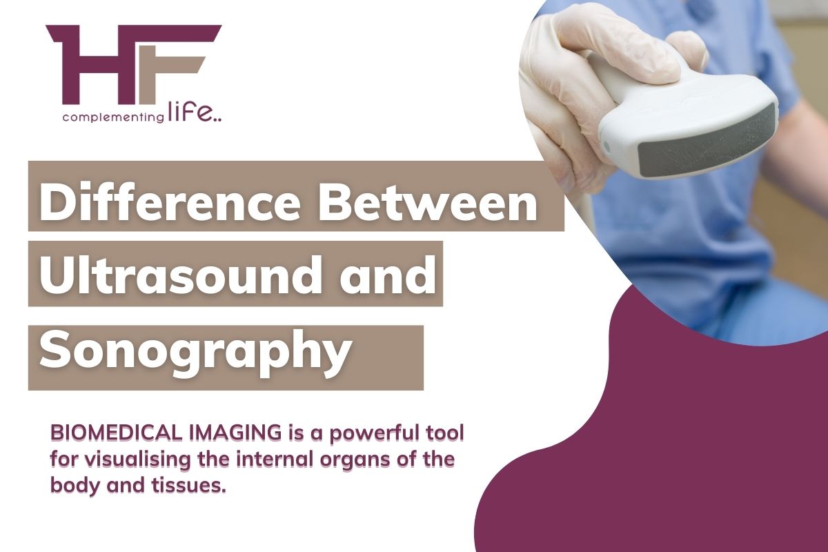
The process begins with a transducer, a handheld device that acts as both a transmitter and receiver of sound waves. This transducer is placed on the skin, typically with a gel medium to ensure good contact and eliminate air gaps, which can interfere with sound transmission. The transducer emits high-frequency sound waves, far beyond the range of human hearing (typically between 2 to 18 megahertz, or MHz). These sound waves travel into the body.
When these sound waves encounter different tissues and structures within the body, they are reflected back towards the transducer. The amount of reflection, or echo, depends on the acoustic properties of the tissue – its density, elasticity, and how it absorbs or transmits sound. For instance, denser tissues like bone reflect sound waves more strongly than softer tissues like muscle or fluid.
Transducer Design and Functionality: From Sound to Image
The transducer is a sophisticated piece of technology comprised of piezoelectric crystals. These crystals have the remarkable property of converting electrical energy into mechanical energy (sound waves) and vice versa. When an electrical current is applied, the crystals vibrate, producing the sound waves. Conversely, when the reflected sound waves (echoes) strike the crystals, they generate a small electrical charge.
The transducer is designed with an array of these piezoelectric crystals. By electronically controlling the timing and frequency of electrical pulses sent to these crystals, the system can steer and focus the sound beam in various directions. This allows for detailed imaging of different depths and angles within the body. The reflected echoes are received by the same crystals, which then convert them back into electrical signals.
Signal Processing and Image Reconstruction: Building the Visual Narrative
The electrical signals generated by the returning echoes are then sent to a sophisticated computer system. This system meticulously processes these signals, analyzing their timing, amplitude, and frequency. The timing of the echo provides information about the depth of the structure from which it originated – the longer it takes for an echo to return, the deeper the structure. The amplitude of the echo indicates the strength of the reflection, correlating with the density and composition of the tissue.
This raw data is then used to construct a real-time image on a monitor. The computer assigns different shades of gray to represent different echo intensities. Brighter areas (hyperechoic) typically represent denser tissues that reflect sound strongly, while darker areas (hypoechoic) represent tissues that absorb or transmit sound more readily, like fluids. This process of converting sound wave reflections into a visual representation is the essence of sonographic imaging.
The Terminology: Ultrasound vs. Sonogram Demystified
While the underlying technology is identical, the terms “ultrasound” and “sonogram” often carry slightly different connotations, primarily in their application and the stage of the process they describe. Understanding these distinctions can refine our technical understanding.
Ultrasound: The Technology and the Process
“Ultrasound” more broadly refers to the technology itself – the use of high-frequency sound waves for imaging or therapeutic purposes. When we speak of “ultrasound technology” or “ultrasound imaging,” we are referring to the entire system, including the transducer, the signal processing unit, and the physics of sound wave propagation. It encompasses the broader field of using sound waves above human hearing for various applications, including medical diagnostics.
In a medical context, “ultrasound” can also refer to the procedure of performing an ultrasound examination. For example, a patient might be told they are “having an ultrasound” of their abdomen. This emphasizes the action of using the technology.

Sonogram: The Resulting Image and its Creation
“Sonogram,” on the other hand, is more specifically used to refer to the image produced by the ultrasound process. The term is derived from “sono” (sound) and “gram” (writing or record), literally meaning “a record of sound.” Therefore, a sonogram is the visual output, the static or dynamic image created by the sonographic equipment.
When a radiologist or technician reviews the images of a patient’s organs, they are looking at a sonogram. The act of creating this image is often referred to as performing a sonography. So, while the technology is ultrasound, the product of that technology is a sonogram. It’s akin to the difference between a camera (the technology) and a photograph (the resulting image).
Interoperability and Common Usage: Bridging the Gap
Despite these subtle differences, the terms are frequently used interchangeably in clinical practice. Many healthcare professionals and patients will use “ultrasound” to refer to both the procedure and the resulting image. This is largely due to the convenience and familiarity of the term.
However, in technical discussions, particularly when describing the specific output or the process of image generation, using “sonogram” for the image itself and “ultrasound” for the technology or procedure can lend greater precision. This distinction is especially relevant in fields like medical engineering, biomedical research, and advanced medical imaging development, where a precise understanding of terminology is crucial for accurate communication and innovation.
Applications of Ultrasound Technology Beyond Diagnostic Imaging
While diagnostic imaging is the most widely recognized application of ultrasound technology, its capabilities extend into various other technological and therapeutic domains. Understanding these diverse uses highlights the versatility and ongoing innovation within this field.
Doppler Ultrasound: Visualizing Blood Flow Dynamics
One significant advancement within ultrasound technology is Doppler ultrasound. This specialized technique utilizes the Doppler effect, a phenomenon where the frequency of a sound wave changes when it is reflected off a moving object. In the context of medical imaging, this allows for the visualization and measurement of blood flow within the body.
The transducer emits sound waves, and when these waves encounter moving red blood cells, the frequency of the reflected waves is altered. The system detects and analyzes these frequency shifts, translating them into color-coded representations of blood flow direction and velocity. Arteries carrying blood away from the heart might be depicted in red, while veins carrying blood towards the heart could be shown in blue, for example. This technology is invaluable for diagnosing conditions such as blood clots, narrowed arteries, and heart valve defects.
Therapeutic Ultrasound: Harnessing Sound for Treatment
Beyond imaging, ultrasound technology also plays a crucial role in therapeutic applications. High-intensity focused ultrasound (HIFU) is a non-invasive treatment modality that uses concentrated beams of ultrasound energy to heat and destroy targeted tissues. This technology is employed in treating conditions like uterine fibroids, essential tremor, and certain types of cancer, where precise targeting and localized heating are beneficial.
The focused energy can cause thermal ablation, effectively destroying unwanted cells without damaging surrounding healthy tissues. Research is also ongoing into using ultrasound for drug delivery, where it can temporarily increase the permeability of cell membranes, allowing therapeutic agents to penetrate more effectively into target areas. This represents a significant area of innovation within medical technology.
Emerging Technologies and Future Directions
The field of ultrasound technology is continuously evolving, with researchers and engineers exploring new frontiers. Innovations include the development of more advanced transducers with higher frequencies for improved resolution, miniaturized ultrasound devices for point-of-care diagnostics, and the integration of artificial intelligence (AI) for automated image analysis and interpretation.
AI algorithms are being trained to identify subtle abnormalities in sonograms that might be missed by the human eye, leading to earlier and more accurate diagnoses. Furthermore, advancements in 3D and 4D ultrasound (which captures real-time movement) are providing increasingly comprehensive views of internal structures, revolutionizing fields like prenatal care and surgical planning. The continued exploration of acoustic wave properties promises to unlock even more groundbreaking applications in the future.
In conclusion, while the terms “ultrasound” and “sonogram” are closely related and often used interchangeably, they highlight different aspects of a singular, powerful technological innovation. “Ultrasound” encompasses the technology and the procedure, while “sonogram” specifically refers to the resulting visual record. Both terms are integral to understanding this vital diagnostic and therapeutic tool that continues to advance at a remarkable pace within the technological landscape.
aViewFromTheCave is a participant in the Amazon Services LLC Associates Program, an affiliate advertising program designed to provide a means for sites to earn advertising fees by advertising and linking to Amazon.com. Amazon, the Amazon logo, AmazonSupply, and the AmazonSupply logo are trademarks of Amazon.com, Inc. or its affiliates. As an Amazon Associate we earn affiliate commissions from qualifying purchases.