
The human heart, a marvel of biological engineering, functions as a tireless pump, circulating blood throughout the body to deliver oxygen and vital nutrients. This intricate process is orchestrated with remarkable precision, and at the core of its efficiency lie the heart valves. These remarkable structures, often overlooked in everyday discourse, are indispensable components of the cardiovascular system. Their primary purpose is deceptively simple yet profoundly crucial: to ensure the unidirectional flow of blood through the heart chambers and into the systemic and pulmonary circulations, preventing backflow and maintaining the pressure gradients necessary for effective pumping. Without properly functioning valves, the heart’s ability to pump blood efficiently would be severely compromised, leading to a cascade of serious health issues.
The Mechanics of Unidirectional Blood Flow
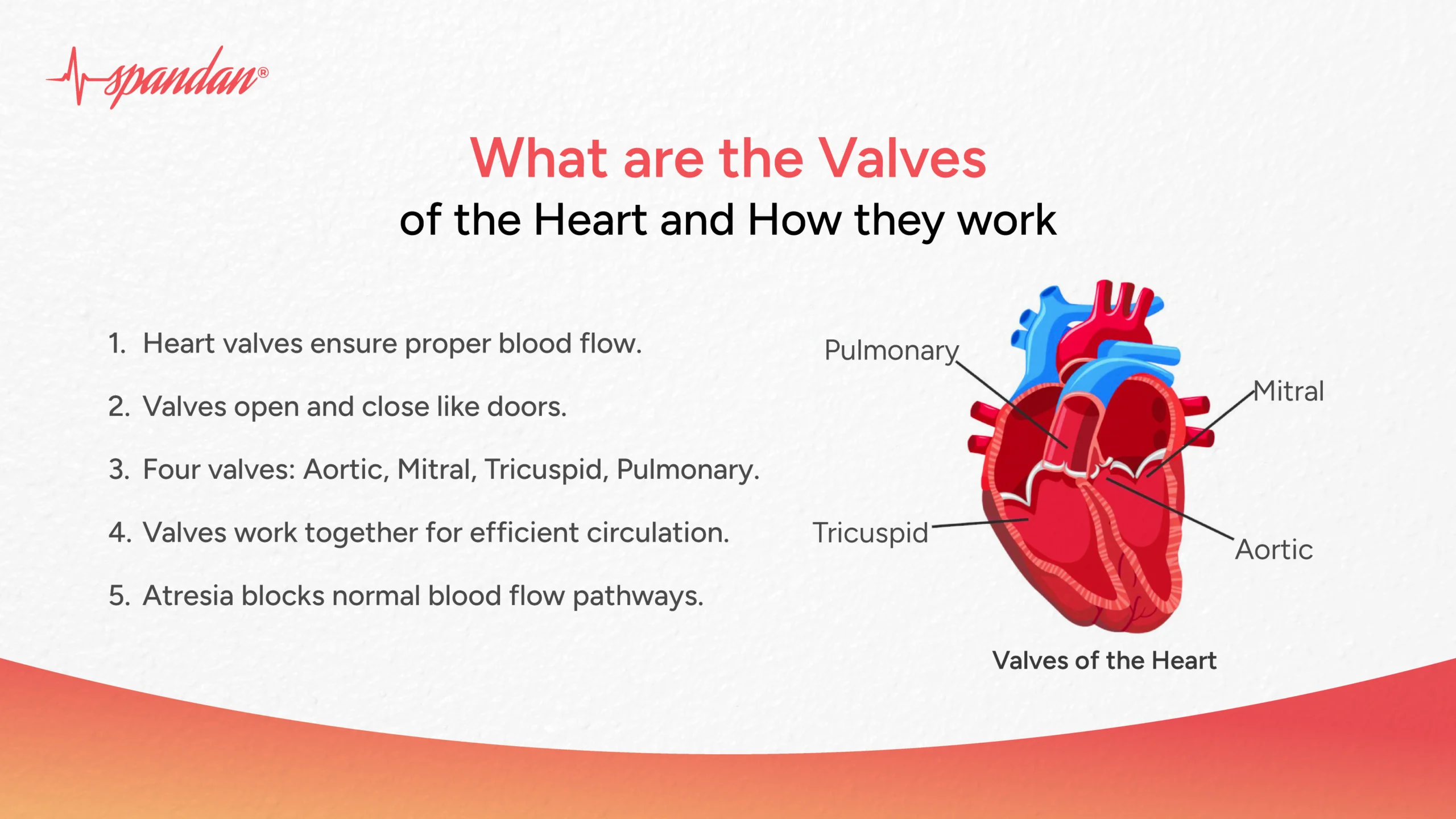
The heart is comprised of four chambers: the right atrium, the right ventricle, the left atrium, and the left ventricle. Blood enters the right atrium, then flows into the right ventricle. From the right ventricle, it is pumped to the lungs for oxygenation. Oxygenated blood then returns to the left atrium, moves into the left ventricle, and is finally pumped to the rest of the body. This sequential filling and emptying of chambers relies heavily on the coordinated opening and closing of the heart valves.
Mitral Valve: The Gateway to the Left Ventricle
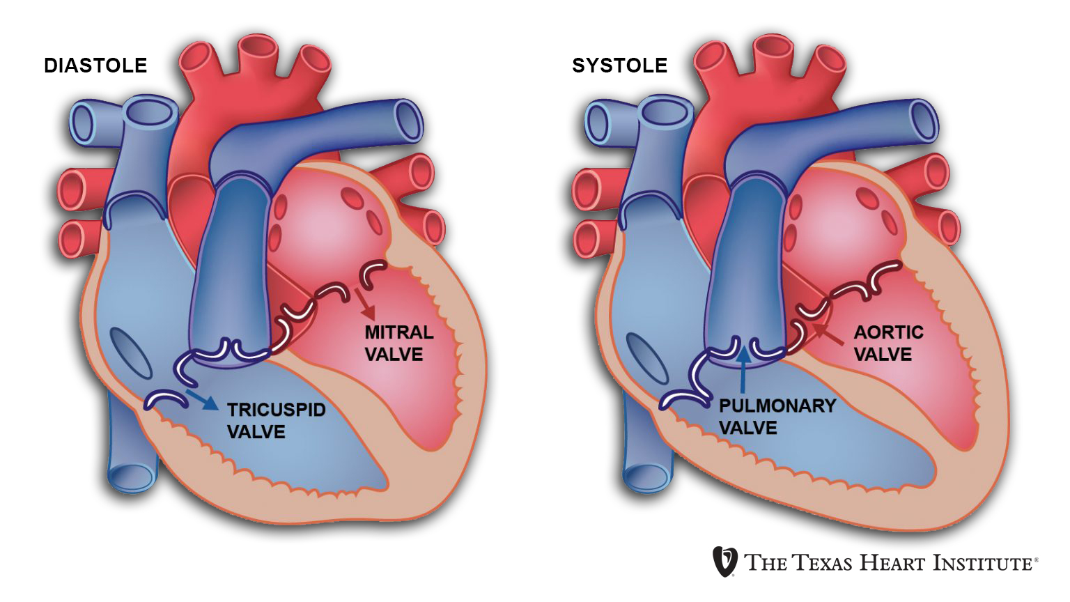
The mitral valve, also known as the bicuspid valve, is situated between the left atrium and the left ventricle. It consists of two leaflets, or cusps, that work in tandem to control blood flow. When the left atrium contracts, the mitral valve opens, allowing oxygenated blood to flow into the left ventricle. As the left ventricle contracts to pump blood out to the body, the mitral valve snaps shut, preventing any blood from regurgitating back into the left atrium. This closure is essential for generating the high pressure required for systemic circulation.
Tricuspid Valve: Regulating Flow into the Right Ventricle
On the right side of the heart, the tricuspid valve separates the right atrium from the right ventricle. Similar to the mitral valve, it possesses leaflets (in this case, three) that facilitate the flow of deoxygenated blood from the right atrium into the right ventricle. Upon contraction of the right ventricle, which pumps blood to the lungs, the tricuspid valve closes tightly to prevent backflow into the right atrium.
Aortic Valve: The Exit to the Body
The aortic valve is a crucial component that controls the flow of oxygenated blood from the left ventricle into the aorta, the body’s largest artery. This valve is a tri-leaflet structure, meaning it has three cusps. When the left ventricle contracts, the aortic valve opens, allowing a surge of blood to enter the aorta and be distributed throughout the body. Immediately after the ventricle relaxes, the aortic valve closes firmly to prevent blood from flowing back into the left ventricle. This closure is vital for maintaining the pressure in the aorta and ensuring a continuous supply of oxygenated blood to the tissues.
Pulmonary Valve: The Path to the Lungs
The pulmonary valve is located between the right ventricle and the pulmonary artery, which carries deoxygenated blood to the lungs. Like the aortic valve, it is a tri-leaflet valve. When the right ventricle contracts, the pulmonary valve opens, allowing blood to be pumped to the lungs for gas exchange. As the right ventricle relaxes, the pulmonary valve closes to prevent blood from flowing back into the ventricle.
The Role of Chordae Tendineae and Papillary Muscles
The proper functioning of the atrioventricular valves (mitral and tricuspid) is further supported by a sophisticated system of fibrous cords called chordae tendineae and muscular projections called papillary muscles. These structures are attached to the valve leaflets. When the ventricles contract, the papillary muscles also contract, pulling on the chordae tendineae. This action prevents the valve leaflets from prolapsing or bulging backward into the atria, which could lead to regurgitation. This intricate mechanism ensures that the valves remain sealed under the high pressures generated during ventricular contraction.
Chordae Tendineae: The Heartstrings
These strong, fibrous cords act like parachute cords, anchoring the valve leaflets to the papillary muscles. They are crucial in preventing the valves from being everted or blown back into the atria during ventricular systole (contraction). Their resilience and tensile strength are critical for withstanding the forces exerted during each heartbeat.
Papillary Muscles: The Controllers of Tension
The papillary muscles are conical muscular projections that extend from the inner walls of the ventricles. Their contraction shortens the chordae tendineae, thereby controlling the tension on the valve leaflets. This precise control is essential for maintaining the integrity of the valve seal and preventing leakage.
The Consequences of Valve Dysfunction
The intricate balance and precise timing of valve opening and closing are paramount. When heart valves malfunction, the consequences can range from mild to life-threatening. Valve dysfunction can occur due to various factors, including aging, infections, rheumatic fever, heart attacks, or congenital defects. The two primary types of valve dysfunction are stenosis and regurgitation.
Valve Stenosis: The Narrowed Passage
Stenosis refers to the narrowing or stiffening of a heart valve, which impedes the flow of blood. For example, aortic stenosis means the aortic valve is narrowed, making it difficult for the left ventricle to pump blood into the aorta. This forces the ventricle to work harder, which can lead to thickening of the heart muscle and eventual heart failure. Symptoms of stenosis can include chest pain, shortness of breath, fatigue, and dizziness.
Valve Regurgitation: The Leaky Seal
Regurgitation, also known as insufficiency or incompetence, occurs when a valve does not close properly, allowing blood to flow backward (leakage). Mitral regurgitation, for instance, means the mitral valve leaks, causing blood to flow back into the left atrium instead of being pumped forward to the body. This backflow increases the volume of blood the heart has to pump with each beat, placing an extra burden on the heart. Symptoms can be similar to stenosis, including shortness of breath, fatigue, and palpitations.
Impact on Cardiac Performance
Both stenosis and regurgitation disrupt the efficient pumping action of the heart. Stenosis creates an obstruction, reducing the amount of blood ejected with each beat and increasing the workload on the upstream chamber. Regurgitation leads to inefficient circulation, as a portion of the blood is pumped backward, requiring the heart to pump the same blood multiple times to achieve adequate forward flow. Over time, these inefficiencies can lead to chamber enlargement, arrhythmias (irregular heartbeats), and ultimately, heart failure.
Technological Advancements in Valve Repair and Replacement
The critical role of heart valves in maintaining cardiovascular health has spurred significant advancements in medical technology aimed at diagnosing, treating, and repairing or replacing faulty valves. From sophisticated imaging techniques to minimally invasive surgical procedures and cutting-edge artificial valve technologies, medicine has made remarkable strides in restoring cardiac function and improving the quality of life for millions of patients.
Diagnostic Tools: Seeing the Unseen
Diagnosing heart valve problems relies on a suite of advanced imaging and diagnostic tools. Echocardiography, often referred to as an “echo,” is the cornerstone of valve assessment. This non-invasive ultrasound technique provides real-time images of the heart’s structure and function, allowing physicians to visualize the valves, assess their movement, and detect any abnormalities like narrowing or leakage. Other imaging modalities, such as cardiac magnetic resonance imaging (MRI) and computed tomography (CT) scans, can offer more detailed anatomical views. Electrocardiograms (ECGs) and stress tests help evaluate the overall electrical activity and functional capacity of the heart, which can be affected by valve disease.
Minimally Invasive Interventions: Less is More
The evolution of surgical techniques has moved towards less invasive approaches, significantly reducing patient recovery times and risks. Transcatheter aortic valve implantation (TAVI) is a prime example. In this procedure, a new valve is delivered to the aortic position through a catheter inserted into an artery, typically in the leg. This method is often used for patients who are at high risk for traditional open-heart surgery. Similarly, transcatheter mitral valve repair techniques, such as MitraClip, involve implanting a small clip to bring the leaky leaflets of the mitral valve closer together, reducing regurgitation without open-heart surgery.
Artificial Valves: Engineered Replacements
When valve repair is not feasible, valve replacement becomes the treatment of choice. Modern artificial heart valves fall into two main categories: mechanical valves and bioprosthetic (tissue) valves. Mechanical valves are made of durable materials like pyrolytic carbon and are designed to last a lifetime. However, they require patients to take lifelong anticoagulant medication (blood thinners) to prevent blood clots from forming on the valve. Bioprosthetic valves are made from animal tissue (porcine or bovine) or human donor tissue. They generally do not require lifelong anticoagulation, but they may degrade over time and require replacement after 10-20 years. The choice between mechanical and bioprosthetic valves depends on a patient’s age, lifestyle, other medical conditions, and personal preferences.
Future Frontiers: Regenerative Medicine and Beyond
The future of heart valve treatment holds immense promise, with ongoing research focusing on regenerative medicine and novel biomaterials. Scientists are exploring the possibility of growing new heart valves from a patient’s own cells, which would eliminate the risk of rejection and the need for lifelong medication. Advanced tissue engineering techniques and the development of “smart” valves that can adapt to the body’s needs are also on the horizon, aiming to further refine the treatment of heart valve disease and improve long-term outcomes for patients. The continuous innovation in this field underscores the profound importance of these often-unseen cardiac structures.
aViewFromTheCave is a participant in the Amazon Services LLC Associates Program, an affiliate advertising program designed to provide a means for sites to earn advertising fees by advertising and linking to Amazon.com. Amazon, the Amazon logo, AmazonSupply, and the AmazonSupply logo are trademarks of Amazon.com, Inc. or its affiliates. As an Amazon Associate we earn affiliate commissions from qualifying purchases.