
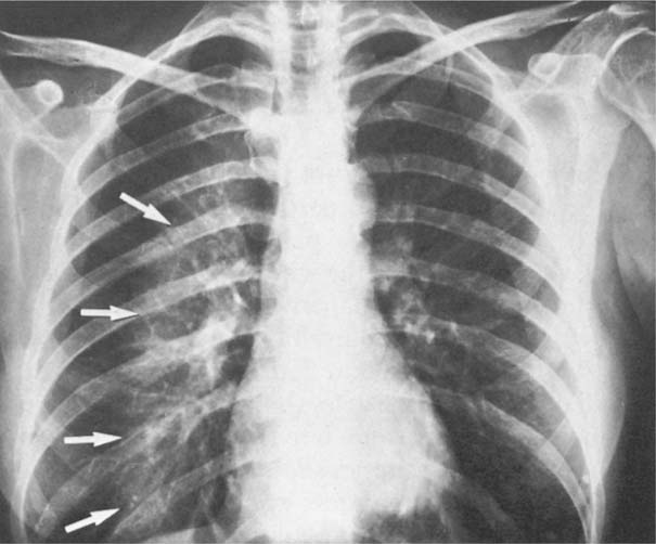
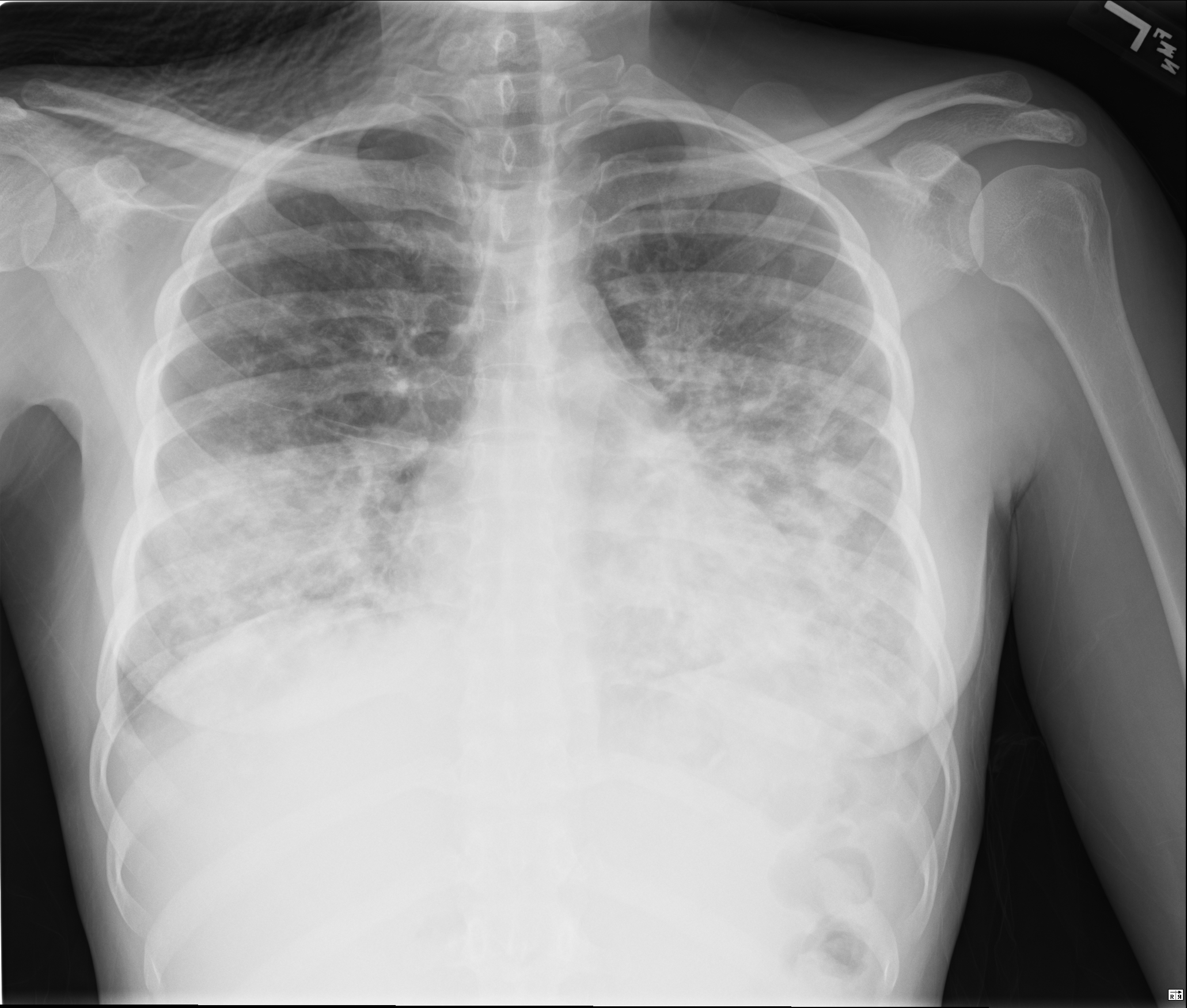
In the rapidly evolving landscape of medical technology, the term “pulmonary infiltrate” has shifted from a purely clinical observation to a complex data point processed by sophisticated algorithms. Traditionally, a pulmonary infiltrate refers to a substance—such as pus, blood, protein, or edema—that has filled the lungs’ air spaces or parenchyma, appearing as a density on a chest radiograph or CT scan. However, for technology professionals, software developers, and data scientists, “pulmonary infiltrate” represents one of the most critical challenges in computer-aided detection (CAD) and medical imaging analytics.
Understanding what a pulmonary infiltrate is within the tech ecosystem requires an exploration of how digital imaging has moved beyond simple visualization toward predictive modeling and automated diagnostic support. As healthcare systems digitize their workflows, the identification of these densities has become a cornerstone of the burgeoning HealthTech sector, driving innovations in artificial intelligence, cloud computing, and cybersecurity.

Decoding Pulmonary Infiltrate through Advanced Diagnostic Algorithms
The identification of pulmonary infiltrates is a primary objective for diagnostic imaging software. In the digital age, a chest X-ray is no longer just a piece of film; it is a matrix of pixels, each containing specific metadata and grayscale values that represent tissue density.
The Shift from Analog to Digital Radiography
The transition from analog film to Digital Radiography (DR) and Computed Radiography (CR) laid the groundwork for modern pulmonary analysis. Digital sensors capture the attenuation of X-ray beams as they pass through the thoracic cavity. When the software detects an area where the beam is significantly weakened—meaning the area is denser than the surrounding air-filled lung tissue—it registers a “density.” In tech terms, identifying an infiltrate involves processing these signals to differentiate between normal anatomical structures, such as blood vessels or ribs, and pathological shadows.
How Computer-Aided Detection (CAD) Identifies Densities
Modern CAD systems utilize sophisticated image processing techniques to flag potential infiltrates for radiologists. These systems employ multi-layered filters to reduce noise and enhance contrast. By applying thresholding algorithms, the software can segment the lung fields from the rest of the image, allowing for a focused analysis of the parenchyma. When an infiltrate is detected, the software highlights the region of interest (ROI), providing a quantitative assessment of the density’s size, shape, and opacity. This level of automated precision is essential in high-volume clinical environments where rapid turnaround is vital for patient outcomes.
The Role of Machine Learning in Radiological Interpretation
While traditional CAD systems relied on pre-defined rules, the modern approach to pulmonary infiltrates is dominated by Machine Learning (ML) and Deep Learning (DL). The tech industry has invested heavily in training neural networks to recognize the subtle patterns of pulmonary pathologies with a degree of accuracy that often rivals human experts.
Neural Networks and Pattern Recognition
Convolutional Neural Networks (CNNs) have become the gold standard for analyzing medical images. To understand a pulmonary infiltrate, a CNN is trained on massive datasets—such as the NIH ChestX-ray14 dataset—containing hundreds of thousands of labeled images. During the training phase, the model learns to identify “features” associated with infiltrates. These features might include ill-defined margins, “air bronchograms” (dark, air-filled bronchi outlined by white, dense infiltrates), or patchy consolidation.
The technical complexity lies in the model’s ability to generalize. A pulmonary infiltrate caused by pneumonia may look different from one caused by pulmonary edema or a hemorrhage. Advanced deep learning architectures, such as ResNet or DenseNet, allow the software to process these nuances through dozens of hidden layers, progressively refining its understanding of what constitutes a “positive” finding.
Reducing False Positives in Infiltrate Detection
One of the greatest hurdles in medical software development is the reduction of false positives. In the context of pulmonary infiltrates, common artifacts—such as patient movement, external leads, or overlapping breast tissue—can be misinterpreted by less sophisticated algorithms as an infiltrate.
To combat this, developers are implementing ensemble learning techniques, where multiple models analyze the same image and their outputs are combined to reach a final consensus. Furthermore, the integration of “Attention Mechanisms” allows the AI to focus on specific regions of the lung that are more likely to contain pathology, effectively ignoring the “noise” of the chest wall and skeletal structure. This leads to higher specificity, ensuring that tech-driven diagnostics are both reliable and actionable.
Emerging Tech Trends in Pulmonary Health Monitoring
The analysis of pulmonary infiltrates is moving out of the radiology suite and into the hands of providers through mobile and decentralized technologies. This democratization of diagnostic power is fueled by the miniaturization of hardware and the expansion of edge computing.
Edge Computing and Handheld Ultrasound Devices
Point-of-Care Ultrasound (POCUS) is a burgeoning field where AI-powered handheld devices are used to detect pulmonary infiltrates at the bedside. Unlike traditional X-rays, ultrasound uses sound waves to visualize the pleural line and the underlying lung tissue. Software integrated into these handheld devices uses edge computing to process images in real-time, identifying “B-lines”—vertical artifacts that indicate interstitial infiltrates.
By processing this data on the device itself rather than in the cloud, medical professionals can receive instantaneous feedback. This is a significant tech milestone, particularly in emergency medicine and rural healthcare, where access to large-scale imaging infrastructure may be limited.
Cloud-Based Image Sharing and Collaborative Diagnostics
The rise of Picture Archiving and Communication Systems (PACS) hosted in the cloud has revolutionized how pulmonary infiltrates are managed. Modern PACS solutions allow for the seamless transfer of high-resolution DICOM (Digital Imaging and Communications in Medicine) files across global networks.
This connectivity enables “Teleradiology,” where a specialist in one time zone can interpret an image containing a pulmonary infiltrate for a clinic in another. From a software perspective, this requires robust APIs, high-speed data compression algorithms that preserve diagnostic quality (lossless compression), and scalable storage solutions. The tech stack supporting these systems must be capable of handling petabytes of data while ensuring that the latency between image upload and clinical review is minimized.
Digital Security and Data Integrity in Pulmonary Diagnostics
As the detection of pulmonary infiltrates becomes increasingly digitized, the security of the associated data becomes a paramount concern. Medical images are highly sensitive pieces of Personal Health Information (PHI), making them prime targets for cyberattacks.
Protecting DICOM Files and Patient Privacy
The DICOM standard is the backbone of medical imaging, but it was not originally designed with modern cybersecurity threats in mind. Tech firms are now developing layers of encryption and obfuscation to protect images of pulmonary infiltrates as they move through the digital pipeline.
End-to-end encryption ensures that even if a data packet is intercepted during transmission to the cloud, the underlying image cannot be reconstructed. Furthermore, de-identification algorithms are used in research settings to strip PHI from images before they are used to train AI models. This process is complex, as it requires the software to scrub text burned into the image pixels as well as the metadata stored in the file headers.
The Future of Blockchain in Health Records
Blockchain technology is being explored as a method to ensure the integrity of pulmonary diagnostic reports. By creating a decentralized, immutable ledger of every time an image was viewed, edited, or interpreted, healthcare providers can create a transparent “audit trail.”
In the event of a diagnostic dispute—such as a missed pulmonary infiltrate—a blockchain-based system provides a verifiable history of the data’s lifecycle. This application of Distributed Ledger Technology (DLT) addresses the “trust” factor in digital medicine, ensuring that the data used to make life-saving decisions has not been tampered with or corrupted.
The Intersection of Virtual Reality and Pulmonary Mapping
The final frontier in the tech-driven understanding of pulmonary infiltrates is the transition from 2D slices to 3D immersive environments. Virtual Reality (VR) and Augmented Reality (AR) are being used to map the exact coordinates of an infiltrate within the bronchial tree.
3D Volumetric Rendering
Using data from high-resolution CT scans, software can now perform volumetric rendering to create a 3D model of a patient’s lungs. This allows surgeons and pulmonologists to visualize an infiltrate not as a flat shadow, but as a physical mass with a specific volume and proximity to major vessels. The tech required for this—high-end GPUs and sophisticated spatial mapping software—is borrowed from the gaming and aerospace industries, demonstrating the cross-disciplinary nature of modern medical technology.
Surgical Navigation and AR Overlays
In procedures like bronchoscopy, AR overlays can guide a robotic probe directly to the site of an infiltrate for a biopsy. By projecting the 3D map onto the physician’s field of view, the software compensates for the lung’s movement during respiration. This real-time “active tracking” is a pinnacle of modern software engineering, combining computer vision, sensor fusion, and low-latency rendering.
As we look forward, the question of “what is a pulmonary infiltrate” will continue to be answered by the code we write and the systems we build. In the hands of tech innovators, it is a challenge to be solved, a pattern to be recognized, and a data point that—when managed correctly—can save countless lives through the power of digital transformation.
aViewFromTheCave is a participant in the Amazon Services LLC Associates Program, an affiliate advertising program designed to provide a means for sites to earn advertising fees by advertising and linking to Amazon.com. Amazon, the Amazon logo, AmazonSupply, and the AmazonSupply logo are trademarks of Amazon.com, Inc. or its affiliates. As an Amazon Associate we earn affiliate commissions from qualifying purchases.