
In the rapidly evolving landscape of medical technology, the term “microadenoma” represents more than just a clinical diagnosis; it serves as a critical benchmark for the capabilities of modern diagnostic hardware and software. By definition, a microadenoma is a noncancerous tumor of the pituitary gland that measures less than 10 millimeters in diameter. While their physical size is minute, the technological requirements to identify, monitor, and treat these lesions are immense. For the tech-driven healthcare sector, the microadenoma is a catalyst for innovation in high-resolution imaging, artificial intelligence (AI) pattern recognition, and robotic-assisted neurosurgery.
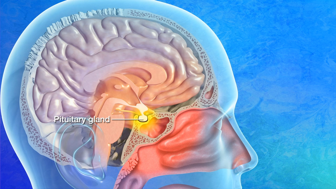
To understand what a microadenoma is from a technological perspective is to understand the limits of current resolution thresholds and the digital tools being developed to push past them. As we shift toward a more data-centric model of care, the management of these small lesions highlights the growing synergy between bio-engineering and digital signal processing.
The Technological Evolution of Pituitary Imaging
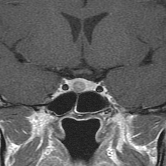
The primary challenge in addressing microadenomas lies in their size. Because they are often only a few millimeters wide, they exist at the very edge of what traditional imaging can reliably capture. The evolution of Magnetic Resonance Imaging (MRI) has been the cornerstone of microadenoma detection, moving from primitive visualization to high-fidelity digital modeling.
High-Field MRI and Resolution Thresholds
The transition from 1.5 Tesla (T) to 3T MRI machines represented a quantum leap in the ability to visualize the pituitary gland. In the context of microadenomas, “Tesla” refers to the strength of the magnetic field. Higher field strengths equate to a higher signal-to-noise ratio, which allows for thinner “slices” during the scanning process.
Current state-of-the-art 7T MRI systems are now entering clinical research phases, offering even greater spatial resolution. These machines allow technologists to visualize the microvasculature of the pituitary, making it easier to distinguish between healthy glandular tissue and a microadenoma. The tech involves complex radiofrequency (RF) pulse sequences designed to highlight the delayed contrast uptake characteristic of these tumors. From a software perspective, this requires sophisticated reconstruction algorithms that can handle massive datasets without introducing motion artifacts or digital noise.
Software-Defined Contrast Enhancement
Hardware is only half of the equation. Software-defined imaging protocols, such as Dynamic Contrast-Enhanced (DCE) MRI, have become the gold standard for microadenoma identification. In this process, a series of rapid images are taken after the injection of a contrast agent.
The technology relies on the fact that microadenomas typically enhance more slowly than the surrounding pituitary tissue. Advanced post-processing software maps these enhancement patterns over time, creating a “time-intensity curve.” Developers are currently working on automated subtraction software that can digitally remove the background signal of the normal gland, leaving only the “footprint” of the microadenoma visible to the technician. This digital subtraction is a feat of computational mathematics, requiring precise alignment of image frames to ensure that even a sub-millimeter shift in the patient’s position doesn’t invalidate the data.
AI and Machine Learning in Microadenoma Detection
As the volume of imaging data grows, the role of the human radiologist is being augmented by Artificial Intelligence. The detection of a microadenoma is a classic “needle in a haystack” problem, making it an ideal use case for machine learning (ML) and computer vision.
Neural Networks and Pattern Recognition
Deep learning models, specifically Convolutional Neural Networks (CNNs), are being trained on thousands of labeled pituitary scans to recognize the subtle textural differences that indicate a microadenoma. Unlike traditional algorithmic programming, where a developer tells the computer exactly what to look for, these neural networks learn to identify features that may be invisible to the human eye.
In a tech-driven clinical environment, AI acts as a “second pair of eyes.” When a patient undergoes an MRI, the AI software scans the raw data in real-time, flagging regions of interest for the radiologist. This reduces the “false negative” rate, particularly in cases where the microadenoma is isointense—meaning its signal strength is nearly identical to the surrounding tissue. The integration of AI into Picture Archiving and Communication Systems (PACS) represents a major step forward in diagnostic workflow automation.
Reducing Radiologist Fatigue with Automated Flagging
One of the most significant trends in MedTech is the drive toward efficiency. Radiologists are often tasked with reviewing hundreds of images per hour. Software tools that can automatically segment the pituitary gland and measure the dimensions of a suspected microadenoma save valuable time.
Current AI tools are moving toward predictive analytics—using the volumetric data of a microadenoma to predict its growth trajectory or its likelihood of responding to specific pharmacological interventions. By analyzing the “Radiomics” of the tumor—the high-dimensional data features extracted from medical images—technology can provide a far more nuanced profile of the lesion than a simple measurement ever could.
Robotic Precision and Minimally Invasive Surgical Tech
When a microadenoma requires intervention, the technological focus shifts from diagnostics to precision hardware. The pituitary gland is located at the base of the brain, a high-stakes environment where a fraction of a millimeter can determine the success of a procedure.
Endoscopic Navigation Systems
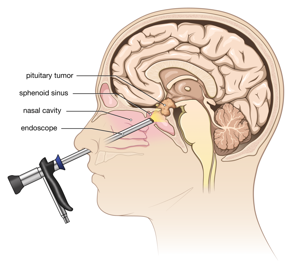
The “Gold Standard” for microadenoma removal is the transsphenoidal approach, which is now heavily reliant on high-definition endoscopes. These devices are marvels of optical engineering, utilizing 4K chip-on-tip technology to provide surgeons with an internal view of the nasal cavity and the sella turcica (the bony pocket housing the pituitary).
The real innovation, however, is the integration of these endoscopes with “Surgical Navigation Systems.” This tech functions much like a GPS for the human body. By uploading the patient’s pre-operative MRI data into the navigation software, the surgeon can see the position of their instruments overlaid on the 3D digital model of the patient’s skull in real-time. This “augmented reality” interface allows for the precise localization of the microadenoma, minimizing trauma to the surrounding optic nerves and carotid arteries.
Real-Time Intraoperative Imaging
The cutting edge of neurosurgical tech involves bringing the imaging to the operating room. Intraoperative MRI (iMRI) allows surgeons to take a scan while the patient is still on the table. This is crucial for microadenomas because, as tissue is removed and pressure is released, the anatomy of the brain shifts slightly—a phenomenon known as “brain shift.”
Software must dynamically update the navigation map to account for these changes. New robotic arms, such as those used in “Digital Exoscopes,” provide stabilized, high-magnification views that can be controlled via foot pedals or hand-trackers, allowing the surgeon to maintain focus on the delicate task of separating the microadenoma from the healthy gland.
The Future of Digital Health and Endocrine Monitoring
The management of a microadenoma doesn’t end with a scan or a surgery; it requires lifelong monitoring of hormonal levels. The future of this field lies in the “Internet of Medical Things” (IoMT) and the digitization of endocrinology.
Wearable Integration and Hormonal Tracking
While we are not yet at the stage where a consumer smartwatch can detect a microadenoma, we are seeing the rise of wearable biosensors that monitor the physiological effects of hormonal imbalances. For example, microadenomas that secrete growth hormone or cortisol can affect heart rate variability (HRV), sleep patterns, and glucose levels.
The next generation of health tech aims to close the loop between these digital markers and clinical diagnostics. We are seeing the development of “digital twins”—virtual models of a patient’s endocrine system that use real-time data from wearables to simulate how a microadenoma might be affecting their systemic health. This data-driven approach allows for personalized medicine, where treatment dosages are adjusted based on a continuous stream of digital feedback rather than a once-a-year blood test.
Big Data and Predictive Analytics in Neurosurgery
As more healthcare providers move their records to the cloud, the opportunity for large-scale data analysis grows. By aggregating anonymized data from thousands of microadenoma patients, researchers can use big data analytics to identify trends that were previously hidden.
Technology is enabling a shift from “reactive” to “proactive” care. Predictive algorithms can now assess the risk of recurrence based on a combination of genetic markers, imaging features, and post-operative hormone levels. For the tech industry, the microadenoma is a microcosm of the broader challenges in healthcare: the need for higher resolution, faster processing, and smarter data interpretation.
In conclusion, a microadenoma is a small clinical entity that necessitates a massive technological infrastructure. From the superconducting magnets of a 3T MRI to the neural networks of diagnostic AI and the sub-millimeter precision of robotic navigation, the story of the microadenoma is the story of modern MedTech. As these technologies continue to converge, the goal remains clear: to turn the “invisible” into the “visible” and to transform complex data into life-saving insights.
aViewFromTheCave is a participant in the Amazon Services LLC Associates Program, an affiliate advertising program designed to provide a means for sites to earn advertising fees by advertising and linking to Amazon.com. Amazon, the Amazon logo, AmazonSupply, and the AmazonSupply logo are trademarks of Amazon.com, Inc. or its affiliates. As an Amazon Associate we earn affiliate commissions from qualifying purchases.