The human body, a marvel of biological engineering, continuously presents complex challenges and opportunities for technological innovation. Among its intricate structures, the mastoid stands out as a critical anatomical region that has seen remarkable advancements in diagnostic, imaging, and surgical technologies. Far from being a mere bony protuberance behind the ear, understanding “what is mastoid” in the 21st century inevitably leads to an exploration of how cutting-edge technology enhances our ability to perceive, analyze, and interact with this vital area.
The Mastoid Process: A Foundation for Technological Insight
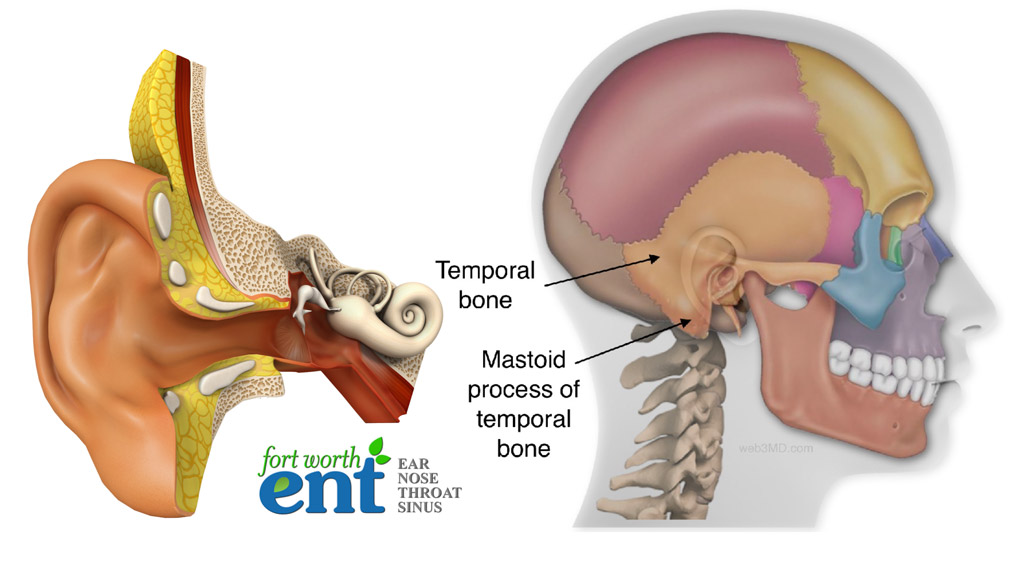
The mastoid process is a conical projection of the temporal bone, located just behind the ear. While its primary biological functions include serving as an attachment point for several muscles of the neck and playing a role in hearing and balance through its internal air cells, its anatomical complexity makes it a significant area of interest for medical technology. Internally, the mastoid contains numerous air cells (mastoid air cells) that connect to the middle ear cavity, making it susceptible to infections and other pathologies that can impact hearing and balance. This vulnerability necessitates precise diagnostic and therapeutic interventions, areas where technology has become indispensable.

The journey from basic anatomical understanding to sophisticated clinical application has been paved by a steady stream of technological innovations. From early X-rays to advanced computed tomography (CT) and magnetic resonance imaging (MRI), technology has continuously refined our ability to visualize the mastoid, moving from macroscopic views to microscopic detail, all while minimizing invasiveness and improving diagnostic accuracy. This evolution underscores the symbiotic relationship between anatomical knowledge and technological capability, where each advances the other.
Advanced Imaging: Unveiling the Mastoid’s Secrets
Diagnostic imaging forms the bedrock of modern understanding and treatment of mastoid-related conditions. The sheer complexity and variability of the mastoid air cell system, coupled with its proximity to critical structures like the facial nerve, brain, and major blood vessels, demand high-resolution, multi-planar imaging. Technological breakthroughs in this domain have transformed the diagnostic landscape.
Computed Tomography (CT) Scans
CT remains the gold standard for evaluating the bony anatomy of the mastoid. Modern CT scanners offer unprecedented detail, allowing clinicians to visualize the mastoid air cells, septa, and any erosions or opacifications indicative of disease (such as mastoiditis, cholesteatoma, or tumors).
- High-Resolution Capabilities: Contemporary multi-detector CT (MDCT) scanners acquire thin slices, often less than 1mm, enabling reconstruction in multiple planes (axial, coronal, sagittal, and oblique) without re-scanning the patient. This volumetric data provides a comprehensive 3D understanding of the mastoid’s intricate architecture.
- Reduced Dose Techniques: Advances in CT technology, including iterative reconstruction algorithms and automatic exposure control, have significantly reduced radiation doses while maintaining diagnostic image quality. This is crucial, especially for pediatric patients or those requiring serial scans.
- 3D Reconstruction and Virtual Endoscopy: Sophisticated software allows for 3D volumetric rendering of CT data, providing a surgeon’s-eye view of the mastoid and surrounding structures. Virtual endoscopy, though less commonly applied to the mastoid directly, offers a simulated internal view, aiding in pre-surgical planning and patient education.
Magnetic Resonance Imaging (MRI)
While CT excels in bony detail, MRI provides superior soft-tissue contrast, making it invaluable for assessing inflammatory processes, fluid collections, and soft-tissue masses within and around the mastoid.
- Detection of Inflammation: MRI is highly sensitive to fluid and inflammation, making it excellent for identifying mastoiditis, abscesses, or granulation tissue that might not be clearly delineated on CT. Specific sequences, such as diffusion-weighted imaging (DWI), can help differentiate cholesteatoma from other fluid collections.
- Evaluation of Intracranial Complications: Given the mastoid’s proximity to the brain, MRI is crucial for detecting potential intracranial complications of mastoid infection, such as meningitis, epidural abscess, or brain abscess, by visualizing the spread of inflammation beyond the bone.
- Functional MRI (fMRI) and Spectroscopy: While less commonly applied directly to the mastoid, advancements in fMRI and MR spectroscopy offer potential future avenues for understanding metabolic changes or neurological impacts related to chronic mastoid conditions, particularly in research settings.
AI and Machine Learning in Mastoid Diagnostics
The sheer volume and complexity of imaging data generated by CT and MRI scans present both a challenge and an opportunity for artificial intelligence (AI) and machine learning (ML). AI-powered tools are beginning to revolutionize how these images are interpreted, leading to faster, more accurate diagnoses and aiding in treatment planning.
Automated Anomaly Detection
ML algorithms, trained on vast datasets of annotated mastoid scans (both healthy and pathological), can identify subtle patterns and anomalies that might be missed by the human eye.
- Early Detection of Mastoiditis: AI can analyze changes in air cell opacification, bone erosion, and fluid accumulation, potentially flagging early signs of mastoiditis or other infections with high sensitivity, leading to earlier intervention.
- Cholesteatoma Identification: Differentiating cholesteatoma (a destructive growth) from other middle ear pathologies is critical. AI models are being developed to assist in this challenging task, leveraging characteristic imaging features across multiple sequences.
- Quantification of Disease Burden: AI can provide quantitative measurements of disease extent, such as the volume of inflamed tissue or the degree of air cell involvement, offering objective metrics for monitoring disease progression and treatment response.
Predictive Analytics and Personalized Treatment
Beyond diagnostics, AI can integrate clinical data, patient history, and imaging findings to predict disease course and optimize treatment strategies.
- Risk Stratification: ML models can identify patients at higher risk for complications based on their mastoid imaging characteristics and other clinical factors, allowing for more aggressive monitoring or prophylactic measures.
- Treatment Response Prediction: By analyzing baseline imaging and subsequent follow-up scans, AI could potentially predict which patients are likely to respond to conservative management versus those who will require surgical intervention, thereby personalizing care pathways.
Surgical Technologies: Precision and Minimally Invasive Approaches
When surgical intervention is necessary for mastoid conditions, technology plays a pivotal role in enhancing precision, reducing invasiveness, and improving patient outcomes. From advanced drills to augmented reality, the operating room for mastoidectomy and related procedures is increasingly a hub of high-tech solutions.
Computer-Assisted Surgery (CAS) and Image-Guided Navigation
CAS systems utilize pre-operative CT or MRI scans to create a 3D model of the patient’s mastoid anatomy. During surgery, tracking systems (optical or electromagnetic) correlate the surgeon’s instruments with this 3D model, providing real-time guidance on a monitor.
- Enhanced Precision and Safety: Surgeons can visualize their instrument’s exact position relative to critical structures (facial nerve, sigmoid sinus, inner ear) even when these are obscured by bone or tissue. This significantly reduces the risk of iatrogenic injury.
- Complex Anatomy Navigation: For cases with distorted or unusual mastoid anatomy, perhaps due to previous surgery or extensive disease, navigation systems provide an invaluable roadmap.
- Minimally Invasive Approaches: By increasing precision, CAS facilitates less invasive surgical approaches, potentially leading to smaller incisions, reduced blood loss, and faster recovery times.
Robotic Assistance and Augmented Reality (AR)
While full robotic autonomy in mastoid surgery is still nascent, robotic assistance and AR are making inroads, promising even greater precision and new capabilities.
- Robotic-Assisted Drills: Research is exploring robotic systems that can assist with bone removal, offering micro-level precision that exceeds human hand steadiness, potentially for critical areas near the facial nerve.
- Augmented Reality Overlays: AR systems project pre-operative imaging data directly onto the patient’s anatomy in the surgeon’s field of view, blending the virtual 3D model with the real-world surgical site. This provides an intuitive, hands-free form of guidance, allowing surgeons to “see through” bone and visualize hidden structures.
- Training and Simulation: AR and virtual reality (VR) are transforming surgical training, allowing residents to practice complex mastoidectomies in a realistic, risk-free environment, improving skills before operating on live patients.
Telemedicine and Remote Monitoring for Mastoid Health
The shift towards digital health has extended the reach of mastoid care beyond the traditional clinic walls. Telemedicine and remote monitoring technologies are improving access, facilitating follow-up, and empowering patients.
Virtual Consultations and Diagnostics
Patients with suspected mastoid issues can undergo initial consultations via video calls, reducing the need for travel.
- Remote Image Review: Radiologists and specialists can remotely review high-resolution CT and MRI scans of the mastoid, offering expert opinions regardless of geographical location. This is particularly beneficial for patients in rural areas or those seeking second opinions.
- Post-Operative Monitoring: Telemedicine can facilitate virtual follow-up appointments after mastoid surgery, allowing clinicians to assess recovery, manage pain, and address concerns without an in-person visit, provided there are no acute complications requiring physical examination.

Wearable Technology and Remote Sensing
While direct mastoid monitoring via wearables is still in early stages, the broader trend in health tech suggests future possibilities.
- Hearing Health Integration: As the mastoid is intimately linked to hearing, future wearable devices focused on auditory health might integrate monitoring for early signs of mastoid-related hearing changes.
- AI-Powered Symptom Checkers: Digital health platforms using AI can help patients understand potential symptoms related to mastoid conditions, guiding them on when to seek professional medical attention.
The question “what is mastoid” today extends far beyond its anatomical definition. It encompasses a dynamic interplay with technology, driving continuous advancements in how we diagnose, treat, and manage conditions affecting this vital part of the temporal bone. From advanced imaging and AI-driven diagnostics to surgical robotics and telemedicine, technology is not merely an adjunct but an integral component in safeguarding mastoid health, promising a future of even greater precision, personalization, and patient empowerment.
aViewFromTheCave is a participant in the Amazon Services LLC Associates Program, an affiliate advertising program designed to provide a means for sites to earn advertising fees by advertising and linking to Amazon.com. Amazon, the Amazon logo, AmazonSupply, and the AmazonSupply logo are trademarks of Amazon.com, Inc. or its affiliates. As an Amazon Associate we earn affiliate commissions from qualifying purchases.