The term “hilar adenopathy” might sound like a purely medical diagnostic term, but within the realm of technology, its significance extends far beyond the confines of a hospital. This article will explore hilar adenopathy not as a disease, but as a diagnostic indicator that increasingly relies on and drives technological innovation. We will delve into how advanced imaging technologies, AI-powered analysis, and sophisticated data management systems are revolutionizing the detection, characterization, and ultimately, the understanding of hilar adenopathy. This exploration will highlight the critical intersection of medical imaging, artificial intelligence, and computational biology, demonstrating how technology is fundamentally reshaping our approach to this significant clinical finding.
The Technological Backbone of Hilar Adenopathy Detection
The ability to accurately identify and characterize hilar adenopathy hinges on cutting-edge imaging modalities and their sophisticated processing. These technologies are not merely tools for visualization; they are increasingly intelligent systems that can extract subtle nuances imperceptible to the human eye.
Advanced Imaging Modalities: Beyond the Static Image
The primary method for detecting hilar adenopathy involves imaging the chest. However, the evolution of these imaging techniques has been a testament to technological progress.
Computed Tomography (CT) Scans: A Revolution in Detail
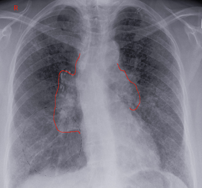
CT scanning has long been the cornerstone for visualizing the hilar region, the area where the bronchi, blood vessels, and nerves enter and exit the lungs. Modern CT scanners boast higher resolution, faster scan times, and lower radiation doses, thanks to advancements in detector technology and reconstruction algorithms. Dual-energy CT (DECT) offers an additional layer of information by acquiring images at two different energy levels, allowing for material decomposition and the differentiation of tissues and contrast agents. This can be particularly useful in distinguishing benign from malignant enlarged lymph nodes, a key aspect of understanding hilar adenopathy. Volumetric CT imaging also enables the creation of detailed 3D reconstructions, offering a comprehensive spatial understanding of the hilar structures and any associated adenopathy.
Magnetic Resonance Imaging (MRI): Unveiling Soft Tissue Nuances
While CT is excellent for visualizing calcifications and bony structures, MRI excels at delineating soft tissues, making it a valuable adjunct in evaluating hilar adenopathy, especially when differentiating between types of enlarged lymph nodes or assessing surrounding structures. Advanced MRI sequences, such as diffusion-weighted imaging (DWI), can provide information about the cellularity of the lymph nodes, which can be indicative of malignancy. Contrast-enhanced MRI further refines visualization, highlighting vascularity patterns that can aid in diagnosis. The increasing speed and improved signal-to-noise ratio of modern MRI scanners have made them more practical for thoracic imaging.
Positron Emission Tomography (PET) Scans: Functional Insights and Metabolic Activity
PET scans, often combined with CT (PET/CT), provide functional and metabolic information. The most common radiotracer used is Fluorodeoxyglucose (FDG), which is taken up by metabolically active cells, including cancerous ones. Enlarged lymph nodes with increased FDG uptake are highly suggestive of malignancy. Technological advancements in PET scanners, such as the development of Time-of-Flight (TOF) PET, have significantly improved sensitivity and image quality, allowing for the detection of smaller lesions and providing more accurate quantitative measurements of metabolic activity. This is crucial in identifying even subtle degrees of hilar adenopathy associated with early-stage disease.
Image Processing and Visualization: Extracting Meaning from Data
Acquiring high-quality images is only the first step. Sophisticated software and algorithms are essential for processing this vast amount of data and presenting it in a clinically actionable format.
Digital Image Archiving and Communication Systems (PACS) and Picture Archiving and Communication Systems (PACS): The Digital Foundation
The widespread adoption of PACS has revolutionized medical imaging. These systems allow for the digital storage, retrieval, and display of medical images, facilitating seamless sharing between radiologists, oncologists, and other specialists. The robust infrastructure of PACS ensures that detailed imaging data related to hilar adenopathy is readily accessible, enabling more efficient and collaborative diagnostic workflows. Advancements in network bandwidth and storage solutions continue to enhance the speed and capacity of these systems.
Advanced Visualization Software: Unlocking Spatial Relationships
Beyond basic viewing, specialized software allows for multiplanar reconstruction (MPR), maximum intensity projection (MIP), and 3D rendering. These tools enable radiologists to navigate through complex anatomical regions like the hilum with unprecedented detail, meticulously examining the size, shape, and distribution of any enlarged lymph nodes. This enhanced visualization is critical for accurately staging diseases and planning treatment strategies, especially when dealing with potentially malignant hilar adenopathy.
The Rise of Artificial Intelligence in Hilar Adenopathy Diagnosis
The integration of Artificial Intelligence (AI) into medical imaging represents a paradigm shift, offering the potential to augment human expertise and improve diagnostic accuracy and efficiency in identifying hilar adenopathy.
AI for Image Analysis: Augmenting the Radiologist’s Eye
AI algorithms, particularly deep learning models, are being trained on massive datasets of medical images to recognize patterns indicative of disease. In the context of hilar adenopathy, AI can assist in several critical areas.

Automated Detection and Segmentation: Finding the Needle in the Haystack
AI-powered tools can automatically scan chest imaging for the presence of enlarged lymph nodes within the hilar regions. This can significantly reduce the time radiologists spend on tedious manual review and help flag subtle abnormalities that might otherwise be overlooked. Machine learning models can be trained to distinguish normal hilar lymph nodes from those that are pathologically enlarged based on size, shape, texture, and enhancement patterns. Furthermore, AI can accurately segment these nodes, precisely delineating their boundaries for quantitative analysis.
Quantitative Feature Extraction: Beyond Simple Size
AI can go beyond simple measurements of lymph node size. It can extract a multitude of quantitative features, such as the degree of irregular borders, the presence of necrosis, and the intensity of contrast enhancement. These features can then be used to predict the likelihood of malignancy, differentiate between various causes of adenopathy (e.g., infection, inflammation, malignancy), and even predict treatment response. This level of detailed, objective analysis is a significant technological advancement in the diagnostic process.
Predictive Modeling and Risk Stratification: Foreseeing the Future
By analyzing a combination of imaging features, clinical data, and potentially genomic information, AI models can be developed to predict the risk of malignancy associated with hilar adenopathy. This can guide further investigations, such as biopsies, and help stratify patients into different management pathways. This proactive approach, driven by data analytics and machine learning, holds immense promise for personalized medicine.
AI in Workflow Optimization: Streamlining the Diagnostic Pathway
Beyond direct image analysis, AI is also being applied to optimize the overall workflow involved in diagnosing and managing hilar adenopathy.
Triage and Prioritization: Ensuring Timely Attention
AI can analyze incoming imaging studies and flag those with a high probability of significant findings, such as suspicious hilar adenopathy, for immediate radiologist review. This intelligent triage system ensures that critical cases are addressed promptly, potentially leading to earlier diagnosis and intervention.
Report Generation Assistance: Accelerating Documentation
AI-powered natural language processing (NLP) can assist in generating radiology reports by automatically populating standard findings and dictation. This can free up radiologists to focus on interpretation and complex cases, improving overall throughput and reducing turnaround times for patient results.
The Future of Hilar Adenopathy Diagnosis: Interconnected Technologies
The ongoing evolution of technology promises even more sophisticated and integrated approaches to understanding hilar adenopathy. The future lies in the seamless interplay of advanced imaging, powerful AI, and robust data management.
Interoperability and Data Integration: A Holistic View
The challenge of hilar adenopathy often extends beyond a single imaging study. Integrating data from various sources – including different imaging modalities, electronic health records (EHRs), pathology reports, and even genomic data – is crucial for a comprehensive understanding. Technologies that promote interoperability between disparate systems are vital for creating a holistic view of the patient and their condition.
Cloud Computing and Big Data Analytics: Scalability and Insight
The exponential growth in medical imaging data necessitates scalable solutions for storage and processing. Cloud computing platforms provide the infrastructure for storing vast amounts of imaging data and running complex AI algorithms. Big data analytics techniques can then be employed to uncover hidden patterns and correlations across large patient populations, leading to a deeper understanding of the factors contributing to hilar adenopathy and its clinical implications.
Federated Learning and Privacy Preservation: Collaborative AI Development
As AI models for medical diagnosis become more sophisticated, the need for diverse and representative training data becomes paramount. Federated learning offers a promising approach, allowing AI models to be trained on decentralized datasets without directly sharing sensitive patient information. This approach addresses privacy concerns while enabling the development of more robust and generalizable AI tools for identifying and characterizing hilar adenopathy.
Personalized Medicine and Precision Diagnostics: Tailored Approaches
Ultimately, the technological advancements in the study of hilar adenopathy are driving towards personalized medicine. By combining detailed imaging analysis, AI-driven insights, and comprehensive patient data, clinicians will be able to tailor diagnostic and treatment strategies to the individual. This could involve more targeted biopsies, personalized treatment regimens, and improved prognostication, all underpinned by sophisticated technological capabilities. The journey from a rudimentary understanding of enlarged lymph nodes to highly precise, AI-assisted diagnostics for hilar adenopathy is a testament to the transformative power of technology in healthcare.
aViewFromTheCave is a participant in the Amazon Services LLC Associates Program, an affiliate advertising program designed to provide a means for sites to earn advertising fees by advertising and linking to Amazon.com. Amazon, the Amazon logo, AmazonSupply, and the AmazonSupply logo are trademarks of Amazon.com, Inc. or its affiliates. As an Amazon Associate we earn affiliate commissions from qualifying purchases.