
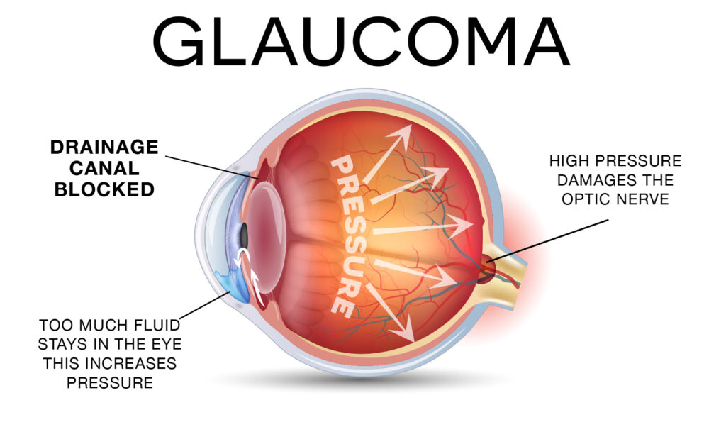
Glaucoma is often described in clinical circles as the “silent thief of sight,” a complex group of ocular disorders characterized by progressive damage to the optic nerve. While the biological roots of the disease lie in intraocular pressure and fluid dynamics within the eye, the modern understanding and management of glaucoma have shifted entirely into the realm of high-technology. In the current landscape of ophthalmology, defining what glaucoma is requires looking beyond the anatomy and exploring the sophisticated AI algorithms, advanced imaging hardware, and digital health ecosystems that now define the standard of care.
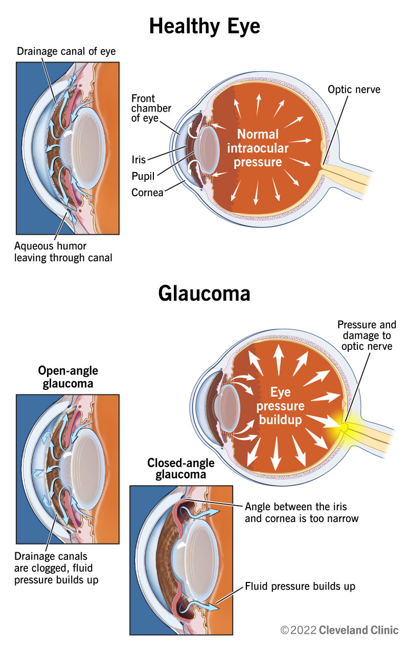
For decades, the diagnosis of glaucoma relied on rudimentary physical examinations and manual pressure checks. Today, the disease is treated as a high-stakes data problem. Because the vision loss associated with glaucoma is irreversible, the tech industry has focused its efforts on early detection and predictive modeling. Understanding glaucoma today means understanding the convergence of biotechnology and digital innovation.
Redefining Diagnosis: AI and Machine Learning in Early Detection
The primary challenge in managing glaucoma is its asymptomatic nature in the early stages. By the time a patient notices a “tunnel vision” effect, significant and permanent damage to the optic nerve has already occurred. This is where Artificial Intelligence (AI) and Machine Learning (ML) have revolutionized the field, turning a biological diagnostic process into a computational one.
Deep Learning and Fundus Photography
The gold standard for identifying glaucoma is the examination of the optic nerve head via fundus photography. However, human interpretation of these images is subjective and prone to variability. Tech giants and specialized med-tech startups have developed deep learning models trained on millions of retinal images to identify microscopic changes in the optic disc that are invisible to the human eye.
These AI tools utilize convolutional neural networks (CNNs) to analyze the “cup-to-disc” ratio and the integrity of the retinal nerve fiber layer. By processing these images through multiple layers of abstraction, the software can assign a probability score for glaucoma with an accuracy that often exceeds that of veteran ophthalmologists. This technological leap allows for mass screening programs where AI can flag “at-risk” patients in remote areas, ensuring that the disease is caught years before the first symptoms manifest.
Automated Perimetry and Data Analytics
Visual field testing, or perimetry, is a crucial metric in determining how much a patient’s vision has been impacted by glaucoma. Traditionally, this was a tedious process prone to patient fatigue and “noise” in the data. Modern perimeters now integrate advanced data analytics to filter out patient errors and provide a “progression analysis” report.
These systems use Bayesian probability models to compare a patient’s current results against their historical data and a massive database of peer demographics. The software can then predict the future rate of vision loss based on current trends, allowing doctors to adjust technological interventions (such as laser settings or drug delivery schedules) before the vision further deteriorates.
The Hardware Revolution: Next-Gen Imaging and Monitoring
While software provides the brain for glaucoma management, the hardware provides the eyes. The evolution of diagnostic hardware has moved from simple magnifying lenses to high-resolution cross-sectional scanning that rivals the precision of aerospace engineering.
Optical Coherence Tomography (OCT) Advancements
Optical Coherence Tomography, or OCT, is arguably the most significant technological advancement in the history of glaucoma management. It functions similarly to ultrasound but uses light waves instead of sound to capture micrometric-resolution, three-dimensional images of the retina.
The latest generation of OCT devices utilizes “Swept-Source” technology, which allows for faster scanning speeds and deeper penetration into the ocular tissues. This hardware can measure the thickness of the retinal nerve fiber layer with sub-micron precision. In the context of tech-driven glaucoma care, OCT serves as a digital “biopsy” of the eye, providing a level of detail that allows clinicians to monitor the death of individual ganglion cells. This shift from qualitative observation to quantitative measurement is a hallmark of the digital transformation in eye care.
Smart Contact Lenses and IoT Tonometer Devices
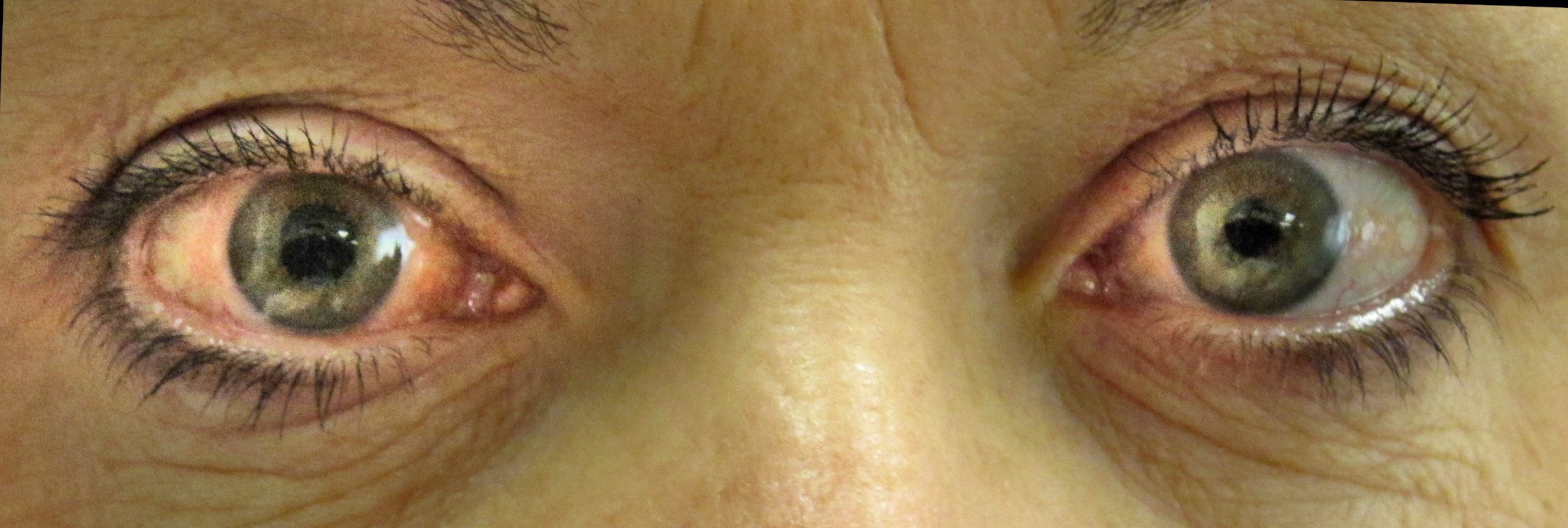
One of the greatest hurdles in treating glaucoma is the fluctuation of intraocular pressure (IOP). Pressure often spikes at night or during activities when the patient is not in a clinic. To solve this, the industry has turned to the Internet of Things (IoT).
The development of “smart” contact lenses represents the cutting edge of wearable tech in medicine. These lenses contain micro-sensors capable of monitoring changes in corneal curvature—a proxy for internal eye pressure—continuously for 24 hours. The data is transmitted wirelessly to a smartphone app or a cloud-based server. This “Big Data” approach to IOP monitoring provides a “pressure profile” that is far more comprehensive than a single “snapshot” reading taken in a doctor’s office. It allows for a personalized tech-driven treatment plan that accounts for the patient’s unique circadian rhythms.
Surgical Innovation: Robotics and Micro-Invasive Technology
When pharmaceutical interventions fail to control the pressure within the eye, technology offers a suite of surgical solutions. The transition from invasive “macro” surgeries to micro-invasive and laser-based procedures is a testament to the miniaturization trends seen across the broader tech sector.
MIGS: The Engineering Behind the Procedure
Micro-Invasive Glaucoma Surgery (MIGS) refers to a group of procedures that use microscopic equipment and stents to improve fluid drainage. These devices, some no larger than a grain of sand, are masterpieces of bio-engineering. Made from surgical-grade titanium or polyethersulfone, these stents are inserted using high-precision delivery systems that require robotic-level stability.
The tech behind MIGS focuses on biocompatibility and fluid dynamics. Engineers use computational fluid dynamics (CFD) software to model how aqueous humor flows through these tiny channels, ensuring that the pressure remains stable without causing hypotony (pressure that is too low). This intersection of mechanical engineering and biology has turned glaucoma surgery into a high-tech “plumbing” fix for the eye.
Femtosecond Lasers and Selective Targeting
Laser technology has also undergone a digital overhaul. Selective Laser Trabeculoplasty (SLT) uses short pulses of low-energy light to target specific pigmented cells in the eye’s drainage meshwork. The latest hardware platforms for these procedures use automated eye-tracking technology—the same tech found in high-end gaming monitors and autonomous vehicles—to ensure the laser hits the exact target even if the patient’s eye moves. This level of precision minimizes collateral damage to surrounding tissues, making the procedure repeatable and significantly safer than traditional cold-steel surgery.
The Future of Glaucoma Management: Telemedicine and Digital Health
As we look toward the future, the definition of glaucoma care is moving out of the clinic and into the digital ecosystem. The integration of SaaS (Software as a Service) platforms and remote monitoring tools is creating a decentralized model of eye care.
Remote Monitoring and SaaS Platforms
The “clinic of the future” for glaucoma is likely a digital platform. Cloud-based SaaS solutions now allow for the seamless integration of data from OCTs, perimeters, and smart tonometers into a single dashboard. This allows for “asynchronous care,” where a technician in a rural area can upload high-resolution scans to the cloud, and a specialist in a major urban hub can review them using AI-assisted decision-support tools.
This technological democratization is crucial for a disease like glaucoma, which requires lifelong monitoring. By reducing the need for frequent in-person visits, digital health platforms ensure better compliance and more frequent data points, which are essential for preventing the “silent” progression of the disease.
Virtual Reality (VR) in Visual Field Testing
Perhaps one of the most exciting trends in glaucoma tech is the adaptation of Virtual Reality (VR) headsets for diagnostic use. Traditional visual field machines are large, immobile, and expensive. New startup ventures are repurposing lightweight VR goggles to conduct these tests.
By using the immersive capabilities of VR, these devices can track a patient’s eye movements and responses to stimuli in a controlled digital environment. This makes the testing process more engaging and less prone to the “boredom errors” that plague traditional perimetry. Furthermore, because these VR units are portable, they can be sent to a patient’s home, allowing for “at-home perimetry.” This represents the ultimate shift in glaucoma management: moving from reactive, clinic-based observation to proactive, tech-enabled home monitoring.
In conclusion, “what is glaucoma” is a question that can no longer be answered solely in biological terms. In the modern era, glaucoma is a data-intensive neurological challenge that is being solved through the application of advanced computing, high-precision hardware, and the ubiquity of the Internet of Things. As these technologies continue to converge, the goal of “zero blindness” from glaucoma moves from a theoretical ambition to a technological certainty.
aViewFromTheCave is a participant in the Amazon Services LLC Associates Program, an affiliate advertising program designed to provide a means for sites to earn advertising fees by advertising and linking to Amazon.com. Amazon, the Amazon logo, AmazonSupply, and the AmazonSupply logo are trademarks of Amazon.com, Inc. or its affiliates. As an Amazon Associate we earn affiliate commissions from qualifying purchases.