
In the realm of medical diagnostics, understanding the intricacies of the human body’s internal systems is paramount. Among the many procedures designed to illuminate these complex systems, the “UGI” stands out as a critical tool for examining the upper digestive tract. While the acronym often refers to either an Upper Gastrointestinal (UGI) Series (a radiological study) or an Upper Gastrointestinal Endoscopy (a direct visualization procedure), both represent significant applications of technology in healthcare. This article delves into what a UGI entails, exploring the underlying technological principles, the evolution of the equipment, and the digital innovations that are shaping its future, firmly rooting our discussion within the domain of medical technology.
The Core Concepts: Understanding UGI Procedures and Their Tech Foundation
At its heart, a UGI procedure aims to diagnose conditions affecting the esophagus, stomach, and duodenum. These conditions can range from acid reflux and ulcers to more serious issues like polyps or cancerous growths. The approach taken—whether radiological or endoscopic—depends on the specific diagnostic questions and clinical context, each leveraging distinct technological paradigms.
Upper Gastrointestinal (UGI) Series: The X-ray Approach
The UGI series, often referred to as a barium swallow or esophagram, is a classic diagnostic imaging technique. It employs X-ray technology to visualize the internal structures and function of the upper digestive tract. The patient swallows a barium-based contrast liquid, which coats the lining of the esophagus, stomach, and duodenum, making them opaque to X-rays. As the barium travels, a radiologist uses a fluoroscope – a specialized X-ray system that produces real-time video images – to observe its passage.
Technologically, the UGI series relies on the interaction of X-rays with matter. Modern fluoroscopy systems have evolved significantly from their early counterparts. Digital fluoroscopy units now incorporate advanced detectors, such as flat-panel detectors, which offer superior image quality, higher resolution, and reduced radiation exposure compared to older image intensifier systems. These digital systems allow for immediate image acquisition, processing, and storage, integrating seamlessly with Picture Archiving and Communication Systems (PACS) for efficient data management and retrieval. Software algorithms play a crucial role in image enhancement, noise reduction, and dose optimization, ensuring diagnostic clarity while prioritizing patient safety.
Upper Gastrointestinal Endoscopy: Direct Visualization and Intervention
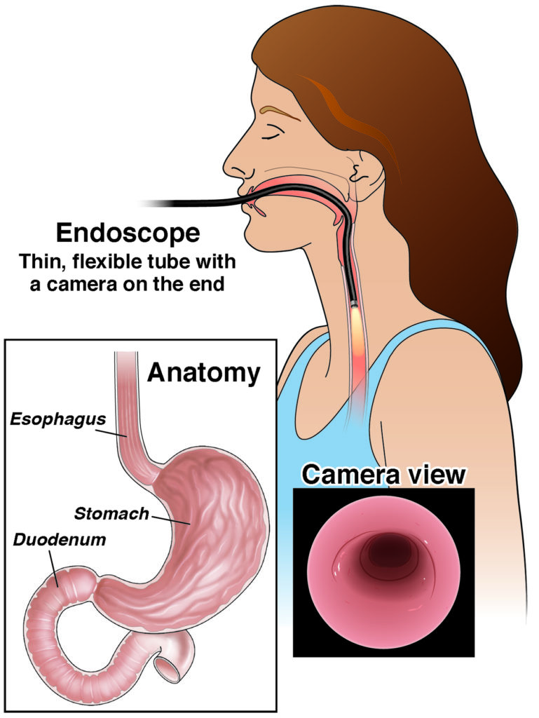
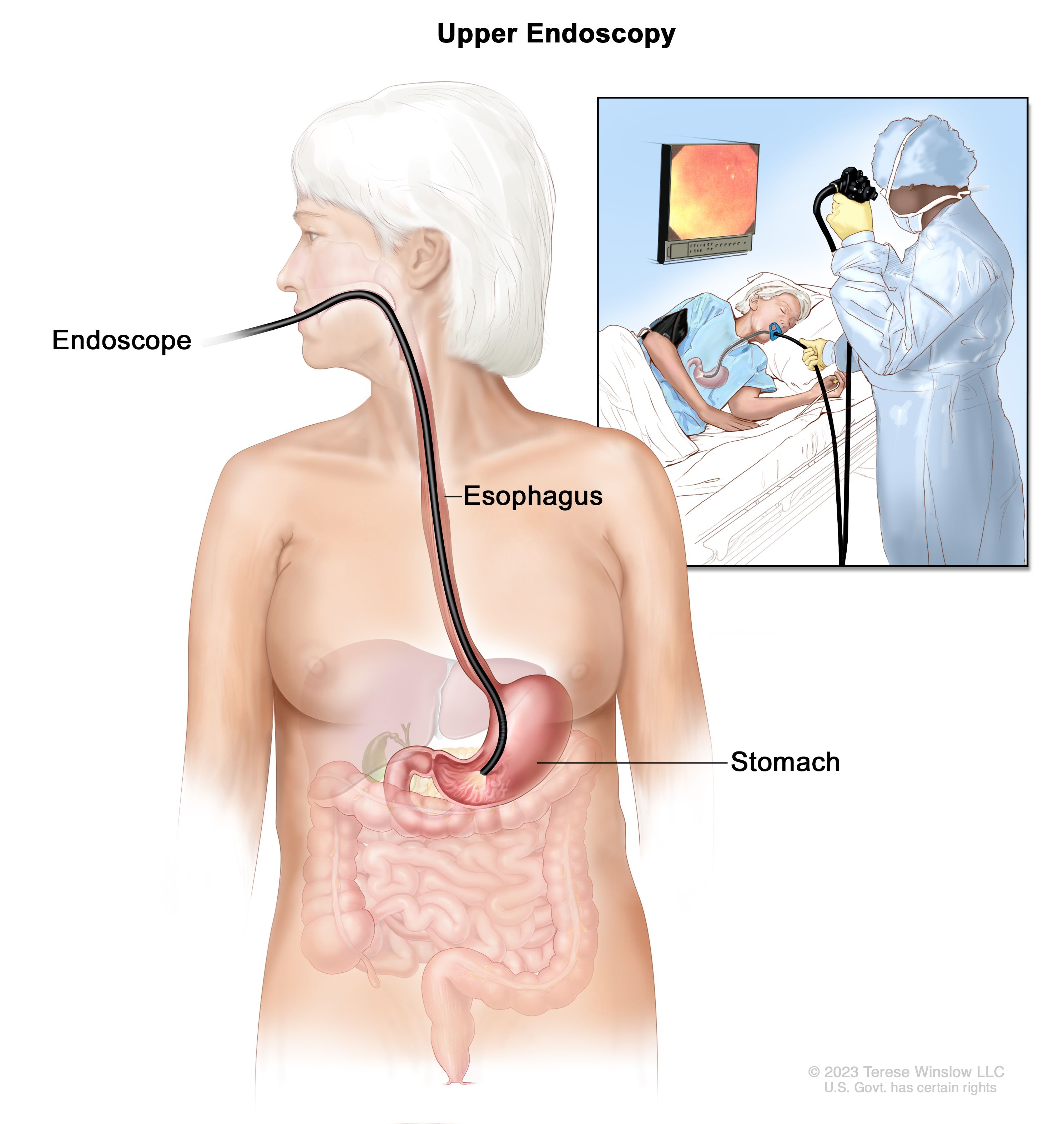
Often simply called an “upper endoscopy” or Esophagogastroduodenoscopy (EGD), this procedure offers a more direct and invasive look at the upper GI tract. It involves inserting a thin, flexible tube called an endoscope into the patient’s mouth, down the esophagus, stomach, and into the duodenum. The endoscope itself is a marvel of miniaturized technology.
Modern endoscopes are equipped with a high-resolution camera at their tip, fiber optics or LED lighting for illumination, and channels for air, water, and instrument insertion. The camera, typically a Charge-Coupled Device (CCD) or Complementary Metal-Oxide-Semiconductor (CMOS) sensor, captures real-time video, which is displayed on a monitor. This allows the gastroenterologist to meticulously examine the mucosal lining for abnormalities. The instrument channel enables the passage of tiny tools, such as forceps for biopsies, snares for polyp removal, or devices for cauterization, making the procedure not just diagnostic but also therapeutic. The flexibility of the scope is controlled by a series of wheels and buttons on the handle, allowing for precise navigation through the convoluted anatomy of the digestive system. The evolution of endoscopes from rigid, cumbersome tubes to highly maneuverable, high-definition flexible instruments represents decades of material science, optics, and digital imaging advancements.
Technological Marvels Driving UGI Diagnostics
The continuous evolution of technology has profoundly impacted both UGI series and endoscopic procedures, enhancing diagnostic accuracy, procedural safety, and patient comfort.
High-Resolution Imaging and Sensor Technology
The quality of images acquired during a UGI procedure is paramount for accurate diagnosis. In fluoroscopy, the shift from film-based X-rays to digital detectors has revolutionized the UGI series. Digital flat-panel detectors provide instant, high-contrast images with a wider dynamic range, allowing radiologists to visualize subtle changes in the mucosal pattern and organ motility. These detectors, often based on amorphous silicon technology, convert X-ray photons directly into digital signals, bypassing intermediate steps and thereby reducing noise and improving signal-to-noise ratio.
Similarly, endoscopic imaging has seen dramatic improvements. Early endoscopes used bundles of fiber optic cables to transmit images, resulting in grainy, limited-resolution views. Today’s video endoscopes employ advanced CCD or CMOS sensors that capture images in full high-definition (HD), and increasingly, ultra-high-definition (4K). These sensors are capable of capturing intricate details of the mucosal surface, helping to identify minute lesions, inflammatory changes, or early signs of malignancy. Furthermore, advancements in narrow-band imaging (NBI) or chromoendoscopy utilize specific light wavelengths to enhance the visualization of vascular patterns and mucosal irregularities, essentially acting as an “optical biopsy” to better characterize tissue without immediate removal.
Miniaturization and Ergonomics in Endoscopy
The physical design of endoscopic equipment has undergone significant refinement. Miniaturization has led to thinner scopes, which are less uncomfortable for patients and easier for clinicians to maneuver. This includes ultra-thin endoscopes that can be passed transnasally, avoiding the need for sedation in some cases, and even pediatric endoscopes tailored for smaller anatomies.
Ergonomics in endoscope design focuses on improving the user experience for the gastroenterologist. Lighter scopes, redesigned control sections, and intuitive button layouts reduce operator fatigue during lengthy procedures. Beyond traditional endoscopes, capsule endoscopy represents a groundbreaking example of miniaturization. Patients swallow a vitamin-sized capsule containing a camera, light source, battery, and radio transmitter. As it passes through the GI tract, it captures thousands of images, wirelessly transmitting them to a receiver worn by the patient. While primarily used for the small bowel, the technology showcases the potential for non-invasive, patient-friendly diagnostic tools, driven by advancements in microelectronics, power management, and wireless communication.
The Role of AI and Software in Modern UGI
The digital revolution has brought artificial intelligence (AI) and sophisticated software into the forefront of medical diagnostics, transforming how UGI procedures are performed, interpreted, and managed.
AI for Enhanced Diagnosis and Lesion Detection
One of the most exciting applications of AI in UGI diagnostics is its ability to assist in lesion detection and characterization. Machine learning algorithms, trained on vast datasets of endoscopic images and radiological scans, can identify subtle abnormalities that might be missed by the human eye, especially during long and complex procedures. For instance, AI systems can automatically highlight suspicious polyps or early-stage cancerous lesions in real-time during an endoscopy, improving the adenoma detection rate, which is a key quality metric. In fluoroscopy, AI can aid in quantifying motility disorders or identifying subtle strictures.
Beyond detection, AI can also help in the characterization of identified lesions, differentiating between benign and malignant findings with high accuracy. This can reduce the need for unnecessary biopsies or guide more targeted biopsies, leading to more efficient and precise patient management. The integration of computer-aided diagnosis (CAD) systems is becoming increasingly common, acting as a second pair of eyes for the clinician and enhancing diagnostic confidence.
Software for Procedure Planning, Data Management, and Reporting
Software forms the backbone of modern UGI units, streamlining everything from patient scheduling to post-procedure reporting. Electronic Health Record (EHR) systems integrate UGI procedure data, including images, videos, and findings, providing a comprehensive patient history accessible across different medical departments. Specialized endoscopy reporting software allows clinicians to quickly document findings, create standardized reports, and track quality indicators.
Advanced imaging software in fluoroscopy units enables sophisticated image manipulation, 3D reconstruction from multiple 2D images, and quantitative analysis of barium flow dynamics. Data management systems ensure secure storage of large image and video files, facilitating easy retrieval for comparison with previous studies or for educational purposes. Furthermore, software tools are increasingly used for procedure planning, risk stratification, and even for simulating endoscopic navigation to train new clinicians, improving skill acquisition and procedural safety.
Future Trends and Innovations in UGI Technology
The landscape of UGI diagnostics is continually evolving, with ongoing research and development promising even more sophisticated and less invasive solutions.
Robotic-Assisted Endoscopy and Remote Procedures
Robotics is poised to revolutionize endoscopy. Robotic-assisted endoscopic systems offer enhanced dexterity, stability, and precision, potentially allowing for more complex therapeutic interventions with greater control. These systems can filter out physician tremors and provide a magnified, stable view, leading to more accurate procedures. Moreover, the concept of remote or “tele-endoscopy” is gaining traction, especially in underserved areas. With robotic systems, a specialist could potentially perform an endoscopy from a remote location, guided by real-time video feeds and haptic feedback systems, significantly expanding access to expert care.
Further innovations include magnetically guided capsule endoscopes, where external magnetic fields can steer the capsule through the GI tract, allowing for more controlled examination than passively moving capsules. These technological leaps are moving UGI diagnostics toward increased automation, precision, and accessibility.
Advanced Biomarkers and Non-Invasive Techniques
While imaging and direct visualization remain central, the future of UGI diagnostics will increasingly integrate molecular and biological insights. Research into advanced biomarkers found in blood, stool, or breath samples could offer non-invasive screening tools that indicate the presence of GI diseases, potentially reducing the need for invasive procedures in some cases. Combining these biomarker profiles with imaging data, enabled by AI-driven analytics, could lead to highly personalized diagnostic and treatment pathways.
Furthermore, continued development in non-invasive imaging techniques, such as advanced forms of ultrasound or functional MRI that can assess GI motility and inflammation without contrast agents, may complement or even replace certain aspects of traditional UGI series, providing a broader, multi-modal diagnostic picture.

Conclusion
The journey into understanding “what is a UGI” reveals a fascinating interplay of medical necessity and technological ingenuity. From the foundational X-ray physics of the UGI series to the intricate optics and robotics of modern endoscopy, technology has continuously pushed the boundaries of what is diagnosable and treatable within the upper gastrointestinal tract. With high-resolution sensors, AI-powered analytics, and the promise of robotic and non-invasive innovations, the future of UGI diagnostics is bright, promising more precise, safer, and patient-friendly ways to maintain digestive health. As technology continues its relentless march forward, UGI procedures will undoubtedly remain at the forefront of medical advancements, ensuring that clinicians have the best possible tools to care for their patients.
aViewFromTheCave is a participant in the Amazon Services LLC Associates Program, an affiliate advertising program designed to provide a means for sites to earn advertising fees by advertising and linking to Amazon.com. Amazon, the Amazon logo, AmazonSupply, and the AmazonSupply logo are trademarks of Amazon.com, Inc. or its affiliates. As an Amazon Associate we earn affiliate commissions from qualifying purchases.