
In the realm of genetic diagnostics and research, the term “karyotype” frequently surfaces, often in discussions surrounding inherited conditions, developmental abnormalities, and even evolutionary biology. But what exactly is a karyotype, and why is this technological process so crucial in understanding the fundamental building blocks of life? At its core, a karyotype is a visual representation, a meticulously organized snapshot, of an individual’s complete set of chromosomes. This isn’t a mere collection of genetic material; it’s a sophisticated technological output that reveals the number, size, shape, and banding patterns of each chromosome. Understanding the karyotype is akin to having a detailed blueprint of our genetic makeup, offering profound insights into health, inheritance, and the very essence of our biological identity.
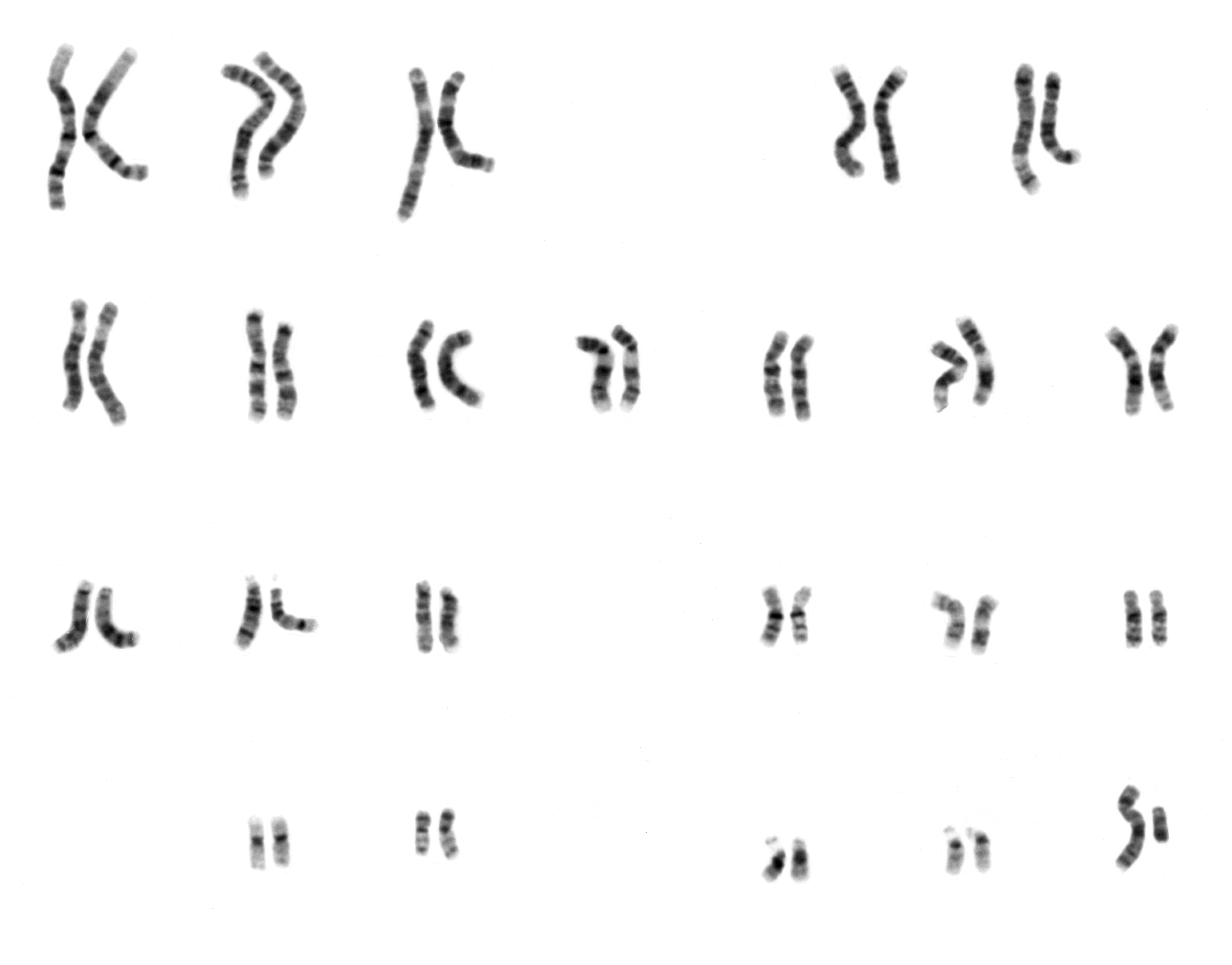
The creation and interpretation of a karyotype represent a significant technological achievement, bridging the gap between abstract genetic code and observable, quantifiable data. It’s a process that has evolved dramatically with advancements in microscopy, staining techniques, and computational analysis, transforming from a rudimentary observation to a high-resolution diagnostic tool. This article delves into the technological underpinnings of karyotyping, exploring how it’s performed, what information it reveals, and its multifaceted applications in modern medicine and scientific research.
The Technological Foundation: From Cell to Chromosome Image
The journey to a visual karyotype is a testament to human ingenuity in harnessing technology to explore the microscopic world. It begins with living cells and culminates in a digital or physical representation of organized chromosomes. This process is not a single technological leap but a series of carefully orchestrated steps, each relying on precise scientific instruments and methodologies.
Cell Collection and Preparation: The Starting Point
The initial stage of karyotype generation involves obtaining a suitable sample containing dividing cells. This can be achieved through various biological sampling methods, but the subsequent processing is where technological intervention becomes paramount. Blood samples are common, but cells can also be derived from amniotic fluid, chorionic villi, or even tissue biopsies.
Once collected, these cells must be cultured to induce division. This culturing process itself relies on sterile laboratory environments, precise temperature control, and specialized growth media – all technological advancements ensuring optimal cell viability and proliferation. Crucially, during the cell cycle, a specific phase known as metaphase is targeted. This is when the chromosomes are most condensed and visible, a prerequisite for accurate observation and analysis. To arrest cells in this specific phase, chemicals like colchicine are used. This chemical agent, acting as a mitotic inhibitor, prevents the formation of the spindle fibers necessary for chromosome separation, effectively freezing the cell in metaphase.
Chromosome Staining and Banding: Revealing the Details
The visual clarity of chromosomes is significantly enhanced through staining techniques. These are not just simple dyes; they are sophisticated chemical agents designed to bind to specific regions of the chromosomes, creating differential staining patterns known as “banding.” The most common banding technique is G-banding (Giemsa banding), which uses the Giemsa stain. This stain differentially stains regions of the chromosome that are rich in adenine-thymine (A-T) base pairs, making them appear darker, and regions rich in guanine-cytosine (G-C) base pairs, which appear lighter.
The resulting banding patterns are not random; they are highly reproducible and specific to each chromosome pair. These bands serve as unique identifiers, much like barcodes, allowing cytogeneticists to distinguish between individual chromosomes and detect structural abnormalities. Other banding techniques, such as Q-banding (quinacrine fluorescence banding) and R-banding (reverse banding), also exist, each offering slightly different visualization characteristics and often used in conjunction with G-banding to provide a comprehensive view. The precision of these staining protocols, combined with advanced microscopy, is what allows for the detailed examination of chromosomal structure.
Microscopy and Imaging: Capturing the Chromosomal Landscape
The culmination of sample preparation is the visualization of these stained chromosomes under a microscope. High-resolution light microscopes, specifically designed for cytogenetic analysis, are essential. These microscopes are equipped with advanced optics and illumination systems to provide sharp, clear images of the condensed chromosomes.
Modern karyotyping heavily relies on digital imaging technology. Instead of solely relying on manual observation through eyepieces, high-resolution digital cameras are attached to the microscopes. These cameras capture images of the chromosomes, which are then processed by specialized computer software. This digital capture is revolutionary, allowing for the easy storage, retrieval, and manipulation of images. It also facilitates the sharing of data among researchers and clinicians, enabling collaborative analysis and faster diagnostic turnaround times. The ability to zoom in on specific chromosomal regions, adjust contrast, and enhance image quality are all technological features that significantly improve the accuracy and efficiency of karyotype analysis.
The Art and Science of Karyotype Analysis: Interpretation and Standardization
Once the chromosomal images are captured, the intricate process of analysis and interpretation begins. This is where the technological output is translated into meaningful biological information. It’s a meticulous task that requires both scientific knowledge and sophisticated computational tools.
Chromosome Sorting and Arrangement: Building the Karyotype
The raw images from the microscope typically show hundreds of chromosomes scattered across the cellular field. The first step in creating a karyotype is to identify and isolate each individual chromosome. This is achieved through a combination of automated or semi-automated computer programs and the expertise of a trained cytogeneticist.
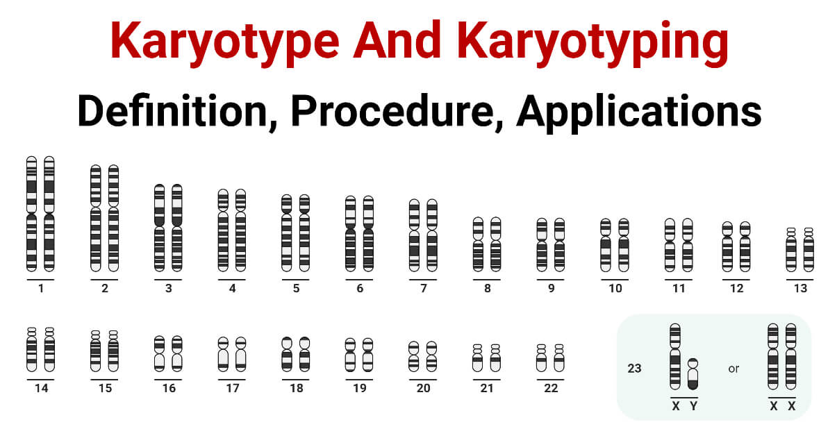
The software identifies chromosomes based on their size, centromere position (the constricted region of a chromosome), and banding patterns. It then “pairs up” homologous chromosomes – chromosomes that carry the same genes, one inherited from each parent. These pairs are then arranged in a standard order, typically from largest to smallest. The sex chromosomes (X and Y) are placed at the end of the arranged set. This standardized arrangement is crucial for consistent interpretation and comparison between individuals and across different laboratories. The technological prowess in image processing and pattern recognition is vital in this stage, minimizing human error and speeding up the process.
Identifying Abnormalities: Decoding the Genetic Blueprint
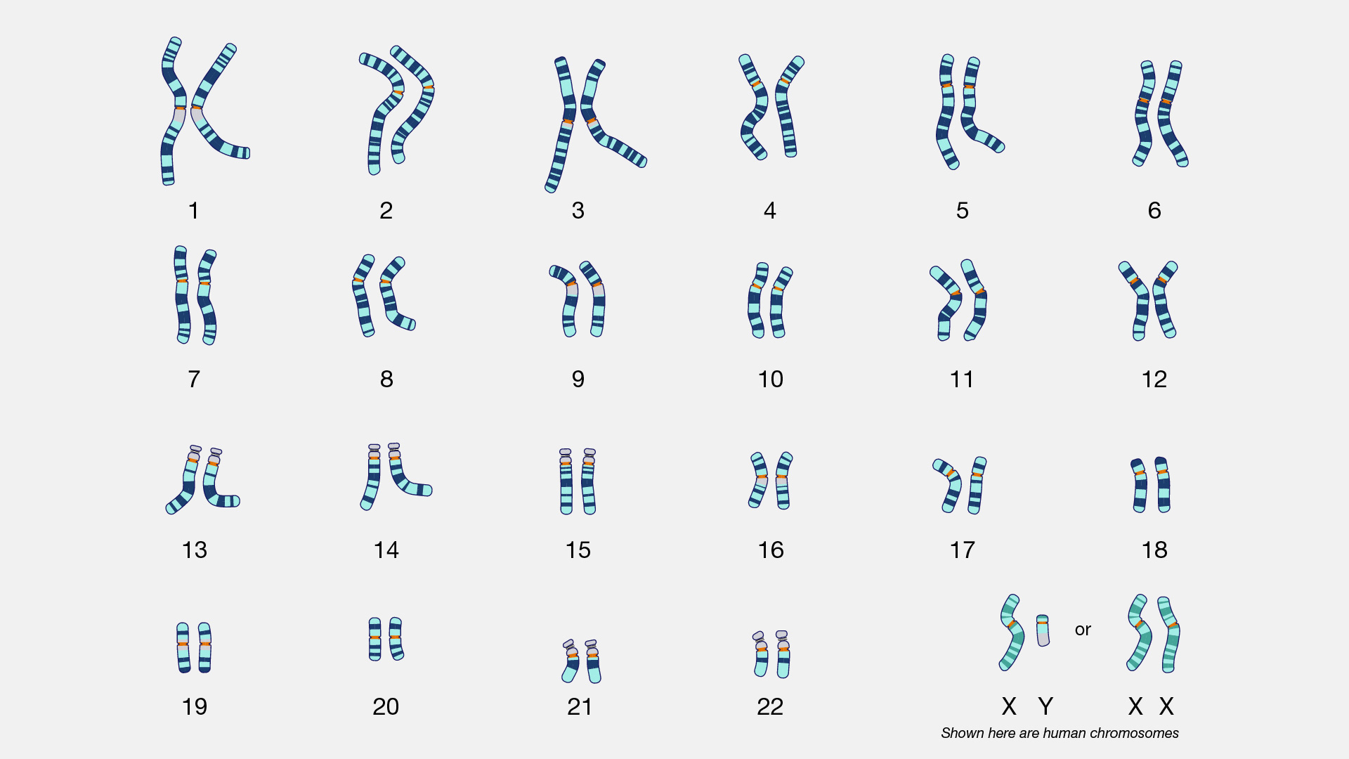
The primary purpose of creating a karyotype is to detect chromosomal abnormalities. These abnormalities can be broadly categorized into numerical abnormalities (aneuploidy) and structural abnormalities.
-
Numerical Abnormalities: These involve an abnormal number of chromosomes. For example, Down syndrome (Trisomy 21) is characterized by an extra copy of chromosome 21, resulting in a total of 47 chromosomes instead of the usual 46. Similarly, Turner syndrome (Monosomy X) in females involves the absence of one X chromosome (45,X), and Klinefelter syndrome in males typically involves an extra X chromosome (47,XXY). The karyotype visually represents these extra or missing chromosomes.
-
Structural Abnormalities: These involve changes in the structure of one or more chromosomes. They can include:
- Deletions: A segment of a chromosome is missing.
- Duplications: A segment of a chromosome is repeated.
- Inversions: A segment of a chromosome is flipped.
- Translocations: A segment of one chromosome breaks off and attaches to another non-homologous chromosome.
- Ring chromosomes: Both ends of a chromosome break and fuse together to form a ring.
The banding patterns are instrumental in identifying these structural changes. A deviation in the size or banding pattern of a chromosome, or the presence of extra genetic material on a chromosome where it shouldn’t be, signals a structural abnormality. The resolution of the karyotype, often expressed as the number of bands visible per haploid set of chromosomes (e.g., 300, 400, 550-band resolution), determines the sensitivity of the technique in detecting smaller structural changes. Higher resolution means more bands are visible, allowing for the identification of finer alterations.
Quality Control and Standardization: Ensuring Accuracy
Given the critical implications of karyotype analysis in diagnosis and treatment, rigorous quality control and standardization are paramount. This is where technological protocols and established guidelines come into play. Laboratories adhere to strict protocols for sample handling, culture, staining, imaging, and analysis to ensure the reliability and reproducibility of results.
The International System for Human Cytogenetic Nomenclature (ISCN) provides a standardized way of describing chromosomal abnormalities. This ensures that geneticists worldwide can understand and compare karyotype reports. Furthermore, advancements in automated metaphase finding and chromosome analysis software are continuously being developed to improve accuracy, reduce turnaround times, and minimize subjective interpretation by technicians. These technological aids, coupled with expert human oversight, form the backbone of reliable karyotype diagnostics.
The Impact of Karyotyping: Applications Across Technology-Driven Fields
The technological process of karyotyping has far-reaching implications, revolutionizing various fields within genetics, medicine, and beyond. Its ability to reveal fundamental chromosomal information makes it an indispensable tool in diagnostics, research, and even in understanding human evolution.
Clinical Diagnostics: Unveiling Genetic Disorders
One of the most significant applications of karyotyping lies in clinical diagnostics. It is a cornerstone in the investigation of:
- Infertility and Recurrent Miscarriage: Karyotyping can identify chromosomal abnormalities in individuals or couples experiencing reproductive challenges, which can be a cause of infertility or recurrent pregnancy loss.
- Congenital Abnormalities: It is routinely used to diagnose genetic disorders in newborns with physical abnormalities or developmental delays.
- Cancer Cytogenetics: In oncology, karyotyping is used to identify specific chromosomal translocations or other abnormalities that are characteristic of certain types of cancer. These abnormalities can aid in diagnosis, prognosis, and guiding treatment decisions. For example, the Philadelphia chromosome (a translocation between chromosomes 9 and 22) is a hallmark of chronic myeloid leukemia (CML).
- Prenatal Diagnosis: Karyotyping of fetal cells obtained through amniocentesis or chorionic villus sampling (CVS) can detect chromosomal abnormalities in a developing fetus, allowing for informed decisions regarding pregnancy management.
The technological advancements in imaging and analysis have made prenatal diagnosis more accessible and accurate, providing crucial information to expectant parents.
Research and Discovery: Advancing Genetic Understanding
Beyond clinical applications, karyotyping is a vital research tool that has significantly advanced our understanding of genetics and evolution.
- Gene Mapping: By studying the location of genes on specific chromosomal bands, researchers can map gene locations and understand their relationships. This is foundational to many genetic studies.
- Evolutionary Biology: Comparing karyotypes across different species can provide insights into evolutionary relationships and the mechanisms of chromosomal evolution. Changes in chromosome number and structure over time can reveal how species have diverged.
- Developmental Biology: Karyotyping helps researchers understand the role of specific chromosomes and genes in embryonic development and the consequences of their abnormalities.
The ability to visualize and analyze entire genomes in a structured format is invaluable for exploring complex genetic questions, from the fundamental mechanisms of cell division to the origins of species.
Emerging Technologies and Future Directions: Enhancing Resolution and Precision
While traditional karyotyping remains a powerful tool, its resolution limitations can sometimes hinder the detection of very small genetic changes. This has spurred the development and integration of newer, higher-resolution technologies.
- Chromosomal Microarray Analysis (CMA): CMA offers a much higher resolution than conventional karyotyping, allowing for the detection of submicroscopic chromosomal imbalances, such as copy number variations (CNVs), which can be associated with intellectual disability, developmental delays, and congenital anomalies. CMA has largely replaced karyotyping for the diagnosis of certain genetic conditions due to its superior sensitivity.
- Fluorescence In Situ Hybridization (FISH): FISH uses fluorescently labeled DNA probes that bind to specific chromosomal regions. This technique allows for the detection of specific chromosomal abnormalities, such as deletions, duplications, or translocations, even at a higher resolution than standard banding. FISH is often used as a targeted approach to investigate suspected abnormalities identified through karyotyping or CMA.
The future of karyotyping likely involves a synergistic integration of these technologies. While conventional karyotyping provides an overview of the entire genome, techniques like CMA and FISH offer detailed insights into specific regions. Computational tools and artificial intelligence are also playing an increasingly significant role, aiding in the automated analysis of large datasets, improving accuracy, and accelerating the discovery of novel genetic insights. This continuous technological evolution ensures that karyotyping, in its various forms, will remain at the forefront of genetic diagnostics and research for years to come.
aViewFromTheCave is a participant in the Amazon Services LLC Associates Program, an affiliate advertising program designed to provide a means for sites to earn advertising fees by advertising and linking to Amazon.com. Amazon, the Amazon logo, AmazonSupply, and the AmazonSupply logo are trademarks of Amazon.com, Inc. or its affiliates. As an Amazon Associate we earn affiliate commissions from qualifying purchases.