
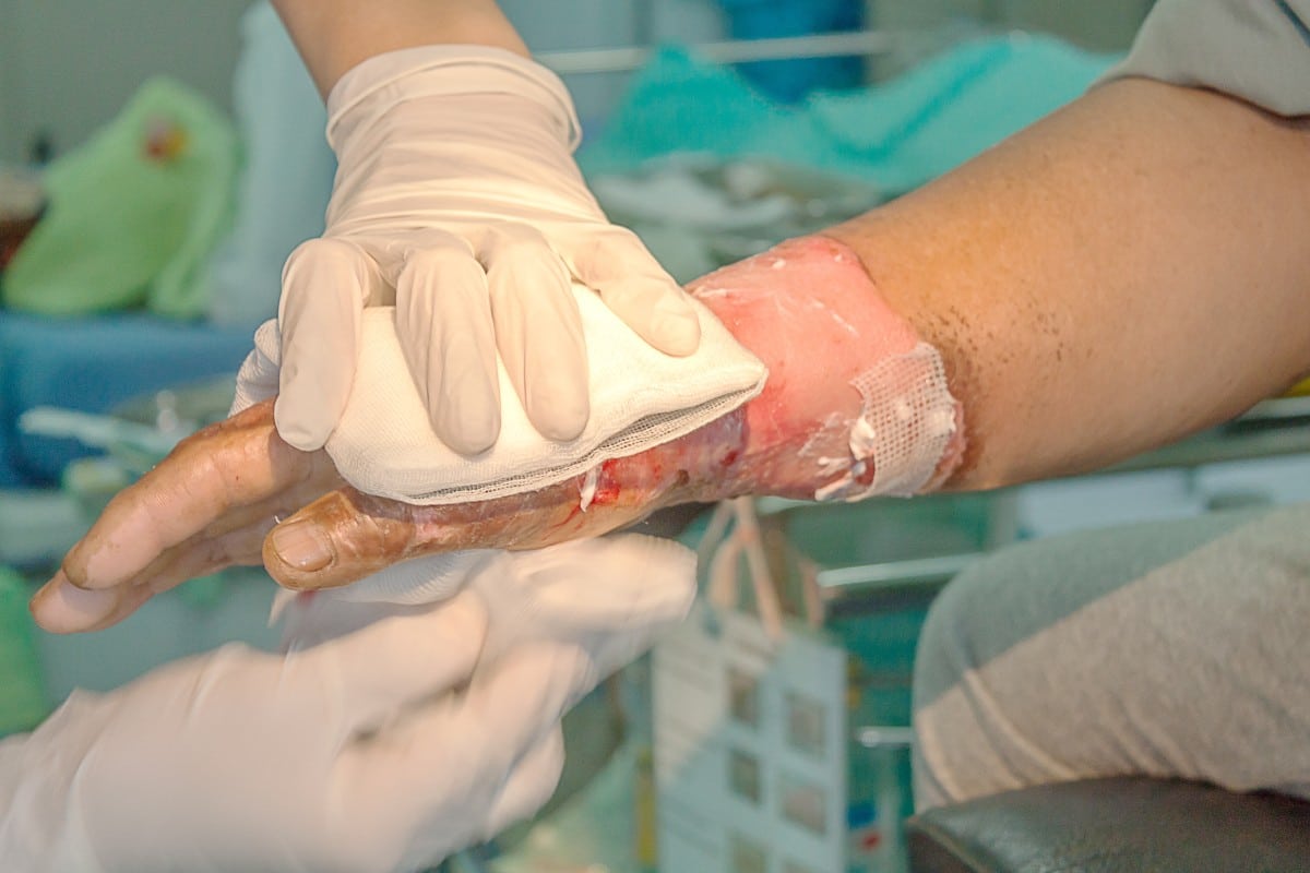
A full-thickness burn, medically known as a third-degree burn, represents the most severe form of thermal injury, extending through all layers of the skin — the epidermis and dermis — and often into subcutaneous tissue, muscle, or even bone. From a technological perspective, understanding and addressing such a profound injury demands the application of cutting-edge innovations across diagnostics, treatment, and long-term care. Technology not only enhances our ability to precisely characterize these complex wounds but also revolutionizes therapeutic interventions, offering unprecedented avenues for healing and functional restoration.
Diagnosing Full-Thickness Burns with Precision Technology
Accurate and early diagnosis of burn depth is paramount for guiding treatment strategies, predicting outcomes, and optimizing resource allocation. Traditional clinical assessment, while fundamental, can be subjective, especially in the immediate aftermath of injury. Modern technology offers objective, non-invasive methods to peer beneath the surface, providing critical data for clinicians.
Advanced Imaging Techniques for Depth and Severity Assessment
The advent of sophisticated imaging technologies has transformed the way clinicians evaluate burn wounds. These tools move beyond visual inspection, offering quantifiable data on tissue viability and perfusion.
- Laser Doppler Imaging (LDI): LDI systems use low-power laser light to measure blood flow in the microvasculature of the skin. By analyzing the Doppler shift of light scattered by moving red blood cells, LDI can create detailed maps of perfusion. Areas of full-thickness burn typically exhibit severely reduced or absent blood flow, making LDI a valuable tool for differentiating between superficial, partial-thickness, and full-thickness injuries, often within the first 24-48 hours. This early, objective assessment significantly reduces diagnostic uncertainty and helps prevent unnecessary surgical interventions or delayed essential care.
- Hyperspectral Imaging (HSI): HSI captures images across a wide spectrum of light wavelengths, from visible to near-infrared. Each tissue type and physiological state (e.g., oxygenation, hydration, hemoglobin concentration) has a unique spectral signature. By analyzing these signatures, HSI can provide detailed maps of tissue composition, oxygen saturation, and perfusion within the burn wound. This technology offers a comprehensive, non-contact assessment that can reveal subtle differences in tissue viability, aiding in the precise demarcation of full-thickness areas that require intervention.
- Optical Coherence Tomography (OCT): Analogous to ultrasound but using light waves, OCT provides high-resolution, cross-sectional images of tissue microstructure up to a few millimeters deep. This allows clinicians to visualize the different layers of the skin, identifying the exact depth of tissue destruction and the presence or absence of dermal structures. OCT is particularly useful for assessing burn depth in equivocal cases and monitoring healing progression.
- Thermal Imaging: While less precise for depth determination, thermal cameras can detect temperature variations across the burn wound. Full-thickness burns, due to compromised blood flow, often present with a lower surface temperature compared to surrounding healthy tissue or more superficial burns. This technique offers a rapid, non-contact screening tool.
AI-Powered Assessment Tools for Enhanced Diagnostics
Artificial intelligence and machine learning are rapidly being integrated into burn care, offering predictive capabilities that augment human expertise. By training algorithms on vast datasets of burn images, patient demographics, and clinical outcomes, AI tools can assist in rapid and accurate diagnosis.
- Automated Image Analysis: AI algorithms can analyze images from digital cameras, LDI, or HSI systems to automatically classify burn depth with high accuracy. These systems can identify patterns and subtle features that might be overlooked by the human eye, providing clinicians with objective, data-driven recommendations.
- Predictive Analytics for Prognosis: Beyond immediate diagnosis, AI models can process a multitude of patient data — including burn characteristics, patient comorbidities, age, and initial treatment response — to predict healing time, the likelihood of complications (e.g., infection, scarring), and the need for surgical intervention. This enables personalized treatment plans and proactive management.
- Decision Support Systems: AI-driven platforms can integrate various diagnostic inputs and clinical guidelines to provide real-time decision support for burn specialists, particularly beneficial in emergency settings or for less experienced personnel. These systems help ensure consistent, evidence-based care.
Revolutionary Treatments: Engineering Solutions for Skin Regeneration
The treatment of full-thickness burns historically relies on surgical debridement (removal of dead tissue) followed by skin grafting. However, technological advancements are pushing the boundaries, offering engineered solutions that aim for more complete and functional skin regeneration, often reducing donor site morbidity and improving aesthetic and functional outcomes.
Bioprinting and Tissue Engineering for Dermal Replacement
The promise of creating functional skin in a lab has moved from science fiction to clinical reality, primarily driven by advances in bioprinting and tissue engineering.
- 3D Bioprinting of Skin: This groundbreaking technology involves depositing “bio-inks” containing living cells (e.g., keratinocytes, fibroblasts) and biomaterials (e.g., collagen, fibrin) layer by layer to construct custom-fit skin grafts. For full-thickness burns, bioprinting can create constructs that mimic the complex architecture of both the epidermis and dermis, offering a potential solution for large burn areas where donor skin is scarce. Research continues into vascularizing these constructs to ensure their long-term viability upon transplantation.
- Cultured Epidermal Autografts (CEA): For patients with extensive full-thickness burns, CEA involves taking a small biopsy of the patient’s healthy skin, culturing the keratinocytes in vitro to expand their numbers, and then producing large sheets of epidermis. These sheets can then be transplanted onto the debrided burn wound. While only addressing the epidermal layer, CEA has been life-saving for patients with massive burns, although challenges remain regarding durability and donor site scarring.
- Dermal Substitutes: Bioengineered dermal scaffolds, often composed of collagen and other extracellular matrix components, are used to reconstruct the dermal layer. These acellular or cellular matrices provide a framework for the patient’s own cells to migrate into, fostering neo-dermis formation. Once the dermal substitute integrates, it can be covered with a thin epidermal graft (such as CEA or a very thin split-thickness skin graft), resulting in more robust and functional skin than an epidermal graft alone.
Smart Wound Dressings and Advanced Drug Delivery Systems
The evolution of wound care has seen a shift towards active, “smart” dressings that do more than simply cover the wound.
- Bioactive Dressings: These dressings are impregnated with growth factors, antimicrobial agents, or anti-inflammatory compounds that are released in a controlled manner directly into the burn wound. Some also incorporate sensors that can monitor pH, temperature, or bacterial load, signaling the need for a dressing change or specific intervention.
- Nanotechnology-Enhanced Dressings: Nanofibers and nanoparticles are being utilized to create dressings with superior mechanical properties, increased surface area for drug loading, and enhanced antimicrobial efficacy. Nanoparticle delivery systems can target specific cells or molecular pathways within the wound, promoting healing and reducing inflammation or scarring.
- Negative Pressure Wound Therapy (NPWT): While not exclusively for burns, NPWT is crucial for managing complex burn wounds. It involves applying controlled sub-atmospheric pressure to the wound bed, which helps remove exudates, reduce edema, increase blood flow, and promote granulation tissue formation, thereby preparing the wound for grafting. Modern NPWT devices are compact and programmable, allowing for precise control and patient mobility.
Robotic Assistance in Debridement and Grafting
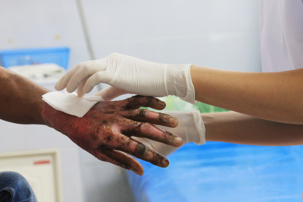
Robotics is beginning to play a role in the intricate and demanding procedures involved in burn surgery.
- Robotic Debridement: Automated systems can provide highly precise and controlled removal of necrotic tissue, minimizing damage to viable surrounding tissue. These systems can integrate with imaging technologies to guide debridement, potentially reducing blood loss and improving surgical efficiency.
- Automated Graft Harvesting and Application: Robots are being developed to assist in harvesting skin grafts with greater consistency and precision, and even to apply skin grafts to recipient sites. This technology aims to standardize procedures, reduce surgeon fatigue, and potentially improve the take of the grafts.
Digital Health and Long-Term Care Management
Managing full-thickness burn patients is a long and arduous process, often extending over months or years. Digital health solutions offer new ways to monitor, support, and rehabilitate patients throughout their recovery journey.
Telemedicine and Remote Monitoring for Burn Patients
Telemedicine has bridged geographical gaps, providing specialized burn care to remote areas and facilitating follow-up without constant hospital visits.
- Virtual Consultations: Patients can connect with burn specialists via video conferencing for initial assessments, post-discharge follow-ups, and scar management consultations. High-resolution cameras and integrated diagnostic tools can allow remote clinicians to assess wound healing and make recommendations.
- Wearable Sensors: Wearable devices can monitor vital signs, activity levels, and skin parameters (e.g., temperature, hydration) in burn patients, transmitting data wirelessly to healthcare providers. This allows for continuous monitoring, early detection of complications like infection, and personalized adjustments to rehabilitation plans.
Virtual Reality for Pain Management and Rehabilitation
VR technology offers immersive experiences that can significantly impact the psychological and physical recovery of burn patients.
- Distraction Therapy for Pain: During painful procedures like dressing changes or physical therapy, VR can transport patients to engaging virtual environments, significantly reducing their perception of pain and anxiety. This reduces the need for pharmacological interventions and improves patient compliance with necessary treatments.
- Rehabilitation and Physical Therapy: Interactive VR games and simulations can make tedious rehabilitation exercises more engaging, motivating patients to improve range of motion, strength, and coordination. Customized VR scenarios can target specific functional deficits related to burn contractures and muscle weakness.
Data Analytics for Personalized Burn Care Protocols
Leveraging big data and analytics is crucial for refining burn care protocols and moving towards truly personalized medicine.
- Outcome Prediction and Risk Stratification: By analyzing historical patient data, AI models can identify risk factors for complications such as hypertrophic scarring, contractures, or infection, allowing for early preventative interventions.
- Treatment Optimization: Data analytics can help identify which treatment regimens, surgical techniques, or rehabilitation protocols yield the best outcomes for specific patient profiles, leading to more evidence-based and tailored care plans.
The Future of Burn Care: Merging Biology and Robotics
The trajectory of burn care is increasingly defined by the convergence of biological understanding with advanced engineering and computational power. The next frontier will likely see even more integrated systems that are proactive, personalized, and minimally invasive.
Nanotechnology in Scar Prevention and Healing
Nanomaterials hold immense potential for not only delivering therapeutic agents but also directly influencing cellular processes involved in wound healing and scar formation. Smart nanoparticles could be engineered to modulate inflammation, promote organized collagen deposition, and inhibit fibroblast proliferation, ultimately leading to scarless or near-scarless healing.

AI-Driven Predictive Models for Outcomes
The continuous evolution of AI will lead to sophisticated models capable of dynamically predicting burn healing, complications, and long-term functional and aesthetic outcomes with unprecedented accuracy. These models, fed by real-time patient data from wearables, imaging, and genomic information, will inform adaptive treatment strategies, making burn care truly personalized and anticipatory.
In conclusion, a full-thickness burn represents a profound challenge to human physiology. However, through relentless innovation in diagnostics, regenerative therapies, digital health, and robotics, technology is continuously redefining the possibilities for understanding, treating, and ultimately restoring the lives of individuals affected by these devastating injuries.
aViewFromTheCave is a participant in the Amazon Services LLC Associates Program, an affiliate advertising program designed to provide a means for sites to earn advertising fees by advertising and linking to Amazon.com. Amazon, the Amazon logo, AmazonSupply, and the AmazonSupply logo are trademarks of Amazon.com, Inc. or its affiliates. As an Amazon Associate we earn affiliate commissions from qualifying purchases.