
Digital radiography (DR) represents one of the most significant technological leaps in the field of diagnostic imaging. By replacing traditional film-based X-ray systems with electronic sensors, DR has revolutionized how medical professionals visualize the internal structures of the human body. This shift from analog to digital has not only improved diagnostic accuracy but has also streamlined clinical workflows, reduced patient radiation exposure, and enhanced the overall quality of patient care.
The Evolution from Film to Digital Imaging
The transition to digital radiography is rooted in the move away from the chemical-based processing required by conventional film X-rays. For decades, radiologists relied on intensifying screens and photographic film, which required a darkroom and various chemical baths to develop the images. This process was not only time-consuming but also prone to environmental inconsistencies and physical degradation.
The Mechanics of Digital Detectors
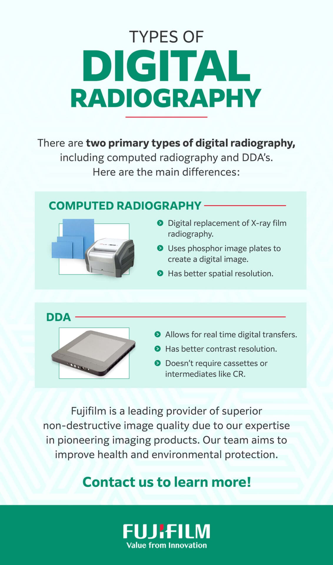
Digital radiography utilizes solid-state detectors—often referred to as digital detector arrays (DDAs)—that convert X-ray energy directly or indirectly into electrical signals. These detectors are essentially sophisticated semiconductors that capture the photon data immediately upon exposure. The result is an instant electronic image that can be viewed on a high-resolution monitor within seconds, eliminating the minutes or hours previously spent in the darkroom.
Direct vs. Indirect Conversion
Understanding digital radiography requires a distinction between the two primary capture technologies:
- Direct Conversion: These systems use a photoconductor, typically amorphous selenium, to convert X-rays directly into an electronic charge. This process offers superior spatial resolution because it minimizes the “blurring” of light signals.
- Indirect Conversion: These systems utilize a scintillator—often made of cesium iodide or gadolinium oxysulfide—which converts X-rays into light. This light is then captured by an amorphous silicon photodiode array and converted into an electrical signal. While slightly less sharp than direct systems, indirect conversion is highly efficient and remains the standard for many general-purpose imaging applications.
Key Advantages of Digital Radiography Systems
The integration of digital technology into radiology suites offers a host of tangible benefits. These advantages extend beyond mere convenience, impacting the financial efficiency of clinics and the long-term health outcomes of patients.
Immediate Image Processing and Workflow Efficiency
In a traditional film environment, an improperly exposed X-ray might not be discovered until the film is developed, necessitating a repeat exposure that inconveniences the patient and delays care. With DR, the image appears on the screen almost instantaneously. If the positioning is slightly off or the technique needs adjustment, the radiographer can retake the image immediately. This real-time feedback loop drastically improves throughput in busy hospital settings.
Enhanced Image Manipulation and Diagnostic Clarity
Digital images provide radiologists with tools that were previously impossible to utilize. Once an image is captured, software allows for:
- Windowing and Leveling: Adjusting the contrast and brightness to highlight specific densities, such as bone structures versus soft tissues.
- Magnification and Zoom: Inspecting minute fractures or subtle abnormalities without a loss of detail.
- Edge Enhancement: Using algorithms to sharpen the borders of objects, which is particularly useful for identifying subtle hairline fractures in pediatric or geriatric imaging.
Significant Reduction in Radiation Dosage
One of the most profound benefits of digital radiography is the concept of ALARA (As Low As Reasonably Achievable). Because digital sensors have a much higher “detective quantum efficiency” (DQE)—meaning they are better at utilizing the photons that reach them compared to traditional film—they require less radiation to produce a high-quality image. This is a critical development for patient safety, particularly in repetitive imaging scenarios.

Infrastructure and Digital Integration
Digital radiography does not exist in a vacuum; it is part of a complex ecosystem of health information technology. The efficacy of a DR system is entirely dependent on its ability to communicate with the broader hospital network.
PACS and DICOM Connectivity
The primary backbone of digital radiology is the Picture Archiving and Communication System (PACS). PACS allows for the storage, retrieval, and distribution of images across a hospital network. All digital radiography systems must comply with the DICOM (Digital Imaging and Communications in Medicine) standard, which ensures that equipment from different manufacturers can share data seamlessly. This interoperability means a patient’s imaging history can be pulled up in an emergency room, a surgeon’s office, or a remote clinic, facilitating collaborative medicine.
Tele-Radiology Capabilities
Because digital images are essentially data files, they can be transmitted over high-speed networks instantly. This has given rise to teleradiology, where images taken in a rural clinic can be analyzed by a sub-specialist radiologist located thousands of miles away. This capability is essential for equitable healthcare, ensuring that advanced diagnostic expertise is accessible to patients regardless of their geographical location.
Future Trends and The Role of AI in Radiography
As digital radiography becomes the global standard, the focus is shifting from simple image capture to intelligent image interpretation. The next wave of innovation is being driven by Artificial Intelligence and machine learning.
AI-Powered Diagnostic Assistance
Artificial Intelligence is currently being integrated into the radiography workflow to act as a “second pair of eyes.” Algorithms are trained to scan images for specific anomalies—such as a small pneumothorax (collapsed lung) or a subtle nodule—before the radiologist even opens the file. By prioritizing these “urgent” cases in the reading queue, hospitals can ensure that critical patients receive attention faster.
Automated Quality Control
Future systems are moving toward autonomous quality control. AI can evaluate an image immediately after capture to determine if it meets diagnostic criteria. If the image is blurred due to motion or if the exposure index is outside the acceptable range, the system can notify the technician automatically, ensuring that only high-quality data enters the patient’s record.
Portable and Bedside Digital Systems
The miniaturization of electronic sensors is leading to an increase in portable, high-quality digital X-ray units. These devices allow imaging to be brought directly to the bedside of critically ill patients who cannot be moved to a radiology department. Modern portable units are now battery-operated and feature wireless image transmission, allowing for seamless integration into intensive care units and emergency settings.
Conclusion
Digital radiography has fundamentally altered the landscape of medical diagnostics. By bridging the gap between hardware and software, it has provided clinicians with the tools to work faster, safer, and with higher precision. The shift from physical film to electronic data has paved the way for advanced connectivity, enabling better patient outcomes through improved accessibility and the burgeoning potential of AI-driven analysis. As technology continues to advance, digital radiography will remain the cornerstone of the diagnostic process, continuously refining the balance between technological innovation and compassionate, evidence-based patient care.
aViewFromTheCave is a participant in the Amazon Services LLC Associates Program, an affiliate advertising program designed to provide a means for sites to earn advertising fees by advertising and linking to Amazon.com. Amazon, the Amazon logo, AmazonSupply, and the AmazonSupply logo are trademarks of Amazon.com, Inc. or its affiliates. As an Amazon Associate we earn affiliate commissions from qualifying purchases.