
At the intersection of biology and advanced technology lies the fundamental understanding of specialized cells that drive innovation in medicine and biotechnology. Among these, the chondrocyte stands out as a critical component, primarily known for its role in cartilage formation and maintenance. Far from being merely a biological curiosity, the study and manipulation of chondrocytes form the bedrock for pioneering technological advancements in regenerative medicine, orthopedics, and diagnostics. Grasping the intricate nature of these cells is not just about biological comprehension; it’s about unlocking the potential for new software algorithms, sophisticated medical devices, and AI-driven therapeutic strategies that promise to revolutionize human health.
The Foundational Cell: Bridging Biology to Biomedical Technology
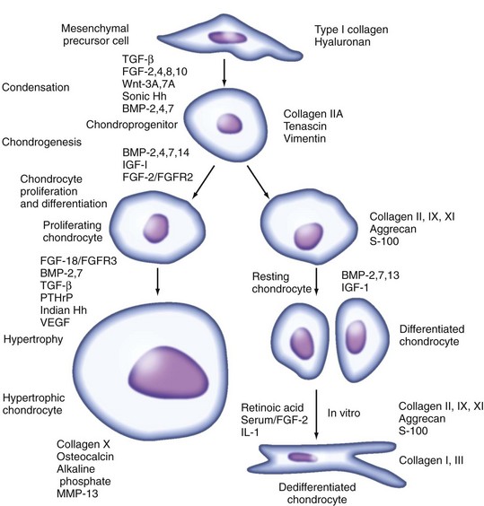
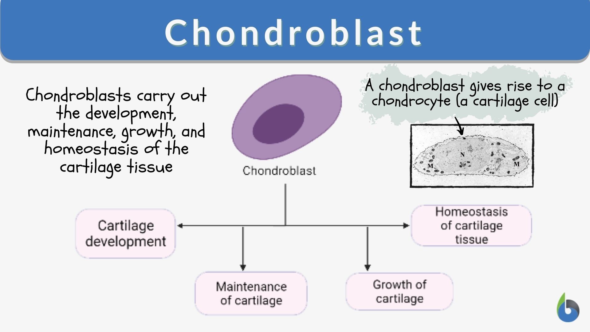
A chondrocyte is the sole cell type found within healthy cartilage, a flexible connective tissue present in various parts of the body, including joints, the rib cage, ears, nose, bronchial tubes, and intervertebral discs. These specialized cells are derived from mesenchymal stem cells and are responsible for producing and maintaining the cartilaginous matrix, which is primarily composed of collagen fibers, proteoglycans, and elastin. This extracellular matrix (ECM) provides cartilage with its unique properties: resilience, shock absorption, and a smooth, low-friction surface for joints. Unlike most tissues, cartilage is avascular, meaning it lacks blood vessels, which significantly impacts its ability to repair itself after injury. Chondrocytes rely on diffusion for nutrient supply and waste removal, making their environment and metabolic activity particularly delicate and complex.
Understanding the specific biological characteristics of chondrocytes – their limited proliferative capacity, sensitivity to mechanical load, and reliance on an anaerobic metabolic pathway – is absolutely crucial for the development of any effective biomedical technology targeting cartilage repair or regeneration. For instance, designing biomaterials for tissue engineering requires intimate knowledge of how chondrocytes interact with different scaffold architectures and biochemical cues. Developing advanced imaging software to detect early signs of osteoarthritis hinges on accurately mapping chondrocyte distribution and health. Furthermore, crafting AI models to predict disease progression or therapeutic efficacy demands comprehensive datasets on chondrocyte gene expression, morphology, and physiological responses. The journey from basic cell biology to cutting-edge medical technology inescapably passes through the detailed study of the chondrocyte.
AI and Computational Tools Revolutionizing Chondrocyte Research
The complexity of chondrocyte behavior and the microenvironment they inhabit makes them an ideal subject for advanced computational analysis and artificial intelligence (AI). Modern biotechnology labs are increasingly leveraging AI tools, machine learning (ML) algorithms, and sophisticated software to unravel the mysteries of cartilage biology and develop targeted interventions.
Predictive Modeling and Machine Learning for Cell Behavior
AI and ML algorithms are being deployed to analyze vast datasets generated from chondrocyte research, including gene expression profiles, proteomic data, and metabolic pathways. These algorithms can identify subtle patterns and correlations that human analysis might miss, leading to a deeper understanding of chondrocyte differentiation, proliferation, and matrix production under various physiological and pathological conditions. For instance, ML models can predict how specific growth factors or pharmacological agents might influence chondrocyte viability or their ability to synthesize collagen, accelerating drug discovery and optimizing cell culture protocols for in vitro cartilage generation. Furthermore, AI can model the biomechanical stress response of chondrocytes, predicting how different physical loads affect cell health and matrix integrity, which is vital for designing prosthetic components or rehabilitation protocols. The iterative learning capacity of these systems allows researchers to refine hypotheses and prioritize experiments, significantly shortening the development cycle for new therapies.
Digital Imaging and Analysis Software
Advanced digital imaging techniques, such as high-resolution microscopy, magnetic resonance imaging (MRI), and computed tomography (CT) scans, generate enormous amounts of visual data on chondrocytes and cartilage tissue. AI-powered image analysis software is transforming how this data is interpreted. Instead of manual quantification, which is time-consuming and prone to human error, algorithms can automatically segment chondrocytes from tissue sections, quantify their density, measure cell morphology, and assess the integrity of the surrounding extracellular matrix. For clinical applications, AI tools are being trained to detect subtle changes in cartilage structure visible in MRI scans, potentially enabling earlier and more accurate diagnosis of conditions like osteoarthritis. These software platforms can identify early signs of cartilage degeneration, quantify lesion size, and even predict disease progression, offering a non-invasive, objective method to monitor treatment effectiveness and improve patient outcomes. The integration of computer vision with deep learning networks allows for unprecedented precision in analyzing the complex microarchitecture of cartilage.

Advanced Engineering and Devices for Chondrocyte-Based Therapies
The insights gained from fundamental chondrocyte biology and computational analysis directly fuel the development of advanced engineering solutions and sophisticated medical devices. These technologies aim to overcome the inherent limitations of cartilage repair and offer innovative therapeutic avenues.
3D Bioprinting and Tissue Engineering
One of the most exciting technological frontiers in regenerative medicine is 3D bioprinting, where chondrocytes are utilized as biological “ink” to create functional cartilage constructs. Using specialized bioprinters, researchers can precisely deposit chondrocytes, often encapsulated within biocompatible hydrogels, layer by layer, to form complex three-dimensional structures that mimic native cartilage. This robotic precision allows for custom-designed implants that match a patient’s specific anatomical defects, promising truly personalized medicine. The technology involves intricate software for designing print patterns, controlling cell viability during the printing process, and integrating growth factors to encourage tissue maturation. Beyond simple scaffolds, the goal is to bioprint fully functional cartilaginous tissue that can be implanted into patients, reducing pain and restoring mobility. The continuous advancement in biomaterials science, combined with engineering prowess, is steadily bringing this vision closer to clinical reality.
Biosensors and Microfluidics in In Vitro Studies
To better understand chondrocyte behavior outside the body, scientists are developing cutting-edge biosensors and microfluidic “lab-on-a-chip” devices. These miniature platforms allow for the precise control of the cellular microenvironment and real-time monitoring of chondrocyte activity. Biosensors can detect subtle changes in pH, oxygen levels, glucose metabolism, or the presence of specific biomarkers released by chondrocytes, providing unprecedented insights into their physiological state. Microfluidic devices, often designed with sophisticated CAD software, can mimic the mechanical loading conditions experienced by chondrocytes in a joint, or create nutrient gradients, enabling researchers to study cell responses under highly controlled and physiologically relevant conditions. These “gadgets” offer high-throughput screening capabilities for various therapeutic compounds, significantly accelerating the process of identifying effective treatments for cartilage disorders while reducing reliance on animal testing.
Gene Editing Technologies and Delivery Systems
The advent of gene editing technologies, particularly CRISPR-Cas9, has opened new frontiers for therapeutic interventions targeting chondrocytes. These powerful molecular “tools” allow scientists to precisely modify the genetic code within chondrocytes to correct mutations that cause genetic cartilage disorders or to enhance the cells’ regenerative capacity. For instance, gene editing could be used to make chondrocytes more resistant to inflammatory processes or to boost their production of crucial extracellular matrix components. However, the successful application of gene editing requires advanced delivery systems. Nanoparticle-based delivery systems, for example, are engineered “gadgets” designed at the nanoscale to safely and efficiently transport gene-editing components into chondrocytes in vivo or ex vivo. Developing these sophisticated delivery vehicles involves advanced materials science, computational chemistry, and precision engineering to ensure targeted and effective cellular uptake without off-target effects.
Data Security and Management in Chondrocyte-Related Biomedical Research
The expansive research and development surrounding chondrocytes generate an immense volume of sensitive data, ranging from genomic sequences and proteomic profiles to high-resolution imaging data and clinical trial results. The proper management and security of this data are paramount within the biotechnology and medical technology sectors.
Secure cloud infrastructure provides the necessary scalability and robust computational power to store and process these large datasets, facilitating collaborative research across institutions while maintaining data integrity. However, the reliance on cloud computing necessitates stringent digital security protocols to protect against unauthorized access, data breaches, and cyber-attacks. Advanced encryption techniques, multi-factor authentication, and intrusion detection systems are critical software components for safeguarding intellectual property and proprietary research data related to novel chondrocyte therapies.
Furthermore, compliance with international data privacy regulations, such as HIPAA in the United States and GDPR in Europe, is non-negotiable, especially when clinical trial data involving patient-derived chondrocytes or related therapeutic outcomes are concerned. Implementing blockchain technology could offer a decentralized, immutable ledger for tracking research data, ensuring transparency and accountability in data provenance and access control. Robust cybersecurity frameworks are not just a regulatory burden; they are an essential foundation for fostering trust, accelerating scientific discovery, and ensuring the ethical translation of chondrocyte research into viable, safe, and effective medical technologies.
aViewFromTheCave is a participant in the Amazon Services LLC Associates Program, an affiliate advertising program designed to provide a means for sites to earn advertising fees by advertising and linking to Amazon.com. Amazon, the Amazon logo, AmazonSupply, and the AmazonSupply logo are trademarks of Amazon.com, Inc. or its affiliates. As an Amazon Associate we earn affiliate commissions from qualifying purchases.