
Cardiac perfusion is a fundamental physiological process, referring to the delivery of oxygenated blood to the myocardial tissue – the heart muscle itself. It is the lifeblood of the heart, ensuring that the cardiac cells receive the oxygen and nutrients necessary to contract rhythmically and efficiently, pumping blood to the rest of the body. Without adequate perfusion, the heart muscle weakens, leading to dysfunction, damage, and ultimately, heart failure or myocardial infarction (heart attack).
While the definition of cardiac perfusion is rooted deeply in biology, our ability to understand, diagnose, monitor, and therapeutically intervene in this critical process is almost entirely dependent on the relentless innovation and application of advanced medical technology. From sophisticated imaging modalities that visualize blood flow to intricate life-support machines that temporarily replace cardiac function, technology serves as the eyes, hands, and extended capacity for clinicians managing cardiac perfusion. This exploration delves into the technological landscape that underpins our modern understanding and management of cardiac perfusion, squarely positioning the discussion within the “Tech” niche by examining the tools, systems, and innovations that make it all possible.

The Criticality of Cardiac Perfusion and Early Diagnostic Technologies
The heart, as the body’s central pump, demands a constant and robust supply of oxygen and nutrients to fuel its incessant work. Any compromise in this supply—known as ischemia—can rapidly lead to cellular damage and death. Historically, assessing cardiac perfusion was a challenge, relying on indirect clinical signs and symptoms. However, technological advancements have revolutionized our diagnostic capabilities, allowing for increasingly precise and non-invasive evaluation of myocardial blood flow.
The Biological Imperative and Indirect Assessment
At its core, cardiac perfusion is about maintaining the delicate balance between oxygen supply and demand within the heart muscle. The coronary arteries are responsible for this vital delivery. When these arteries narrow or become blocked, perfusion is compromised, leading to conditions like angina or myocardial infarction. Early diagnostic approaches relied heavily on patient history, physical examination, and basic electrocardiography (ECG). While the ECG could detect electrical changes indicative of ischemia or infarction, it provided only an indirect snapshot and lacked the ability to directly visualize blood flow or quantify perfusion deficits. These initial technological steps, though rudimentary by today’s standards, laid the groundwork for more sophisticated methods by confirming the electrical consequences of impaired perfusion.
Imaging the Invisible: Non-Invasive Perfusion Assessment
A major leap in understanding cardiac perfusion came with the development of imaging technologies that could either directly or indirectly visualize blood flow to the heart muscle. These non-invasive techniques have become cornerstones of modern cardiology.
-
Myocardial Perfusion Imaging (MPI): SPECT and PET: These nuclear medicine techniques revolutionized the assessment of perfusion.
-
Single-Photon Emission Computed Tomography (SPECT): Patients are injected with a small amount of a radioactive tracer (e.g., Thallium-201, Technetium-99m Sestamibi) which is taken up by healthy heart muscle cells in proportion to blood flow. A special camera then detects the emitted gamma rays, creating 3D images of tracer distribution. By performing scans both at rest and after stress (pharmacological or exercise-induced), clinicians can compare perfusion patterns and identify areas of reduced blood flow (ischemia) or scar tissue (infarction).
-
Positron Emission Tomography (PET): Offering superior resolution and quantification capabilities compared to SPECT, PET uses different radioactive tracers (e.g., Rubidium-82, Ammonia N-13). PET provides more accurate absolute quantification of myocardial blood flow, which can be critical for precise diagnosis and guiding treatment, especially in complex cases or microvascular dysfunction.
-
-
Cardiac Magnetic Resonance Imaging (cMRI) with Perfusion Sequences: MRI technology, particularly its cardiac applications, has become indispensable. With dedicated perfusion sequences, cMRI can visualize the first pass of a gadolinium-based contrast agent through the myocardium. This allows for a detailed assessment of regional myocardial blood flow, identifying areas of hypoperfusion, often with superior spatial resolution to nuclear methods, and without ionizing radiation.
-
Contrast Echocardiography: This technique involves injecting microscopic gas-filled bubbles (microbubbles) into the bloodstream. These bubbles enhance the ultrasound signal, allowing sonographers to visualize blood flow within the myocardial capillaries. Contrast echo can detect perfusion defects, especially in patients where traditional echo views are suboptimal, providing real-time assessment of myocardial viability.
These imaging technologies have transformed cardiac care by allowing clinicians to non-invasively identify, localize, and quantify perfusion abnormalities, leading to more targeted interventions and improved patient outcomes.
Advanced Monitoring and Real-time Perfusion Analysis Systems
Beyond initial diagnosis, maintaining optimal cardiac perfusion, especially in critically ill patients or during complex surgical procedures, requires continuous, real-time monitoring. Modern technology provides a suite of tools for assessing systemic and regional perfusion, offering clinicians critical data to guide immediate decision-making.
Hemodynamic Monitoring Devices
These devices measure various parameters related to blood flow and pressure, providing indirect but crucial insights into the adequacy of cardiac perfusion.
-
Invasive Hemodynamic Monitoring: Technologies like arterial lines provide continuous blood pressure measurements, while pulmonary artery (Swan-Ganz) catheters offer more comprehensive data, including cardiac output, pulmonary artery pressures, and central venous pressure. These parameters, when interpreted in conjunction, help assess the overall circulatory status and indirectly infer the adequacy of perfusion to vital organs, including the heart.
-
Non-Invasive Cardiac Output Monitors: Advances have led to non-invasive alternatives for measuring cardiac output, such as devices utilizing bioreactance or impedance cardiography. These technologies estimate blood flow using external sensors, reducing the risks associated with invasive lines while still providing continuous trend data for perfusion assessment.
Tissue-Level Perfusion Monitoring
While systemic hemodynamic monitoring provides a macro-level view, understanding perfusion at the tissue level offers more granular insights.
-
Near-Infrared Spectroscopy (NIRS): NIRS is a non-invasive technology that measures regional tissue oxygen saturation (rSO2) by emitting near-infrared light into the tissue and detecting its absorption. It is commonly used for cerebral oximetry to monitor brain perfusion during cardiac surgery, but can also be applied to other tissues to assess regional oxygen delivery and consumption, providing direct insight into tissue perfusion status.
-
Microcirculatory Imaging: Emerging technologies, such as handheld vital microscopes, are used primarily in research settings to directly visualize blood flow in the microcirculation (the smallest blood vessels) of accessible tissues (e.g., sublingual area). While not yet routine for cardiac perfusion, this technology highlights the ongoing push to understand and monitor perfusion at its most fundamental level.
Integrated Monitoring Platforms and Predictive Analytics
Modern intensive care units and operating rooms are equipped with sophisticated patient monitoring systems that integrate data from multiple sources—ECG, blood pressure, oxygen saturation, NIRS, capnography, and more. These platforms consolidate information, often displaying it graphically, to provide a comprehensive and easily interpretable overview of a patient’s physiological status.
Furthermore, advancements in computing power and artificial intelligence are leading to predictive analytics tools. These systems analyze vast amounts of real-time data to identify subtle patterns or trends that may indicate impending perfusion compromise, allowing clinicians to intervene proactively before a crisis develops. This integration of data and intelligent analysis represents a significant technological leap in maintaining optimal cardiac perfusion.
Technological Interventions: Supporting and Restoring Perfusion

When cardiac perfusion is severely compromised, diagnostic and monitoring technologies are complemented by powerful interventional devices designed to support or completely take over the heart’s pumping function, thereby restoring essential blood flow to the myocardium and systemic circulation.
Cardiopulmonary Bypass (CPB) Machines
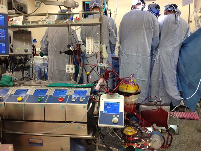
Perhaps the most iconic piece of perfusion technology is the cardiopulmonary bypass (CPB) machine, often referred to as a heart-lung machine. This complex system is indispensable during open-heart surgery, allowing surgeons to operate on a still, bloodless heart.
-
Functionality: A CPB machine temporarily assumes the functions of both the heart and lungs. It consists of:
-
A Pump: To circulate blood throughout the body.
-
An Oxygenator: To add oxygen to the blood and remove carbon dioxide.
-
A Heat Exchanger: To regulate body temperature.
-
Filters: To remove emboli and debris.
-
-
Technological Evolution: Modern CPB machines are highly sophisticated, featuring advanced controls for flow rates, pressure, gas exchange, and temperature. Ongoing research focuses on miniaturization, biocompatibility (to reduce inflammatory responses), and automation to improve patient outcomes and reduce complications associated with bypass. These machines are a testament to engineering excellence, allowing for life-saving surgeries that would otherwise be impossible.
Mechanical Circulatory Support (MCS) Devices
For patients experiencing acute or chronic heart failure, where the heart cannot adequately perfuse the body, mechanical circulatory support (MCS) devices provide crucial assistance.
-
Intra-Aortic Balloon Pumps (IABP): The IABP is a temporary device inserted into the aorta. It inflates during diastole (when the heart relaxes), increasing coronary blood flow (thus improving cardiac perfusion), and deflates during systole (when the heart contracts), reducing afterload and making it easier for the heart to pump.
-
Ventricular Assist Devices (VADs): These are mechanical pumps surgically implanted to support the failing ventricles. VADs can be short-term bridges to recovery or transplantation, or long-term destination therapy. Modern VADs are small, durable, and battery-powered, continuously pumping blood from the ventricle into the aorta or pulmonary artery, ensuring systemic and, by extension, coronary perfusion.
-
Extracorporeal Membrane Oxygenation (ECMO): ECMO is an advanced form of life support that provides full cardiopulmonary support for patients with severe heart and/or lung failure. Similar to CPB but designed for longer-term use (days to weeks), ECMO circulates blood outside the body through an oxygenator and pump, returning oxygenated blood to the patient, thereby maintaining systemic and cardiac perfusion when the heart and lungs cannot.
Drug Delivery Systems and Smart Infusion Pumps
While not devices that directly pump blood, the precise delivery of pharmacological agents is critical for optimizing cardiac perfusion. Advanced infusion pumps and drug delivery systems ensure accurate and controlled administration of medications (e.g., inotropes to improve contractility, vasopressors to maintain blood pressure, vasodilators to improve blood flow). “Smart” infusion pumps incorporate drug libraries and safety features to prevent medication errors, thereby indirectly supporting perfusion by maintaining optimal physiological conditions.
The Future Landscape: AI, Robotics, and Personalized Perfusion
The trajectory of technology in cardiac perfusion is one of increasing sophistication, integration, and personalization. The future promises even more precise, less invasive, and highly individualized approaches, driven by artificial intelligence, robotics, and advanced biological insights.
Artificial Intelligence in Perfusion Management
AI is poised to transform various aspects of perfusion management:
-
Predictive Analytics: AI algorithms can analyze continuous streams of patient data to predict the likelihood of perfusion compromise, allowing for preemptive interventions. This includes forecasting the risk of cardiac events or optimizing parameters during CPB or ECMO based on real-time physiological responses.
-
Automated Interpretation: AI can assist in the automated interpretation of complex perfusion imaging studies, identifying subtle abnormalities that might be missed by the human eye and improving diagnostic consistency.
-
Treatment Optimization: AI could guide the optimal titration of medications or the fine-tuning of mechanical support devices, tailoring interventions precisely to an individual patient’s needs and dynamic physiological state.
Robotics and Precision Surgery
Robotic-assisted cardiac surgery (e.g., using systems like the Da Vinci Surgical System) is already a reality. Robotics enable surgeons to perform complex procedures with enhanced precision, dexterity, and visualization through small incisions. This minimally invasive approach can lead to reduced trauma, faster recovery times, and potentially improved post-operative myocardial perfusion by minimizing surgical stress and inflammation.
Advanced Imaging and Multi-Omics Integration
Future imaging technologies will likely offer even higher resolution and more detailed physiological information, such as quantifying microvascular perfusion at an unprecedented level. Furthermore, integrating perfusion data with multi-omics data (genomics, proteomics, metabolomics) will allow for a truly personalized understanding of an individual’s cardiac perfusion status, identifying genetic predispositions to coronary artery disease or specific metabolic pathways that impact myocardial blood flow.
Wearable and Remote Monitoring
The development of sophisticated wearable sensors and remote monitoring technologies holds promise for continuous, non-invasive assessment of cardiac perfusion parameters in at-risk populations. Imagine smart patches or integrated devices that can detect early signs of ischemia or changes in cardiac output, alerting patients and clinicians long before symptoms become severe, thus enabling proactive management and prevention of acute events.
Conclusion
Cardiac perfusion, the continuous supply of life-sustaining blood to the heart muscle, stands as a cornerstone of cardiovascular health. While inherently a biological phenomenon, our profound understanding and sophisticated management of this process are inextricably linked to the relentless march of technological innovation. From the early days of indirect assessment to today’s high-resolution imaging, real-time monitoring, and advanced mechanical support systems, technology has consistently pushed the boundaries of what’s possible in cardiac care.
The journey continues, with artificial intelligence, robotics, and integrated data analytics promising even more personalized, predictive, and preventive approaches to maintaining and restoring cardiac perfusion. In essence, the narrative of cardiac perfusion is not just a biological one, but a compelling testament to the interdisciplinary synergy between life sciences and cutting-edge engineering, constantly evolving to save lives and enhance the quality of life for millions.
aViewFromTheCave is a participant in the Amazon Services LLC Associates Program, an affiliate advertising program designed to provide a means for sites to earn advertising fees by advertising and linking to Amazon.com. Amazon, the Amazon logo, AmazonSupply, and the AmazonSupply logo are trademarks of Amazon.com, Inc. or its affiliates. As an Amazon Associate we earn affiliate commissions from qualifying purchases.