
The C-arm stands as a cornerstone in modern medical imaging, an indispensable piece of technology that empowers clinicians with real-time, high-resolution X-ray images during a vast array of diagnostic and interventional procedures. Its distinctive “C” shape, which gives the device its name, allows for unparalleled flexibility and positioning around a patient, providing dynamic views that static X-rays cannot offer. Essentially, a C-arm is a mobile fluoroscopic device used primarily in surgery, orthopedics, pain management, and emergency medicine, bridging the gap between static imaging and continuous visual guidance.
The Core Technology: How C-Arms Work
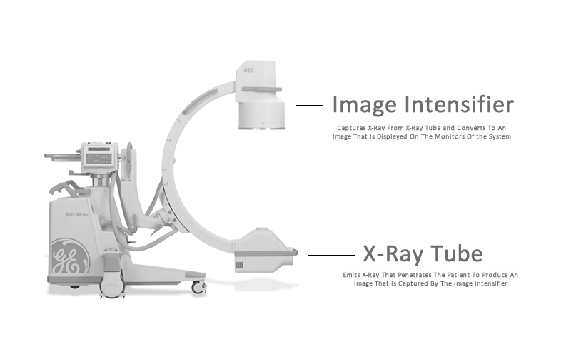
At its heart, a C-arm operates on the fundamental principles of X-ray imaging, but with crucial enhancements that facilitate its real-time capabilities. The device comprises two primary components mounted at opposite ends of the C-shaped arc: an X-ray source and an image intensifier or flat panel detector.
X-Ray Generation and Image Intensification
The X-ray source, typically located on one end of the “C,” generates a controlled beam of radiation that passes through the patient’s body. As these X-rays traverse different tissues, bones, and organs, they are attenuated (absorbed or scattered) to varying degrees. On the opposite end of the C-arm, the image intensifier (or detector) captures the attenuated X-rays.
An image intensifier, a traditional component in older C-arms, converts the X-ray photons into light photons, which are then amplified and converted into an electronic signal. This signal is subsequently displayed as a real-time image on an attached monitor. This process allows medical professionals to visualize internal structures and movements instantaneously, making it invaluable for guiding instruments or confirming anatomical positions.
Digital Imaging and Processing
Modern C-arms often utilize flat panel detectors (FPDs) instead of image intensifiers. FPDs represent a significant technological leap, offering several advantages. These detectors directly convert X-ray photons into a digital signal, eliminating several conversion steps inherent in image intensifiers. This direct conversion results in superior image quality, higher spatial resolution, and a wider dynamic range, meaning they can capture a broader spectrum of tissue densities with greater clarity.
Furthermore, FPDs are less susceptible to image distortion, which can sometimes occur at the periphery of image intensifiers. The digital data captured by FPDs is then processed by sophisticated computer algorithms. These algorithms can enhance image clarity, reduce noise, and allow for various post-processing functions like magnification, contrast adjustment, and digital subtraction angiography (DSA). DSA, for instance, involves subtracting a “mask” image (taken before contrast injection) from subsequent images (taken after contrast injection), thereby highlighting blood vessels by removing overlapping bone and soft tissue structures.
Design and Mobility: The “C” Shape Advantage
The defining characteristic of a C-arm, its iconic “C” shape, is not merely aesthetic but a critical design element that underpins its functionality and versatility in clinical environments. This design allows for a unique range of motion and positioning capabilities.
Ergonomics and Positioning
The C-shaped gantry enables the X-ray source and detector to rotate independently around the patient, providing oblique, lateral, and anteroposterior (AP) views without repositioning the patient. This flexibility is crucial in surgical settings where patient movement might be impractical or contraindicated. Clinicians can adjust the angulation and rotation of the C-arm to obtain the precise projection needed to visualize intricate anatomical details or guide delicate instruments, all while maintaining sterile fields. The ability to “orbit” the C-arm around a fixed point of interest, known as isocentric rotation, ensures that the target remains centered in the image regardless of the C-arm’s angle, significantly simplifying navigation.
Movement and Flexibility in Clinical Settings
Beyond its rotational capabilities, the C-arm is designed for high mobility. Most units are equipped with wheels, allowing them to be easily transported between operating rooms, emergency departments, or imaging suites. This portability is a key advantage, bringing advanced imaging directly to the point of care rather than requiring patients to be moved to a fixed imaging facility. Advanced models feature motorized movements for height adjustment, horizontal slide, and orbital rotation, all controllable via intuitive user interfaces or even foot pedals, allowing surgeons to operate the device while keeping their hands free. This ease of movement and precise positioning minimizes procedural delays and enhances workflow efficiency.
Key Applications Across Medical Specialties
The versatility of the C-arm makes it an indispensable tool across a wide spectrum of medical disciplines, providing real-time imaging guidance that enhances accuracy, safety, and outcomes.
Orthopedic Surgery and Traumatology
In orthopedics, C-arms are foundational. They are extensively used for fracture reduction and fixation, allowing surgeons to visualize bone alignment and the placement of plates, screws, and pins in real-time. This ensures optimal positioning and stability, minimizing the need for subsequent corrective surgeries. For complex spinal surgeries, a C-arm assists in precisely placing pedicle screws and verifying spinal alignment, critical for preventing neurological damage. Similarly, in joint replacements, it aids in component positioning and confirming prosthetic stability.
Pain Management and Anesthesiology

C-arms are vital for interventional pain management procedures. They guide the accurate placement of needles for nerve blocks, epidural injections, facet joint injections, and radiofrequency ablations. Real-time visualization ensures that medication is delivered precisely to the target nerve or joint, maximizing therapeutic effect and minimizing the risk of complications such as nerve damage or unintended tissue penetration. The ability to confirm needle tip location before injection significantly improves patient safety and procedural efficacy.
Vascular Procedures and Cardiology
In cardiology and vascular surgery, C-arms are used for diagnostic angiograms and interventional procedures like stent placement, angioplasty, and embolization. Digital Subtraction Angiography (DSA) capabilities are particularly useful here, providing clear images of blood vessels by removing distracting background anatomy. This allows clinicians to accurately assess blockages, visualize blood flow, and precisely deploy devices within the vascular system, crucial for treating conditions like peripheral artery disease, aneurysms, and coronary artery disease.
Emergency Medicine and Critical Care
In emergency settings, the rapid deployment and real-time imaging capabilities of a C-arm are invaluable. It aids in quickly diagnosing fractures, locating foreign bodies, and guiding emergency procedures such as central line placements or thoracostomy tube insertions. Its mobility allows it to be brought directly to a patient in trauma bays or intensive care units, facilitating immediate assessment and intervention without transporting critically ill or injured patients.
Types of C-Arms: From Standard to Specialized
The evolution of C-arm technology has led to the development of various types, each tailored to specific clinical needs and procedural demands.
Full-Size C-Arms
Full-size C-arms are the most common and versatile, found in operating rooms, emergency departments, and imaging suites. They feature larger image intensifiers or flat panel detectors, typically ranging from 9 to 12 inches (or more), offering a wider field of view. These systems are capable of handling a broad spectrum of procedures, from intricate orthopedic surgeries to complex vascular interventions, requiring high-resolution imaging and comprehensive anatomical coverage. Their robust design accommodates more powerful X-ray generators, providing deeper penetration for denser anatomy.
Mini C-Arms
Mini C-arms are smaller, more compact versions designed specifically for imaging extremities—hands, wrists, feet, and ankles. They feature smaller detectors (typically 4-6 inches) and lower X-ray dose outputs. Their compact footprint and enhanced maneuverability make them ideal for outpatient clinics, sports medicine facilities, and specialized orthopedic practices where only small anatomy needs to be visualized. While their field of view is limited, their portability, ease of use, and reduced radiation exposure for localized imaging make them highly efficient for specific applications.
Flat Panel Detector (FPD) C-Arms
FPD C-arms represent the cutting edge of the technology. As discussed, they replace traditional image intensifiers with digital flat panel detectors. These systems offer superior image quality, higher contrast resolution, and a larger dynamic range. They are also less bulky than image intensifier systems and are typically designed for lower radiation dose operation due to their higher detection efficiency. FPD C-arms are increasingly becoming the standard, especially for demanding procedures in vascular surgery, cardiology, and neurosurgery where image fidelity is paramount. Their digital nature also facilitates seamless integration into hospital Picture Archiving and Communication Systems (PACS) and allows for advanced image processing features.
Advantages and Considerations in Modern Healthcare
The integration of C-arms into medical practice has profoundly impacted patient care, offering numerous advantages while also necessitating careful consideration of certain factors.
Real-time Imaging Benefits
The primary advantage of C-arms is their ability to provide real-time, dynamic imaging. This immediate feedback loop is invaluable for guiding surgical instruments, confirming anatomical alignment, monitoring the flow of contrast agents, and verifying the successful completion of a procedure. This immediacy reduces the need for multiple static X-rays, decreasing overall procedure time and improving efficiency. For patients, this often translates to shorter anesthesia times, reduced risk, and potentially faster recovery. For clinicians, it means greater confidence in precision and outcomes.
Radiation Dose Management
While immensely beneficial, C-arms utilize ionizing radiation, which necessitates careful radiation dose management. Modern C-arms are equipped with various dose-saving features, including pulsed fluoroscopy, virtual collimation, dose reduction filters, and automatic dose rate control (ADRC). Pulsed fluoroscopy delivers X-rays in short bursts rather than continuously, significantly reducing exposure without compromising image quality for most procedures. Virtual collimation allows the operator to define the imaging area without exposing the patient to radiation during the collimation process. Despite these advancements, adherence to the ALARA (As Low As Reasonably Achievable) principle is paramount, requiring operators to use the lowest possible dose for the shortest possible time. Proper shielding for both patients and medical staff is also a critical consideration.
Integration with Navigation Systems
A significant technological trend is the integration of C-arms with advanced surgical navigation systems. These systems use optical or electromagnetic tracking technology to overlay real-time instrument positions onto pre-operative CT or MRI scans, or even directly onto the C-arm’s live fluoroscopic images. This fusion of imaging modalities creates a highly precise “GPS-like” system for surgeons, particularly in complex spinal, cranial, or orthopedic procedures. The C-arm provides the real-time bone and instrument position, while the navigation system correlates this with a comprehensive 3D anatomical map, enhancing accuracy, reducing risks, and potentially expanding the scope of minimally invasive techniques. This synergy represents a powerful advancement in image-guided surgery, pushing the boundaries of what is possible in precision medicine.
aViewFromTheCave is a participant in the Amazon Services LLC Associates Program, an affiliate advertising program designed to provide a means for sites to earn advertising fees by advertising and linking to Amazon.com. Amazon, the Amazon logo, AmazonSupply, and the AmazonSupply logo are trademarks of Amazon.com, Inc. or its affiliates. As an Amazon Associate we earn affiliate commissions from qualifying purchases.