In the rapidly evolving landscape of medical imaging, diagnostic technologies continue to push the boundaries of what is possible, offering increasingly detailed and insightful views into the human body. Among these innovations, 5D ultrasound stands out as a significant leap forward, building upon the foundations laid by its 2D, 3D, and 4D predecessors. Far from being a mere numerical increment, 5D ultrasound represents a sophisticated integration of automation, intelligent data processing, and advanced visualization, revolutionizing prenatal care, gynecological diagnostics, and various other medical applications.
The Evolution of Ultrasound Technology: A Technological Trajectory
To truly grasp the significance of 5D ultrasound, it’s essential to understand the journey of this diagnostic tool, from its rudimentary beginnings to its current state of sophisticated automation. Ultrasound technology has consistently leveraged advancements in computing power and transducer design to provide progressively richer diagnostic information.

From 2D to 3D and 4D: Adding Dimensions to Diagnostics
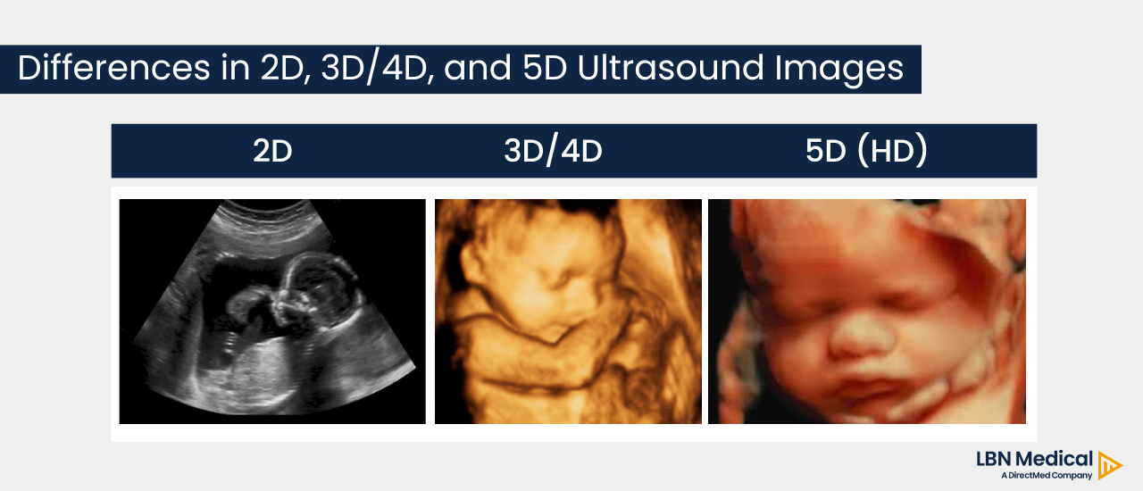
The foundational 2D ultrasound image, which has been a cornerstone of medical diagnostics for decades, provides flat, cross-sectional views of internal structures. These images, though invaluable, require a skilled operator to mentally reconstruct a three-dimensional understanding from a series of two-dimensional slices.
The advent of 3D ultrasound marked a substantial technological improvement. By acquiring multiple 2D images from different angles and then digitally compiling them, 3D ultrasound creates static, volumetric renderings of organs and tissues. This capability offered a more intuitive understanding of complex anatomical relationships, particularly beneficial in visualizing fetal structures and surface features.
Building upon 3D, 4D ultrasound introduced the element of time, adding motion to the volumetric images. Essentially, 4D ultrasound is a real-time 3D scan, allowing clinicians and expectant parents to observe fetal movements, facial expressions, and organ function in live video. This dynamic aspect significantly enhanced diagnostic capabilities, especially for assessing cardiac function and behavioral patterns. While 3D and 4D ultrasounds were groundbreaking, they often required considerable operator skill and time to acquire and reconstruct optimal images, and their analysis could still be somewhat subjective.
The Leap to 5D: A New Paradigm in Automation
The transition to 5D ultrasound is not simply about adding another dimension in the traditional sense; rather, it signifies an unprecedented integration of automation and intelligent software algorithms into the ultrasound workflow. The “5D” primarily refers to the inclusion of automated volume acquisition, intelligent navigation, and standardized measurement and analysis tools, which work in real-time to provide comprehensive diagnostic data with minimal operator input. This paradigm shift moves beyond mere visualization, entering the realm of automated quantification and streamlined diagnosis. It’s a leap that significantly enhances efficiency, reproducibility, and diagnostic accuracy, particularly in complex medical scenarios.
Deciphering 5D Ultrasound: How It Works
The core innovation of 5D ultrasound lies in its ability to automate traditionally manual and time-consuming tasks, transforming raw volumetric data into clinically actionable insights with remarkable speed and precision. This is achieved through sophisticated software algorithms that interact seamlessly with advanced transducer technology.
Automated Volume Calculation and Intelligent Navigation
At the heart of 5D ultrasound is its capability for automated volume calculation. Unlike previous generations where operators manually selected regions of interest or laboriously traced structures, 5D systems employ intelligent software to automatically detect and delineate specific anatomical structures within a captured 3D volume. For instance, in fetal imaging, the system can automatically identify and measure fetal organs like the brain, heart, or long bones from a single sweep, significantly reducing measurement variability and scan time.
This automation extends to intelligent navigation. Once a 3D volume is acquired, the 5D system can automatically generate a series of standardized 2D planes and 3D views that are most relevant for diagnostic assessment. For example, in fetal heart assessment, the system can automatically derive the standard cardiac views (e.g., four-chamber view, outflow tracts) from a single volume acquisition, guiding the clinician through a comprehensive examination protocol. This intelligent guidance system ensures that all necessary measurements and views are obtained, even by less experienced operators, thereby standardizing the diagnostic process.
Beyond Simple Visualization: Quantification and Reporting
While visualization remains a critical component, 5D ultrasound extends its utility far beyond simply seeing structures. It integrates advanced algorithms for automated quantification and reporting. Once structures are identified and measured, the system can automatically compare these measurements against normative databases, flagging any deviations that might indicate pathology. This immediate feedback assists clinicians in making rapid and informed decisions.
For example, in obstetrics, 5D systems can automatically measure fetal biometry (head circumference, abdominal circumference, femur length), amniotic fluid index, and even estimate fetal weight, presenting the data in a clear, standardized report. This not only saves time but also reduces the potential for human error inherent in manual measurements and calculations. The system essentially transforms complex raw data into understandable, clinically relevant information with minimal manual intervention.
Key Applications and Benefits Across Specialties
The enhanced capabilities of 5D ultrasound translate into substantial benefits across various medical disciplines, fundamentally altering diagnostic workflows and improving patient outcomes. Its automation and precision make it an invaluable tool in areas where detailed and reproducible imaging is critical.
Enhanced Fetal Diagnostics and Prenatal Care
Perhaps the most prominent application of 5D ultrasound is in enhanced fetal diagnostics and prenatal care. For obstetricians and maternal-fetal medicine specialists, 5D offers unparalleled efficiency and accuracy in assessing fetal development and screening for anomalies. The automated measurement of biometrics, the standardized evaluation of the fetal heart (e.g., 5D Heart, a feature that provides nine standard fetal cardiac views from a single volume), and the detailed analysis of the central nervous system (e.g., 5D CNS) significantly improve the detection rate of congenital malformations.
Furthermore, the ability to generate reproducible measurements helps in monitoring fetal growth and well-being over time with greater consistency. For expectant parents, the highly detailed, real-time images provide an emotionally engaging experience, fostering a stronger bond with their unborn child, while simultaneously offering clinicians robust data for diagnosis and management.
Gynecological Imaging Advancements
Beyond obstetrics, 5D ultrasound has made significant inroads into gynecological imaging. It excels in the detailed visualization and analysis of the uterus, ovaries, and fallopian tubes. For instance, 5D technologies can automatically delineate the uterine wall, measure endometrial thickness, and map uterine fibroids or polyps with greater precision than traditional 2D methods.
In fertility assessment, 5D ultrasound can precisely measure ovarian follicles, aiding in ovulation induction and in-vitro fertilization (IVF) procedures. The volumetric data also allows for more accurate characterization of ovarian masses, helping to distinguish between benign and malignant conditions, which is crucial for timely intervention. The automation reduces scan time for complex gynecological evaluations, making the procedure more comfortable for patients and more efficient for clinicians.
Cardiac and Other Specialized Uses
While primarily known for its impact in obstetrics and gynecology, the principles of 5D ultrasound are being adapted for cardiac imaging and other specialized areas. In cardiology, automated volume analysis can precisely quantify cardiac chambers, evaluate ejection fraction, and assess wall motion abnormalities with high reproducibility, aiding in the diagnosis and management of heart disease.
The automation of specific measurement protocols in areas like vascular imaging or musculoskeletal assessment is also under active development. The overarching benefit across all these specialties is the reduction in operator dependency, leading to more standardized, consistent, and accurate diagnostic information, ultimately contributing to better patient care.
Technological Underpinnings and AI Integration
The power of 5D ultrasound stems from a sophisticated synergy of advanced hardware and intelligent software, with artificial intelligence (AI) playing an increasingly crucial role in its capabilities.
Advanced Probes and Processing Power
The physical foundation of 5D ultrasound lies in its advanced transducer technology. These probes are designed to acquire high-resolution 3D volumetric data rapidly and efficiently. Coupled with powerful processing units within the ultrasound system, they can handle the immense amount of raw data generated during a scan in real-time. This processing power is critical for the instant reconstruction of volumes and the immediate application of automated algorithms, ensuring a seamless user experience. The transducers often incorporate wider bandwidths and higher element counts, contributing to superior image clarity and penetration.
The Role of Artificial Intelligence and Machine Learning
The “intelligence” in 5D ultrasound is largely driven by Artificial Intelligence (AI) and Machine Learning (ML) algorithms. These algorithms are trained on vast datasets of medical images to recognize anatomical patterns, identify specific structures, and perform measurements with accuracy comparable to, or even exceeding, human experts. AI enables the automated segmentation of organs, the detection of anomalies, and the intelligent navigation features that guide the user.
ML models constantly learn and improve, making the systems more robust and adaptable to variations in patient anatomy or image quality. This deep learning capability allows 5D systems to perform complex tasks like automatically identifying the plane of optimal visualization for a specific heart chamber or accurately distinguishing between different tissue types, even in challenging scanning conditions.
Data Analysis and Predictive Capabilities
Beyond immediate diagnosis, the integration of AI also opens doors to advanced data analysis and predictive capabilities. By consistently collecting standardized, high-quality data, 5D systems can contribute to large-scale datasets that fuel further research. This data can be used to refine normative growth curves, identify subtle biomarkers for early disease detection, and even assist in predicting disease progression or treatment response. The move towards quantitative ultrasound, where precise measurements rather than subjective visual assessments dominate, is heavily reliant on the consistent data processing capabilities inherent in 5D technology.
Challenges and Future Outlook
Despite its impressive capabilities, 5D ultrasound, like any cutting-edge technology, faces certain challenges while simultaneously charting an exciting course for the future of diagnostic imaging.
Accessibility and Cost Implications
One significant challenge is the accessibility and cost implication. 5D ultrasound systems are complex and represent a substantial investment for healthcare providers. This cost can limit their adoption, particularly in regions with resource constraints. Furthermore, the specialized nature of the technology means that proper maintenance and software updates also contribute to ongoing operational expenses. Ensuring equitable access to these advanced diagnostics remains a crucial hurdle to overcome.
Training and Expertise Requirements
While 5D ultrasound automates many tasks, it still requires a degree of training and expertise to operate effectively and interpret the results accurately. Clinicians and sonographers need to understand the underlying principles of the technology, how to optimize image acquisition, and how to effectively utilize the automated tools. The shift from purely manual scanning to supervised automation requires a new skillset, emphasizing critical review of AI-generated data rather than just direct acquisition. Continuous education and training programs are essential to maximize the benefits of these sophisticated systems.
The Future Landscape of Diagnostic Imaging
Looking ahead, the future landscape of diagnostic imaging is undoubtedly influenced by the trajectory of 5D ultrasound. We can anticipate further integration of AI, leading to even more autonomous and intelligent scanning systems. The development of context-aware AI that can adapt to individual patient characteristics and clinical questions will enhance diagnostic precision. Remote diagnostics, powered by telemedicine and advanced imaging, could also leverage 5D capabilities to extend specialized care to underserved areas.
Furthermore, the convergence of ultrasound with other imaging modalities, leveraging AI for multi-modal data fusion, holds immense promise for even more comprehensive diagnostic insights. The trend is towards making ultrasound not just a visualization tool but a powerful, quantitative, and predictive diagnostic platform, continuously evolving to provide deeper, more reliable insights into human health.
aViewFromTheCave is a participant in the Amazon Services LLC Associates Program, an affiliate advertising program designed to provide a means for sites to earn advertising fees by advertising and linking to Amazon.com. Amazon, the Amazon logo, AmazonSupply, and the AmazonSupply logo are trademarks of Amazon.com, Inc. or its affiliates. As an Amazon Associate we earn affiliate commissions from qualifying purchases.