
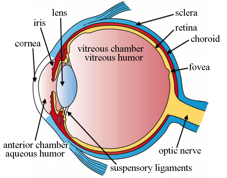
The human eye is an engineering marvel, a sophisticated biological camera that translates light into complex neural impulses. Yet, beyond the lenses and the retinas, the physical integrity of this delicate organ depends on a specific substance: the vitreous humor. This transparent, gelatinous mass fills the posterior segment of the eye, providing the necessary internal pressure to maintain the globe’s spherical shape. In the world of biotechnology and medical imaging software, understanding this substance is not just a physiological curiosity—it is a cornerstone of modern diagnostic tech and surgical robotics.
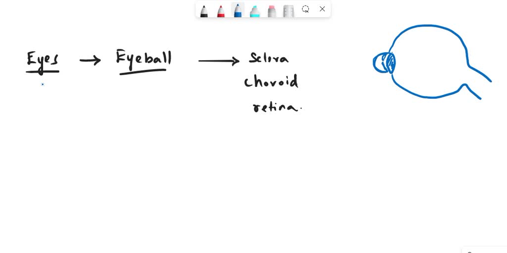
The Biomechanics of the Vitreous Humor
At its core, the vitreous humor is a hydrogel—a complex network of water, collagen fibers, and hyaluronic acid. While it appears simple, its structural properties are critical for the optical health of the eye. From a materials science perspective, the vitreous body acts as a shock absorber, protecting the delicate retinal tissue from mechanical stress while remaining perfectly clear to allow light transmission.
Composition and Structural Integrity
The vitreous is composed of approximately 99% water. The remaining 1% consists of a fine scaffold of collagen type II fibrils and glycosaminoglycans, primarily hyaluronic acid. These components work in tandem to create a viscous, gel-like state. This structure provides the “turgor pressure” required to hold the retina against the choroid, ensuring that the light-sensitive cells are positioned exactly where they need to be to capture focused images.
In digital modeling and simulation software used by ophthalmologists, the vitreous is often treated as a viscoelastic medium. Engineers developing next-generation intraocular surgical robots must account for this fluid density. If the vitreous is too viscous or experiences liquefaction, the entire mechanics of the eyeball change, potentially leading to retinal detachment—a condition that modern diagnostic AI tools are now tasked with identifying through high-resolution OCT (Optical Coherence Tomography) scans.
The Role of Vitreous Health in Diagnostic AI
The advancement of Artificial Intelligence in medical diagnostics has shifted the focus from human observation to algorithmic pattern recognition. The vitreous humor, being a clear medium, allows for unobstructed imaging of the posterior eye. However, changes in the vitreous—such as the formation of floaters or the progression of synchysis (the liquefaction of the gel)—can be subtle.
Machine Learning and Retinal Mapping
Modern AI diagnostic platforms utilize convolutional neural networks to map the relationship between the vitreous gel and the retinal surface. By training algorithms on millions of fundus images, developers have created software capable of detecting the early markers of vitreous detachment. This is particularly valuable in telemedicine, where remote clinicians may lack access to high-end slit-lamp microscopy.
The tech industry is currently pivoting toward “smart” contact lenses and wearable ocular monitors. These devices aim to measure intraocular pressure in real-time, relying on the fact that the vitreous mass transmits pressure fluctuations throughout the eye. By digitizing these physical properties, software engineers are creating predictive models that warn patients of high-pressure states before permanent damage occurs.
Surgical Robotics and the Vitreous Interface
One of the most complex challenges in robotic surgery is performing procedures within the posterior segment of the eye. The vitreous humor represents a unique obstacle: it is a physical medium that must be navigated, manipulated, or in some cases, replaced during vitrectomy procedures.
Precision Engineering in Ophthalmology
In a standard vitrectomy, surgeons must remove the vitreous to gain access to the retina. The specialized software controlling the robotic arms used in these surgeries must compensate for the gel’s resistance. If the robotic software does not account for the density of the vitreous, the tool movement could cause unintended traction on the retina.
The evolution of these robotic interfaces relies heavily on haptic feedback sensors. As the surgeon operates remotely, the robotic system converts the physical resistance of the vitreous gel into digital data, which is then fed back to the surgeon’s control interface. This “digital twin” approach allows for microscopic movements that would be impossible for the human hand to perform with equal stability, significantly improving outcomes for patients with diabetic retinopathy and severe ocular trauma.
Future Tech: Synthetic Vitreous and Digital Optimization
As we look toward the future of biotechnology, the focus is moving from repairing the eye to engineering solutions that mimic or supplement the natural vitreous. This intersection of material science and digital technology is paving the way for synthetic hydrogels that are more resilient than natural biology.
Simulation-Driven Innovation
The development of synthetic vitreous substitutes involves extensive digital simulation. Before a new polymer can be tested in vivo, it undergoes rigorous stress-test modeling. Software platforms simulate how these substances will interact with the lens, the retina, and the light entering the eye. By modeling the refractive index and the viscoelastic properties in a virtual environment, researchers can fine-tune the chemical composition of these substances to perfectly match the original biological mass.
Furthermore, the rise of cloud-based medical databases allows for the aggregation of “vitreous profiles” across different demographics. By analyzing how the vitreous gel ages across various populations using big data analytics, tech firms can predict when an individual is likely to experience vitreous-related vision degradation. This shift toward predictive ocular health relies on the seamless integration of hardware (sensors) and software (analytical engines).

The Convergence of Optical and Digital Systems
The maintenance of the eyeball’s shape via the vitreous is not just a biological fact—it is a design parameter for everything from augmented reality (AR) headset lenses to advanced telescopic camera arrays. The vitreous gel provides the perfect geometry for light to converge, a principle that optical engineers have studied for decades to improve the resolution of digital displays.
As we integrate more digital devices into our daily lives, the strain on our ocular physiology becomes more apparent. Technology is now tasked with monitoring the health of the vitreous as part of a holistic wellness ecosystem. From AI apps that measure eye fatigue to sophisticated surgical systems that preserve the integrity of the vitreous chamber, the study of this gelatinous mass remains at the cutting edge of technological advancement.
In summary, the vitreous humor is more than just a biological filler. It is a critical component of the ocular system that sustains the structural integrity necessary for vision. Through the lens of modern technology, we are learning that the better we understand the physics of this gel, the more accurately we can diagnose disease, develop advanced surgical tools, and innovate the next generation of ocular health solutions. As software continues to evolve, the digital representation of the eye’s internal environment will undoubtedly lead to breakthroughs that preserve human sight in increasingly complex environments.
aViewFromTheCave is a participant in the Amazon Services LLC Associates Program, an affiliate advertising program designed to provide a means for sites to earn advertising fees by advertising and linking to Amazon.com. Amazon, the Amazon logo, AmazonSupply, and the AmazonSupply logo are trademarks of Amazon.com, Inc. or its affiliates. As an Amazon Associate we earn affiliate commissions from qualifying purchases.