The eyepiece, often called the ocular lens, is the part of a microscope that you look through. While it might seem like a simple window into the microscopic world, its function is far more complex and critical to the overall magnification and viewing experience. In the realm of technology, understanding the nuances of optical instruments like microscopes is fundamental. The eyepiece is not merely a passive component; it actively contributes to image formation and can significantly impact the quality and detail that a user can perceive. Its design and specifications are a testament to optical engineering, playing a pivotal role in translating the magnified image from the objective lens into a viewable form for the human eye or a digital sensor.
The Eyepiece’s Role in Magnification and Image Relay
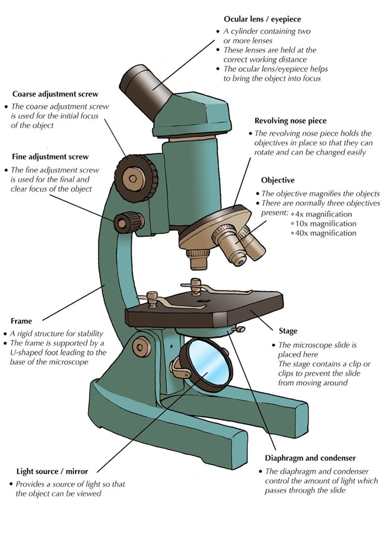
The primary function of the eyepiece is to further magnify the image that has already been enlarged by the objective lens. This two-stage magnification process is fundamental to achieving the high levels of detail required in scientific observation. Understanding this interplay is crucial for anyone utilizing optical technologies, from students learning basic microscopy to researchers pushing the boundaries of nanotechnology. The eyepiece acts as a secondary magnifying system, effectively enlarging the intermediate image formed by the objective lens. Without the eyepiece, the intermediate image, while magnified, would be too small and often not in a suitable format for direct viewing.
Magnification: The Primary Purpose
The total magnification of a microscope is calculated by multiplying the magnification of the objective lens by the magnification of the eyepiece. For example, if a microscope has a 40x objective lens and a 10x eyepiece, the total magnification is 400x. This simple equation highlights the direct and significant contribution of the eyepiece to the overall power of the microscope. Different eyepieces offer varying magnification levels, typically ranging from 5x to 30x, allowing users to select the appropriate magnification for their specific observation needs. The selection of an eyepiece is not arbitrary; it’s a deliberate choice that influences the level of detail resolvable, impacting diagnostic accuracy in pathology, identification precision in materials science, and discovery in biological research.
Image Relay: Bridging the Gap
The eyepiece’s role extends beyond simple magnification. It acts as an “image relay” system, taking the real, inverted, and magnified image produced by the objective lens and transforming it into a virtual, erect image that the observer’s eye can comfortably focus on. This is a sophisticated optical feat, involving precisely ground lenses within the eyepiece assembly. This internal lens system is designed to present the image at a comfortable viewing distance and to correct for aberrations introduced by the objective lens, ensuring a clear and distortion-free view. The quality of these internal lenses directly impacts the sharpness and clarity of the final image. Modern eyepieces, especially those integrated into advanced digital microscopy systems, can also incorporate features like field flatteners, which ensure that the entire field of view is in focus, not just the center.
Optical Design and Construction of Eyepieces
The internal construction of an eyepiece is a marvel of optical engineering. It typically consists of a system of two or more lenses, carefully designed and arranged to achieve the desired magnification and image correction. The materials used, the curvature of the lenses, and their spacing all play a crucial role in the performance of the eyepiece.
Lens Systems and Aberration Correction
Eyepieces are not monolithic. They are comprised of a “field lens” and an “eye lens,” or more complex multi-lens configurations. The field lens collects the light from the objective and directs it into the eye lens, which then magnifies the intermediate image. Each lens element is crafted from specific types of optical glass and precisely shaped to minimize optical aberrations. Common aberrations include chromatic aberration (where different colors of light are not focused at the same point) and spherical aberration (where light rays striking the edge of a lens are not focused at the same point as rays striking the center). Advanced eyepiece designs incorporate apochromatic or achromatic lens elements to significantly reduce these distortions, resulting in sharper, more color-accurate images. The design choices made in the eyepiece directly influence the “resolution” and “clarity” of the final image.
Field of View and Eye Relief
Two critical specifications for eyepieces are the “field of view” (FOV) and “eye relief.” The field of view refers to the diameter of the circular area that can be seen through the microscope at a given magnification. A wider field of view allows the user to see more of the specimen at once, which can be beneficial for locating specific areas of interest or for observing the spatial relationships between different structures. Eye relief is the distance between the eyepiece lens and the point where the eye can comfortably view the entire field of view without obstruction. This is particularly important for users who wear glasses, as a longer eye relief allows them to keep their glasses on while still seeing the full image. Eyepieces with generous eye relief are often described as “long eye relief eyepieces” and are preferred for extended viewing sessions or by individuals who require corrective lenses.
Advanced Eyepiece Technologies and Digital Integration
In today’s technologically driven world, the humble eyepiece is evolving. While traditional eyepieces remain vital for many applications, new innovations are integrating digital capabilities and enhancing the user experience in ways previously unimaginable. This evolution is key to the advancement of optical technologies and their applications across various scientific and industrial fields.
Digital Eyepieces and Camera Integration
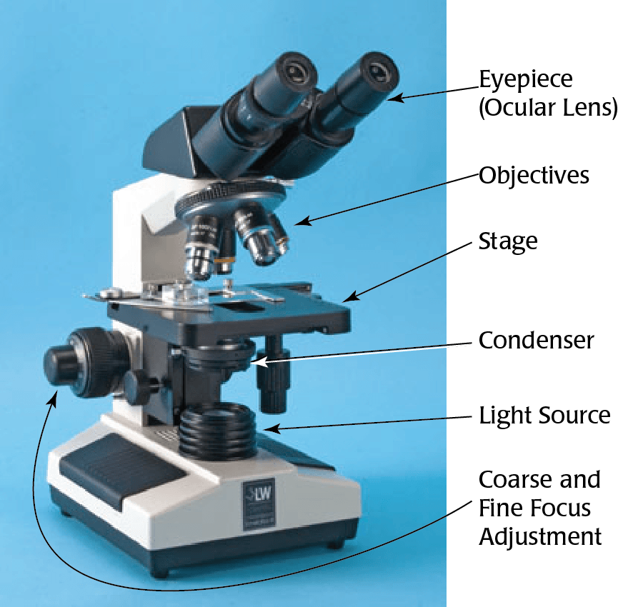
The advent of digital cameras has revolutionized microscopy. “Digital eyepieces” are essentially small cameras that can be inserted into the eyepiece tube of a microscope. These devices capture the magnified image and transmit it to a computer screen or other display device. This allows for easy sharing of images, video recording, and the integration of advanced image analysis software. These cameras often replace the traditional eyepiece entirely, providing a direct digital output. Furthermore, many microscopes now feature trinocular heads, which have a dedicated port for attaching a camera, allowing simultaneous viewing through the eyepieces and digital capture. This seamless integration of imaging hardware with optical systems is a hallmark of modern technological development in microscopy.
Reticles and Measurement Capabilities
Many eyepieces can be fitted with “reticles” or “graticules.” These are small glass discs etched with precise scales or patterns that are superimposed onto the field of view. Reticles are invaluable for quantitative measurements. For instance, a reticle with a millimeter scale can be used to measure the size of microscopic objects. Calibration is essential for reticles; they must be precisely matched to the magnification of the objective lens and the eyepiece to ensure accurate measurements. In scientific and industrial contexts, the ability to accurately measure microscopic features is paramount for quality control, research, and development. Specialized reticles can also be used for counting cells, assessing particle distribution, or identifying specific features within a sample. The precision offered by reticles, when properly calibrated, underscores the role of the eyepiece in delivering not just a magnified image, but also actionable data.
The Eyepiece’s Impact on User Experience and Ergonomics
Beyond its purely optical functions, the eyepiece significantly influences how a user interacts with the microscope and the overall comfort of the viewing experience. Ergonomics and intuitive design are increasingly important in the development of scientific instruments.
Viewing Comfort and Eyepiece Design
The design of the eyepiece, particularly its barrel diameter and the shape of the eye lens, contributes to viewing comfort. Eyepieces are typically available in standard diameters, such as 23.2mm, 30mm, or 30.5mm, which dictate their compatibility with different microscope models. The “eyecup,” a rubber or plastic attachment that fits over the eyepiece, can further enhance comfort by blocking ambient light and providing a softer surface for the eye. For prolonged microscopy sessions, the comfort afforded by a well-designed eyepiece can make a significant difference in a user’s ability to maintain focus and concentration, thus improving their productivity and the quality of their observations.

Interfacing with the User and Digital Outputs
The eyepiece is the primary interface between the observer and the microscopic world. Its design dictates the ease with which a user can bring the specimen into focus and scan the field of view. In advanced digital microscopy, the eyepiece can be a gateway to a wealth of information. As discussed with digital eyepieces, the user is no longer limited to just seeing; they can interact with the image on a screen, manipulate it, annotate it, and save it for future analysis. This shift from a purely visual experience to an interactive digital one highlights the evolving role of the eyepiece in modern scientific and technological workflows. The continued development of eyepieces aims to make microscopy more accessible, efficient, and informative, bridging the gap between the microscopic realm and the digital age.
aViewFromTheCave is a participant in the Amazon Services LLC Associates Program, an affiliate advertising program designed to provide a means for sites to earn advertising fees by advertising and linking to Amazon.com. Amazon, the Amazon logo, AmazonSupply, and the AmazonSupply logo are trademarks of Amazon.com, Inc. or its affiliates. As an Amazon Associate we earn affiliate commissions from qualifying purchases.